Herpes keratitis in the absence of anterograde transport of virus from sensory ganglia to the cornea
- PMID: 16055558
- PMCID: PMC1183562
- DOI: 10.1073/pnas.0503230102
Herpes keratitis in the absence of anterograde transport of virus from sensory ganglia to the cornea
Abstract
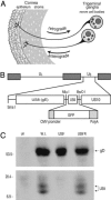
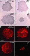
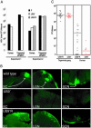
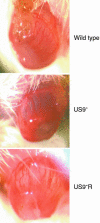
Herpes stromal keratitis is an immunopathologic disease in the corneal stroma leading to scarring, opacity, and blindness, and it is an important problem in common corneal surgeries. Paradoxically, virus antigens are largely focused in the epithelial layer of the cornea and not in the stromal layer, and viral antigens are eliminated before stromal inflammation develops. It is not clear what drives inflammation, whether viral antigens are necessary, or how viral antigens reach the stroma. It has been proposed that herpes simplex virus (HSV) travels from the corneal epithelium to sensory ganglia then returns to the stroma to cause disease. However, there is also evidence of HSV DNA and infectious virus persistent in corneas, and HSV can be transmitted to transplant recipients. To determine whether HSV resident in the cornea could cause herpes stromal keratitis, we constructed an HSV US9- mutant that had diminished capacity to move in neuronal axons. US9- HSV replicated and spread normally in the mouse corneal epithelium and to the trigeminal ganglia. However, US9- HSV was unable to return from ganglia to the cornea and failed to cause periocular skin disease, which requires zosteriform spread from neurons. Nevertheless, US9- HSV caused keratitis. Therefore, herpes keratitis can occur without anterograde transport from ganglia to the cornea, probably mediated by virus persistent in the cornea.
Figures


Similar articles
-
A Central Role for Sympathetic Nerves in Herpes Stromal Keratitis in Mice.Invest Ophthalmol Vis Sci. 2016 Apr;57(4):1749-56. doi: 10.1167/iovs.16-19183. Invest Ophthalmol Vis Sci. 2016. PMID: 27070108 Free PMC article.
-
Expression of herpes virus entry mediator (HVEM) in the cornea and trigeminal ganglia of normal and HSV-1 infected mice.Curr Eye Res. 2009 Oct;34(10):896-904. doi: 10.3109/02713680903184250. Curr Eye Res. 2009. PMID: 19895317
-
A replication competent HSV-1(McKrae) with a mutation in the amino-terminus of glycoprotein K (gK) is unable to infect mouse trigeminal ganglia after cornea infection.Curr Eye Res. 2014 Jun;39(6):596-603. doi: 10.3109/02713683.2013.855238. Epub 2014 Jan 8. Curr Eye Res. 2014. PMID: 24401006
-
Herpes simplex epithelial and stromal keratitis: an epidemiologic update.Surv Ophthalmol. 2012 Sep;57(5):448-62. doi: 10.1016/j.survophthal.2012.01.005. Epub 2012 Apr 28. Surv Ophthalmol. 2012. PMID: 22542912 Free PMC article. Review.
-
Management of herpes simplex virus stromal keratitis: an evidence-based review.Surv Ophthalmol. 2009 Mar-Apr;54(2):226-34. doi: 10.1016/j.survophthal.2008.12.004. Surv Ophthalmol. 2009. PMID: 19298901 Review.
Cited by
-
Characterization of the Herpes Simplex Virus (HSV) Tegument Proteins That Bind to gE/gI and US9, Which Promote Assembly of HSV and Transport into Neuronal Axons.J Virol. 2020 Nov 9;94(23):e01113-20. doi: 10.1128/JVI.01113-20. Print 2020 Nov 9. J Virol. 2020. PMID: 32938770 Free PMC article.
-
Microtubule-Dependent Trafficking of Alphaherpesviruses in the Nervous System: The Ins and Outs.Viruses. 2019 Dec 17;11(12):1165. doi: 10.3390/v11121165. Viruses. 2019. PMID: 31861082 Free PMC article. Review.
-
Pseudorabies Virus Fast Axonal Transport Occurs by a pUS9-Independent Mechanism.J Virol. 2015 Aug;89(15):8088-91. doi: 10.1128/JVI.00771-15. Epub 2015 May 20. J Virol. 2015. PMID: 25995254 Free PMC article.
-
Directional transneuronal spread of α-herpesvirus infection.Future Virol. 2009 Nov 1;4(6):591. doi: 10.2217/fvl.09.62. Future Virol. 2009. PMID: 20161665 Free PMC article.
-
A single-amino-acid substitution in herpes simplex virus 1 envelope glycoprotein B at a site required for binding to the paired immunoglobulin-like type 2 receptor alpha (PILRalpha) abrogates PILRalpha-dependent viral entry and reduces pathogenesis.J Virol. 2010 Oct;84(20):10773-83. doi: 10.1128/JVI.01166-10. Epub 2010 Aug 4. J Virol. 2010. PMID: 20686018 Free PMC article.
References
-
- Pepose, J. S., Leib, D. A., Stuart, P. M. & Easty, D. L. (1996) in Ocular Infection and Immunity, eds., Pepose, J. S., Holland, G. & Wilhelmus, K. (Mosby, St. Louis), pp. 905-932.
-
- Streilein, J. W., Dana, M. R. & Ksander, B. R. (1997) Immunol. Today 18, 443-449. - PubMed
-
- Hendricks, R. L. (1999) Chem. Immunol. 73, 120-136. - PubMed
-
- Deshpande, S. P., Zheng, M., Lee, S. & Rouse, B. T. (2002) Vet. Microbiol. 86, 17-26. - PubMed
-
- Zhao, Z. S., Granucci, F., Yeh, L., Schaffer, P. A. & Cantor, H. (1998) Science 279, 1344-1347. - PubMed
Publication types
MeSH terms
Substances
Grants and funding
LinkOut - more resources
Full Text Sources
Other Literature Sources

