Human {beta}-defensin 2 is expressed and associated with Mycobacterium tuberculosis during infection of human alveolar epithelial cells
- PMID: 16040961
- PMCID: PMC1201238
- DOI: 10.1128/IAI.73.8.4505-4511.2005
Human {beta}-defensin 2 is expressed and associated with Mycobacterium tuberculosis during infection of human alveolar epithelial cells
Abstract
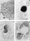
To determine the role of human beta-defensin 2 (HBD-2) in human tuberculosis, we studied the in vitro induction of HBD-2 gene expression by Mycobacterium tuberculosis H37Rv infection in the human lung epithelial cell line A549, in alveolar macrophages (AM), and in blood monocytes (MN) by reverse transcription-PCR. We also studied the induction of HBD-2 gene expression by mannose lipoarabinomannan (manLAM) from M. tuberculosis. Intracellular production of HBD-2 peptide was detected by immunocytochemistry and electron microscopy. Our results demonstrated that there was induction of HBD-2 mRNA in A549 cells after infection with M. tuberculosis at various multiplicities of infection (MOI) and that there was stimulation with manLAM. AM expressed the HBD-2 gene only at a high MOI with M. tuberculosis. MN did not express HBD-2 at any of the experimental M. tuberculosis MOI. Immunostaining revealed the presence of intracellular HBD-2 peptide in A549 cells following infection with M. tuberculosis, and the staining was more intense in areas where there were M. tuberculosis clusters. By using electron microscopy we also demonstrated production of HBD-2 after M. tuberculosis infection and adherence of HBD-2 to the membranes of M. tuberculosis. Alveolar epithelial cells are among the first cells to encounter M. tuberculosis following aerogenic infection. As HBD-2 has been shown to control growth of M. tuberculosis and has chemotactic activity, our results suggest that HBD-2 induction by M. tuberculosis may have a role in the pathogenesis of human tuberculosis.
Figures


Similar articles
-
Air pollution particulate matter alters antimycobacterial respiratory epithelium innate immunity.Infect Immun. 2015 Jun;83(6):2507-17. doi: 10.1128/IAI.03018-14. Epub 2015 Apr 6. Infect Immun. 2015. PMID: 25847963 Free PMC article.
-
The role and regulation of Moraxella catarrhalis-induced human beta-defensin 3 expression in human pulmonary epithelial cells.Biochem Biophys Res Commun. 2015 Nov 6;467(1):46-52. doi: 10.1016/j.bbrc.2015.09.126. Epub 2015 Sep 28. Biochem Biophys Res Commun. 2015. PMID: 26417692
-
Modulation of human beta-defensin-2 transcription in pulmonary epithelial cells by lipopolysaccharide-stimulated mononuclear phagocytes via proinflammatory cytokine production.J Immunol. 2003 Apr 15;170(8):4226-36. doi: 10.4049/jimmunol.170.8.4226. J Immunol. 2003. PMID: 12682256
-
Induction of nitric oxide release from the human alveolar epithelial cell line A549: an in vitro correlate of innate immune response to Mycobacterium tuberculosis.Immunology. 2004 Jul;112(3):471-80. doi: 10.1046/j.1365-2567.2004.01905.x. Immunology. 2004. PMID: 15196216 Free PMC article.
-
Vitamin D induces interleukin-1β expression: paracrine macrophage epithelial signaling controls M. tuberculosis infection.PLoS Pathog. 2013;9(6):e1003407. doi: 10.1371/journal.ppat.1003407. Epub 2013 Jun 6. PLoS Pathog. 2013. PMID: 23762029 Free PMC article. Clinical Trial.
Cited by
-
Air pollution particulate matter alters antimycobacterial respiratory epithelium innate immunity.Infect Immun. 2015 Jun;83(6):2507-17. doi: 10.1128/IAI.03018-14. Epub 2015 Apr 6. Infect Immun. 2015. PMID: 25847963 Free PMC article.
-
Potentials of Host-Directed Therapies in Tuberculosis Management.J Clin Med. 2019 Aug 3;8(8):1166. doi: 10.3390/jcm8081166. J Clin Med. 2019. PMID: 31382631 Free PMC article. Review.
-
1,25-dihydroxyvitamin D3 induces LL-37 and HBD-2 production in keratinocytes from diabetic foot ulcers promoting wound healing: an in vitro model.PLoS One. 2014 Oct 22;9(10):e111355. doi: 10.1371/journal.pone.0111355. eCollection 2014. PLoS One. 2014. PMID: 25337708 Free PMC article.
-
Expression of cathelicidin LL-37 during Mycobacterium tuberculosis infection in human alveolar macrophages, monocytes, neutrophils, and epithelial cells.Infect Immun. 2008 Mar;76(3):935-41. doi: 10.1128/IAI.01218-07. Epub 2007 Dec 26. Infect Immun. 2008. PMID: 18160480 Free PMC article.
-
Antimicrobial Peptides and Physical Activity: A Great Hope against COVID 19.Microorganisms. 2021 Jun 30;9(7):1415. doi: 10.3390/microorganisms9071415. Microorganisms. 2021. PMID: 34209064 Free PMC article. Review.
References
-
- Ashitani, J., H. Mukae, T. Hiratsuka, M. Nakazato, K. Kumamoto, and S. Matsukura 2001. Plasma and BAL fluid concentrations of antimicrobial peptides in patients with Mycobacterium avium-intracellulare infection. Chest 119:1131-1137. - PubMed
-
- Birchler, T., R. Seibl, K. Buchner, S. Loeliger, R. Seger, J. P. Hossle, A. Aguzzi, and R. P. Lauener 2001. Human Toll-like receptor 2 mediates induction of the antimicrobial peptide human beta-defensin 2 in response to bacterial lipoprotein. Eur. J. Immunol. 31:3131-3137. - PubMed
Publication types
MeSH terms
Substances
Grants and funding
LinkOut - more resources
Full Text Sources
Research Materials

