. 2005 Jul 18;170(2):317-25.
doi: 10.1083/jcb.200503059.

Actin- and myosin-driven movement of viruses along filopodia precedes their entry into cells

Affiliations

Actin- and myosin-driven movement of viruses along filopodia precedes their entry into cells

Maik J Lehmann et al. J Cell Biol. .

Abstract

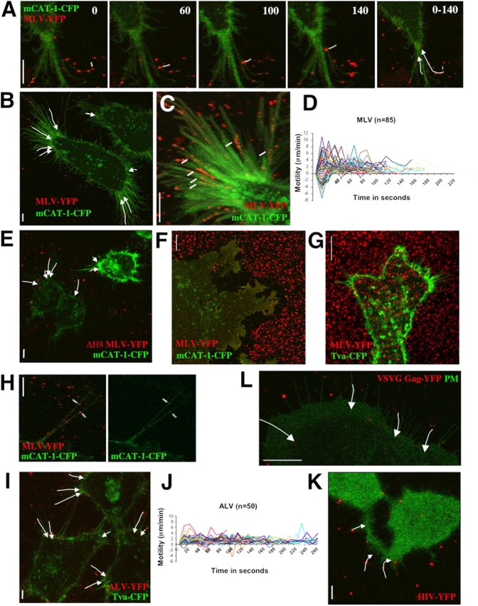
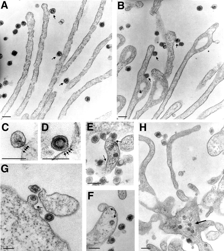
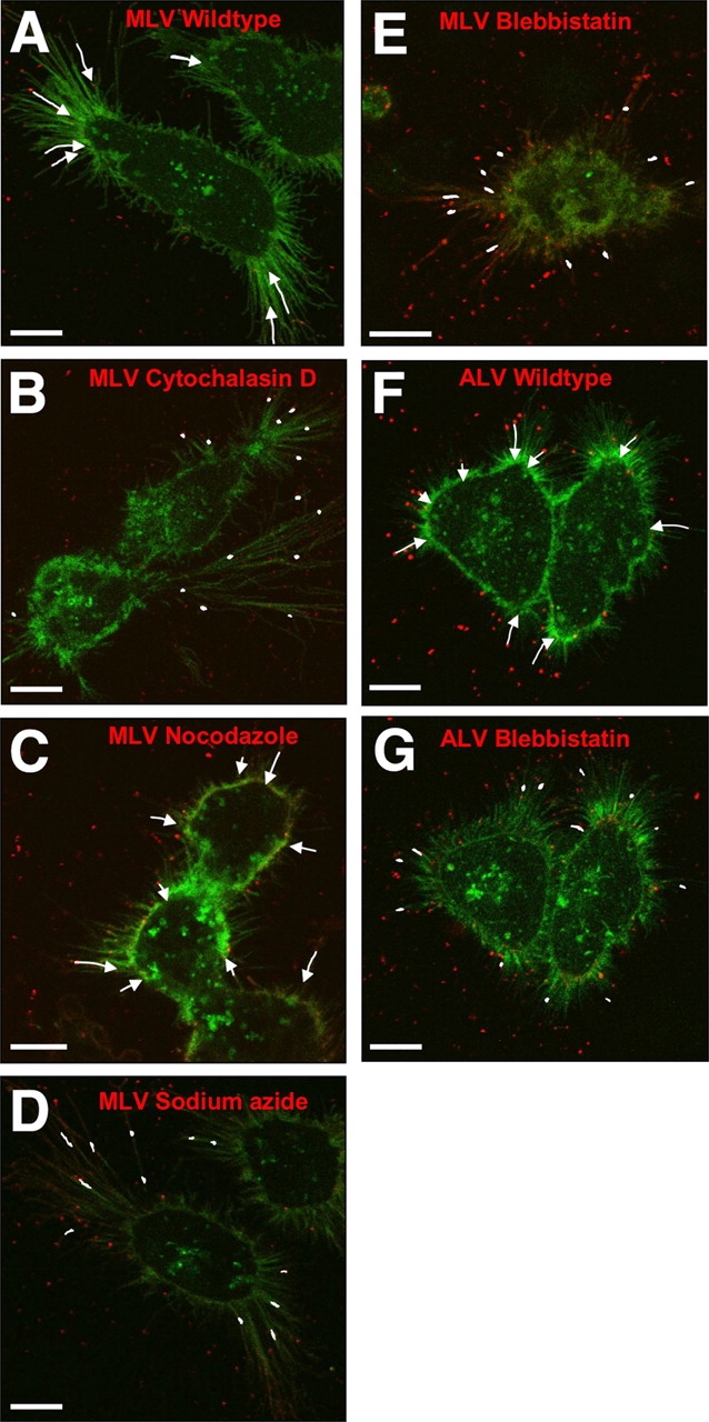
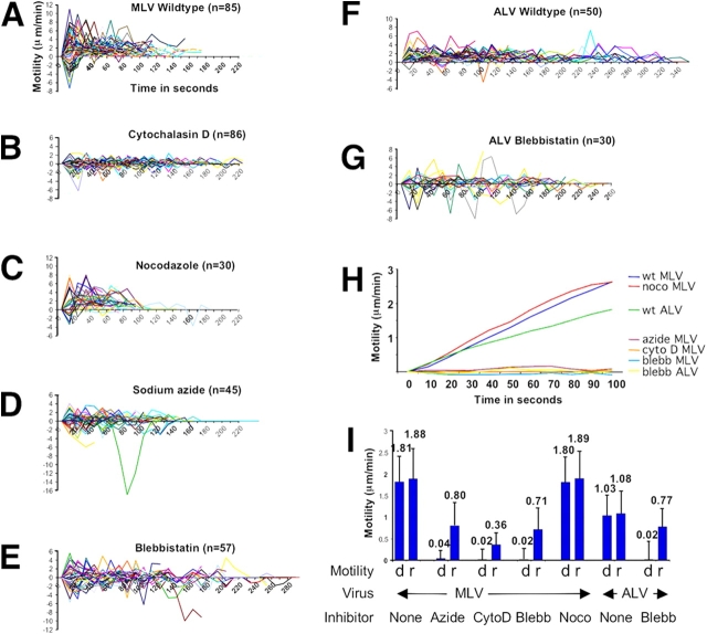
Viruses have often been observed in association with the dense microvilli of polarized epithelia as well as the filopodia of nonpolarized cells, yet whether interactions with these structures contribute to infection has remained unknown. Here we show that virus binding to filopodia induces a rapid and highly ordered lateral movement, "surfing" toward the cell body before cell entry. Virus cell surfing along filopodia is mediated by the underlying actin cytoskeleton and depends on functional myosin II. Any disruption of virus cell surfing significantly reduces viral infection. Our results reveal another example of viruses hijacking host machineries for efficient infection by using the inherent ability of filopodia to transport ligands to the cell body.


Figures

Figure 1.
Figure 1.
MLV is associated with filopodia. HEK 293 cells expressing receptor mCAT-1 were incubated with infectious MLV for 10 min and then prepared for analysis by SEM. Arrows indicate virions associated with filopodia. Bars, 1 μm.
Figure 2.
Figure 2.
Virus cell surfing along filopodia. (A) An individual MLV particle fluorescently labeled with envelope–YFP (MLV–YFP, pseudocolored in red, marked in white) surfs along the filopodium of a HEK 293 cell transduced with mCAT-1–CFP (pseudocolored in green). The time in seconds is presented in each frame. The overall movements of the two particles are summarized by arrows in the far right panel (0–140 s). (B) The image summarizes the overall movement of selected particles on filopodia of a single HEK 293 cell as observed in Video 1. (C) To visualize the gradual loss of fluorescence of moving particles (marked in white), 31 frames from a region of interest of Video 1 were superimposed. (D) To quantify virus cell surfing, the motility (μm/min) of 85 individual MLV particles was plotted over time (in seconds), with time point 0 representing the moment of virus attachment to a filopodium. Positive values of motility represent particles moving toward the cell body whereas negative numbers represent viruses moving toward the tip of the fiber. (E) MLV bearing a fusion-defective envelope protein also surfs. The image, generated as in B, visually summarizes a time-lapse video not shown. (F) Virus capture by short-lived filopodia and ruffles. HEK 293 cells expressing mCAT-1–CFP were allowed to spread for 30 min on a glass coverslip containing prebound MLV–YFP. Cells formed a large syncytium as a consequence of viral infection. (G) Virus capture is receptor dependent. An experiment as in F was performed with MLV–YFP and HEK 293 cells expressing Tva–CFP, the receptor for the ALV. Note that cells are unable to take up prebound virus from beneath the cell and its surroundings. (H) Receptor mCAT-1–CFP (green) is recruited to MLV particles labeled with Env–YFP (red) when they attach to filopodia of XC cells. (I) ALV surfs on HEK 293 cells expressing Tva–CFP. The image visualizes the movement of individual particles as shown in Video 4. (J) A quantitative analysis as in D was performed for 50 ALV particles. (K) HIV (red) surfs on HEK 293 cells expressing CD4 and CXCR4. To identify receptor-expressing cells, cytoplasmic CFP (green) was coexpressed. The image summarizes the movement of three particles as shown in Video 5. (L) MLV Gag–YFP-labeled virions containing the VSVG protein (red) surf along filopodia of XC cells expressing mCAT-1–CFP (green) as a plasma membrane marker. The entire sequence is shown in Video 6. In all panels, vertical size bars correspond to 5 μm.
Figure 3.
Figure 3.
MLV fuses with the plasma membrane at the base of filopodia or at the cell body. (A and B) HEK 293 cells infected with MLV for 30 min were prepared for TEM and cut en face within 100 nm from the glass coverslip to visualize viruses and filopodia. Circular nodules as observed in B are indicative of retraction fibers (Mitchison, 1992). Viruses engaged with filopodia or retraction fibers are marked with arrows. (C and D) Two examples of frequently observed spikes (marked by arrows) between virus and membrane are shown. (E–G) Fusion sites (marked by arrows) were observed at the widening base of fibers or at the cell body. (H) Viral capsids accumulate inside the cytoplasm at the base of fibers. In all panels, size bars correspond to 200 nm.
Figure 4.
Figure 4.
Virus cell surfing is actin and myosin II dependent. (A–G) Images visually summarize the extent of MLV (A–E) and ALV (F and G) surfing in the absence and presence of inhibitors as indicated; cytochalasin D (50 μM), nocodazole (50 μM), sodium azide (10 mM), and blebbistatin (30 μM). The data are based on time-lapse movies, some of them provided as supplemental data (Videos 1, 4, and 9). To compare the surfing activity visually, the motility observed over 120 s (MLV) or 200 s (ALV) is displayed. Bars, 10 μm.
Figure 5.
Figure 5.
Quantitative analysis of the effects of inhibitors on virus cell surfing. (A) The motility (μm/min) of 85 individual MLV particles as presented in Fig. 2 D in the absence of any inhibitor (Wildtype) and plotted over time (in seconds). (B-E) An analysis as in (A) was performed in the presence of the inhibitors cytochalasin D (50 μM), nocodazole (50 μM), sodium azide (10 mM), and blebbistatin (30 μM), respectively. The number of particles (n) analyzed in each experiment is indicated. (F and G) Experiments as described in A and E, respectively, were analyzed for ALV movement on 293 cells expressing Tva–CFP. (H) Progressive average movement (μm) was determined using the mean motility for all analyzed particles at each given time point in the absence and presence of inhibitors as indicated. (I) The average directed motility toward the cell body (d, directed) was compared with the overall random motility (r, random) including forward as well as backward movement for all of the above experiments. To exclude the observed initial random motility seen in A, only the period between 30 and 90 s was included in the analysis. Cyto D, Blebb, and Noco are abbreviations for cytochalasin D, blebbistatin, and nocodazole, respectively.
Figure 6.
Figure 6.
MLV enters polarized epithelial cells via myosin II–dependent surfing along microvilli. (A) Individual steps of MLV infection of polarized MDCK cells were visualized by SEM in the presence and absence of blebbistatin as indicated. Arrows indicate virions associated with microvilli. Bars, 500 nm. (B) To analyze the nature of clusters seen in A TEM was performed revealing multivalent interactions between viruses and microvilli. Viruses similar in size to microvilli are recognized by their electron dense capsid core. (C) Parallel TEM of infected MDCK cells shows that at 30 min viruses are internalized. Bars: (B and C) 200 nm.

Similar articles

Cited by

References

    1. Abercrombie, M., J.E. Heaysman, and S.M. Pegrum. 1970. The locomotion of fibroblasts in culture. 3. Movements of particles on the dorsal surface of the leading lamella. Exp. Cell Res. 62:389–398. - PubMed
    1. Albrecht-Buehler, G., and R.M. Lancaster. 1976. A quantitative description of the extension and retraction of surface protrusions in spreading 3T3 mouse fibroblasts. J. Cell Biol. 71:370–382. - PMC - PubMed
    1. Albritton, L.M., L. Tseng, D. Scadden, and J.M. Cunningham. 1989. A putative murine ecotropic retrovirus receptor gene encodes a multiple membrane-spanning protein and confers susceptibility to virus infection. Cell. 57:659–666. - PubMed
    1. Bates, P., J.A. Young, and H.E. Varmus. 1993. A receptor for subgroup A Rous sarcoma virus is related to the low density lipoprotein receptor. Cell. 74:1043–1051. - PubMed
    1. Bomsel, M., and A. Alfsen. 2003. Entry of viruses through the epithelial barrier: pathogenic trickery. Nat. Rev. Mol. Cell Biol. 4:57–68. - PMC - PubMed

Publication types

MeSH terms