. 2005 Aug;79(15):9912-25.
doi: 10.1128/JVI.79.15.9912-9925.2005.

Kaposi's sarcoma-associated herpesvirus K-bZIP represses gene transcription via SUMO modification

Affiliations

Kaposi's sarcoma-associated herpesvirus K-bZIP represses gene transcription via SUMO modification

Yoshihiro Izumiya et al. J Virol. 2005 Aug.

Abstract

Kaposi's sarcoma-associated herpesvirus (KSHV) is a human gammaherpesvirus implicated in AIDS-related neoplasms. Previously, we demonstrated that the early lytic gene product K-bZIP is a transcriptional repressor that affects a subset of viral gene transcriptions mediated by the viral transactivator K-Rta (Y. Izumiya et al. J. Virol. 77:1441-1451, 2003). Sumoylation has emerged as an important posttranslational modification that affects the location and function of cellular and viral proteins and also plays a significant role in transcriptional repression along with Ubc9, the E2 SUMO conjugation enzyme. Here, we provide evidence that K-bZIP is sumoylated at the lysine 158 residue and associates with Ubc9 both in a cell-free system and in virus-infected BCBL-1 cells. Reporter assays showed that the expression of SUMO-specific protease 1 attenuated the transcriptional repression activity of K-bZIP. The expression of a K-bZIPK158R mutant, which was no longer sumoylated, exhibited the reduced transcriptional repression activity. This indicates that sumoylation plays an important part in the transcriptional repression activity of K-bZIP. Finally, chromatin immunoprecipitation experiments demonstrated that K-bZIP interacts with and recruits Ubc9 to specific KSHV promoters. Thus, our data indicate that K-bZIP is a SUMO adaptor, which recruits Ubc9 to specific viral target promoters, thereby exerting its transcriptional repression activity.


Figures

FIG. 1.
FIG. 1.
(A) SUMO conjugation in vitro. K-bZIP but not K-Rta is efficiently modified by SUMO. In vitro-translated (IVT)-K-bZIP was incubated with purified recombinant proteins E1 (SAE1/SAE2), E2 (Ubc9), and each activated form of SUMO protein (SUMO-1GG, SUMO-2GG, or SUMO-3GG). The reaction was carried out in the presence of the ATP regenerating system. Reactions without Ubc9 served as negative controls. (B) Mapping of SUMO-modified region. Deletion mutants of IVT-K-bZIP were incubated in the SUMO reaction. Basic region (amino acids 122 to 189) of K-bZIP is important for the sumoylation (*). (C) Sumoylation of K-bZIP in vivo. K-bZIP transfected into 293T cells displays a high-molecular-weight band when probed with anti-K-bZIP antibody, and which disappeared upon cotransfection with increasing amounts of SUMO-specific protease (SENP) but not an SENP mutant (SENPmut) lacking protease activity. (D) Sumoylated K-bZIP was detected in TREx-K-Rta BCBL-1 activated by K-Rta expression. Protein extract (100 μg/lane) was loaded. K-bZIP was detected with anti-K-bZIP antibody. KSHV negative B-cell line (GA10) was the negative control. An alternatively spliced K-bZIP (K-bZIPΔLZ) and sumoylated form of K-bZIP was also observed. Actin protein served as an internal control for the amount of protein on membrane and was detected by anti-actin goat serum. (E) K-bZIP is modified by both SUMO-1 and SUMO-2/3. BCBL-1 cells (1 mg) induced viral reactivation by K-Rta expression and lysed in SDS and were immunoprecipitated (IP) with 4 μg of preinoculated rabbit IgG (Pre) or anti-K-bZIP rabbit IgG (Kz). Immunoprecipitates were separated on a SDS-9% PAGE and immunoblotted (W.B.) with indicated antibodies. Anti-SUMO-3 polyclonal antibody detects both SUMO-2 and SUMO-3 proteins because of significant identity.
FIG. 2.
FIG. 2.
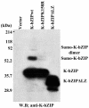
Identification of sumoylation site. K-bZIP wild type, K-bZIP K158R, or K-bZIPΔLZ was transfected into 293T cells. Protein extract (50 μg/lane) lysed in SDS was loaded. K-bZIP was detected with anti-K-bZIP antibody. W.B., Western blotting.
FIG. 3.
FIG. 3.
Localization of K-bZIP wt (a) and K-bZIPK158R (b) with PML in 293 cells. Immunofluorescence analysis was performed by using anti-K-bZIP rabbit serum and anti-PML mouse monoclonal antibody. K-bZIP (red) and PML (green) were detected with Alexa Fluor 555-conjugated anti-rabbit IgG and Alexa Fluor 488-conjugated anti-mouse IgG. Fluorescence images were collected separately and overlaid by a computer. The rightmost panel shows enlarged images of indicated cells. These panels are representative of 10 different fields. (magnification, ×600).
FIG. 4.
FIG. 4.
Determination of the stability of the K-bZIP wild type and the K-bZIPK158R mutant. (A) K-bZIP wild type and K-bZIPK158R were transfected into 293 cells and cells were labeled with [35S]methionine and [35S]cysteine for a 4-h pulse from 24 to 28 h posttransfection. Cells were chased with medium containing nonlabeled amino acids for the indicated time periods. K-bZIP proteins were immunoprecipitated with anti-K-bZIP antibody and subjected to SDS-PAGE followed by autoradiography. (B) Quantification of the labeled protein. Protein amount was quantified by PhosphorImager analysis of the dried SDS-PAGE gel with Quantity One (Bio-Rad). Black indicates the K-bZIP wild type, while gray refers to K-bZIPK158R.
FIG. 5.
FIG. 5.
(A) Repression via sumoylation. K-Rta activation of the ORF57 promoter (leftmost bar) was repressed by K-bZIP wild type but had less repression by K-bZIP K158R. Cotransfection with the SENP1 wild type but not an inactive mutant diminished the repression. 293 cells were cotransfected with K-Rta, ORF57 promoter, and the indicated plasmids. K-Rta and K-bZIP amounts were examined by immunoblotting with anti-K-Rta or anti-K-bZIP antibody. (B) Ubc9 dominant-negative relived K-bZIP repression. 293 cells were cotransfected with indicated plasmids. Protein amounts of K-Rta and K-bZIP were examined by immunoblotting with indicated antibodies.
FIG.6.
FIG.6.
Identification of the K-bZIP binding sites on the KSHV chromosome by the ChIP method. (A) Reactivation by K-Rta induction. K-bZIP expression was analyzed by immunofluorescence assay using anti-K-bZIP antibody (red). DNA was stained with TOPRO-3 (Molecular Probes; blue). (B) Schematic diagram of the KHSV genome. The five cosmids (GB11, GA29, Not33, Not39, and GA2) spanning the KSHV genome are indicated. (C) Scanning of K-bZIP binding sites. Cosmids were digested with the restriction enzymes and separated on agarose gel (0.8%). The gel was stained with ethidium bromide (EtBr; right panel) and Southern blotted with radiolabeled probes derived from ChIP before (Dox, 0 h) and after 48 h of KSHV reactivation (Dox 48 h). The DNA associated with K-bZIP chromatin was radiolabeled as described in Materials and Methods. (D) Schematic diagram of K-bZIP binding sites. Putative major K-bZIP binding sites are summarized in the diagram. (E) PCR verification of K-bZIP binding sites. PCR primers were designed for the K-bZIP associated regions. Other KSHV promoters and coding region served as controls (ORF36 promoter, PAN promoter, ORF57 coding region). For each primer set, a PCR with the total input DNA (In) before immunoprecipitation was carried out. ChIP fragments of preimmune serum (Pre) or anti-K-bZIP (Kz) after 48 h of KSHV reactivation were subjected to PCR analyses to confirm Southern blotting results.
FIG.6.
FIG.6.
Identification of the K-bZIP binding sites on the KSHV chromosome by the ChIP method. (A) Reactivation by K-Rta induction. K-bZIP expression was analyzed by immunofluorescence assay using anti-K-bZIP antibody (red). DNA was stained with TOPRO-3 (Molecular Probes; blue). (B) Schematic diagram of the KHSV genome. The five cosmids (GB11, GA29, Not33, Not39, and GA2) spanning the KSHV genome are indicated. (C) Scanning of K-bZIP binding sites. Cosmids were digested with the restriction enzymes and separated on agarose gel (0.8%). The gel was stained with ethidium bromide (EtBr; right panel) and Southern blotted with radiolabeled probes derived from ChIP before (Dox, 0 h) and after 48 h of KSHV reactivation (Dox 48 h). The DNA associated with K-bZIP chromatin was radiolabeled as described in Materials and Methods. (D) Schematic diagram of K-bZIP binding sites. Putative major K-bZIP binding sites are summarized in the diagram. (E) PCR verification of K-bZIP binding sites. PCR primers were designed for the K-bZIP associated regions. Other KSHV promoters and coding region served as controls (ORF36 promoter, PAN promoter, ORF57 coding region). For each primer set, a PCR with the total input DNA (In) before immunoprecipitation was carried out. ChIP fragments of preimmune serum (Pre) or anti-K-bZIP (Kz) after 48 h of KSHV reactivation were subjected to PCR analyses to confirm Southern blotting results.
FIG. 7.
FIG. 7.
K-bZIP association with Ubc9. K-bZIP expressed in BCBL-1 was coprecipitated with Ubc9 by Ubc9 antibodies. Anti-HA antibody was used as a negative control. (B) Association between K-bZIP and Ubc9 in cotransfected 293T cells. 293T cells were cotransfected with the indicated plasmids. Cell lysates were precipitated with Flag antibody-conjugated agarose, and coimmunoprecipitation of K-bZIP was detected by using anti-T7 tag antibody. The expression of T7-tagged K-bZIP in one-tenth of total cell lysates used in coimmunoprecipitation is shown in the same blot as a control. (C) K-bZIP associated with Ubc9 in vitro. GST-Ubc9 but not GST precipitates in vitro-translated (IVT)-K-bZIP. The inputs are one-tenth of the lysates used for the binding study.
FIG. 8.
FIG. 8.
Recruitment of Ubc9 to K-bZIP binding sites. ChIP assay was performed by using anti-Ubc9 antibody or rabbit normal serum (control serum). PCR showed recruitment of Ubc9 to K-bZIP-associated chromatin.

Similar articles

Cited by

References

    1. Adamson, A. L., and S. Kenney. 2001. Epstein-barr virus immediate-early protein BZLF1 is SUMO-1 modified and disrupts promyelocytic leukemia bodies. J. Virol. 75:2388-2399. - PMC - PubMed
    1. Adamson, A. L., and S. C. Kenney. 1998. Rescue of the Epstein-Barr virus BZLF1 mutant, Z(S186A), early gene activation defect by the BRLF1 gene product. Virology 251:187-197. - PubMed
    1. Ahn, J.-H., Y. Xu, W. J. Jang, M. J. Matunis, and G. S. Hayward. 2001. Evaluation of interactions of human cytomegalovirus immediate-early IE2 regulatory protein with small ubiquitin-like modifiers and their conjugation enzyme Ubc9. J. Virol. 75:3859-3872. - PMC - PubMed
    1. AuCoin, D. P., K. S. Colletti, S. A. Cei, I. Papouskova, M. Tarrant, and G. S. Pari. 2003. Amplification of the Kaposi's sarcoma-associated herpesvirus/human herpesvirus 8 lytic origin of DNA replication is dependent upon a cis-acting AT-rich region and an ORF50 response element and the trans-acting factors ORF50 (K-Rta) and K8 (K-bZIP). Virology 318:542-555. - PubMed
    1. Chang, L. K., Y. H. Lee, T. S. Cheng, Y. R. Hong, P. J. Lu, J. J. Wang, W. H. Wang, C. W. Kuo, S. S. Li, and S. T. Liu. 2004. Post-translational modification of Rta of Epstein-Barr virus by SUMO-1. J. Biol. Chem. 279:38803-38812. - PubMed

Publication types

LinkOut - more resources