Lamellipodial contractions during crawling and spreading
- PMID: 16006627
- PMCID: PMC1366668
- DOI: 10.1529/biophysj.105.066720
Lamellipodial contractions during crawling and spreading
Erratum in
- Biophys J. 2006 Jan 15;90(2):708
Abstract
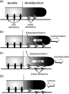
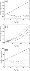
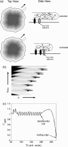
Most eukaryotic cells can crawl over surfaces. In general, this motility requires three distinct actions: polymerization at the leading edge, adhesion to the substrate, and retraction at the rear. Recent experiments with mouse embryonic fibroblasts showed that during spreading and crawling the lamellipodium undergoes periodic contractions that are substrate-dependent. Here I show that a simple model incorporating stick-slip adhesion and a simplified mechanism for the generation of contractile forces is sufficient to explain periodic lamellipodial contractions. This model also explains why treatment of cells with latrunculin modifies the period of these contractions. In addition, by coupling a diffusing chemical species that can bind actin, such as myosin light-chain kinase, with the contractile model leads to periodic rows and waves in the chemical species, similar to what is observed in experiments. This model provides a novel and simple explanation for the generation of contractile waves during cell spreading and crawling that is only dependent on stick-slip adhesion and the generation of contractile force and suggests new experiments to test this mechanism.
Figures


Similar articles
-
Stick-slip model for actin-driven cell protrusions, cell polarization, and crawling.Proc Natl Acad Sci U S A. 2020 Oct 6;117(40):24670-24678. doi: 10.1073/pnas.2011785117. Epub 2020 Sep 21. Proc Natl Acad Sci U S A. 2020. PMID: 32958682 Free PMC article.
-
Periodic lamellipodial contractions correlate with rearward actin waves.Cell. 2004 Feb 6;116(3):431-43. doi: 10.1016/s0092-8674(04)00058-3. Cell. 2004. PMID: 15016377
-
Actin disassembly clock determines shape and speed of lamellipodial fragments.Proc Natl Acad Sci U S A. 2011 Dec 20;108(51):20394-9. doi: 10.1073/pnas.1105333108. Epub 2011 Dec 9. Proc Natl Acad Sci U S A. 2011. PMID: 22159033 Free PMC article.
-
How nematode sperm crawl.J Cell Sci. 2002 Jan 15;115(Pt 2):367-84. doi: 10.1242/jcs.115.2.367. J Cell Sci. 2002. PMID: 11839788 Review.
-
The comings and goings of actin: coupling protrusion and retraction in cell motility.Curr Opin Cell Biol. 2005 Oct;17(5):517-23. doi: 10.1016/j.ceb.2005.08.004. Curr Opin Cell Biol. 2005. PMID: 16099152 Review.
Cited by
-
Tissue Augmentation in Wound Healing: the Role of Endothelial and Epithelial Cells.Med Arch. 2018 Dec;72(6):444-448. doi: 10.5455/medarh.2018.72.444-448. Med Arch. 2018. PMID: 30814778 Free PMC article. Review.
-
Mechanosensitive Adhesion Explains Stepping Motility in Amoeboid Cells.Biophys J. 2017 Jun 20;112(12):2672-2682. doi: 10.1016/j.bpj.2017.04.033. Biophys J. 2017. PMID: 28636923 Free PMC article.
-
Stick-slip model for actin-driven cell protrusions, cell polarization, and crawling.Proc Natl Acad Sci U S A. 2020 Oct 6;117(40):24670-24678. doi: 10.1073/pnas.2011785117. Epub 2020 Sep 21. Proc Natl Acad Sci U S A. 2020. PMID: 32958682 Free PMC article.
-
The Moving Boundary Node Method: A level set-based, finite volume algorithm with applications to cell motility.J Comput Phys. 2010 Sep 20;229(19):7287-7308. doi: 10.1016/j.jcp.2010.06.014. J Comput Phys. 2010. PMID: 20689723 Free PMC article.
-
Emergent complexity of the cytoskeleton: from single filaments to tissue.Adv Phys. 2013 Jan;62(1):1-112. doi: 10.1080/00018732.2013.771509. Epub 2013 Mar 6. Adv Phys. 2013. PMID: 24748680 Free PMC article. Review.
References
-
- Abercrombie, M. 1980. The Croonian lecture, 1978. The crawling movement of metazoan cells. Proc. R. Soc. Lond. B. Biol. Sci. 207:129–147.
-
- Lauffenburger, D. A., and A. F. Horwitz. 1996. Cell migration: a physically integrated molecular process. Cell. 84:359–369. - PubMed
-
- Mitchison, T. J., and L. P. Cramer. 1996. Actin-based cell motility and cell locomotion. Cell. 84:371–379. - PubMed
Publication types
MeSH terms
Substances
Grants and funding
LinkOut - more resources
Full Text Sources

