. 2005 Jun 29;25(26):6105-18.
doi: 10.1523/JNEUROSCI.1432-05.2005.

Integrin-mediated dendrite branch maintenance requires Abelson (Abl) family kinases

Affiliations

Integrin-mediated dendrite branch maintenance requires Abelson (Abl) family kinases

Eva Marie Yang Moresco et al. J Neurosci. .

Abstract

Dendrite arbor structure is a critical determinant of nervous system function that must be actively maintained throughout life, but the signaling pathways that regulate dendrite maintenance are essentially unknown. We report that the Abelson (Abl) and Abl-related gene (Arg) nonreceptor tyrosine kinases are required for maintenance of cortical dendrites in the mouse brain. arg-/- cortical dendrites initially develop normally and are indistinguishable from wild-type dendrites at postnatal day 21. Dendrite branches are not efficiently maintained in arg-/- neurons, leading to a reduction in dendrite arbor size by early adulthood. More severe dendrite loss is observed in abl-/-arg-/- neurons. Elevation of Arg kinase activity in primary cortical neurons promotes axon and dendrite branching. Activation of integrin receptors by adhesion to laminin-1 or Semaphorin 7A also promotes neurite branching in cortical neurons, but this response is absent in arg-/- neurons because of the reduced dynamic behavior of mutant neurite branches. These data suggest that integrin signaling through Abl and Arg support cortical dendrite branch maintenance by promoting dendrite branch dynamics in response to adhesive cues.


Figures
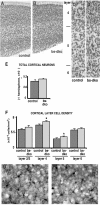
Figure 1.
Figure 1.
Neuronal density is increased in bs-dko cortices. A, B, Sagittal sections (50 μm) from 6-week-old abl+/+arg+/- (control) (A) and bs-dko (B) littermate mice are stained with antibodies to the neuronal marker NeuN. Sections show a portion of the cortex and are oriented with pial surface at the top. C, D, Magnified view of control (C) and bs-dko (D) sections. Cortical layer divisions are indicated on the left. E, Quantitation of the number of cortical neurons in control or bs-dko mice. Mean ± SE; n = 5 mice; 9-12 sections each per genotype. F, Quantitation of cortical neuron density by cortical layer in control or bs-dko mice. Mean ± SE; n = 5 mice per genotype. *p ≤ 0.05, Student's t test of bs-dko relative to control. G, H, Coronal sections (50 μm) from 6-week-old abl+/+arg+/- (control) (G) and bs-dko (H) littermate mice are stained with antibodies to the glial marker S100-β. Sections show a portion of cortical layer 5.
Figure 2.
Figure 2.
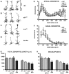
Dendrite arbors are reduced in adult abl-/-, arg-/-, and bs-dko mice. A-D, Camera lucida drawings of representative wild-type (A), abl-/- (B), arg-/- (C), and bs-dko (D) layer 5 pyramidal neurons from 6- to 8-week-old mice. Apical and basal dendrite arbors are indicated for a wild-type neuron in A. E, F, Sholl analysis of apical (E) and basal (F) dendrites from wild-type, abl-/-, arg-/-, and bs-dko layer 5 pyramidal neurons. Sholl analysis measures dendrite complexity: concentric three-dimensional shells of increasing radius are centered on the cell body of the neuron; the number of intersections of the dendrite arbor with a given shell is plotted versus the shell radius. Each point represents mean ± SE. For all data presented in this figure, analysis was performed on the following: apical dendrites, wild type, n = 6 mice, 16 neurons; abl-/-, n = 4 mice, 15 neurons; arg-/-, n = 4 mice, 16 neurons; bs-dko, n = 6 mice, 17 neurons; basal dendrites, wild type, n = 6 mice, 16 neurons; abl-/-, n = 4 mice, 16 neurons; arg-/-, n = 4 mice, 17 neurons; bs-dko, n = 6 mice, 16 neurons. G, H, Mean total length (G) and branchpoint number (H) of apical (left) and basal (right) dendrite arbors from wild-type, abl-/-, arg-/-, and bs-dko layer 5 pyramidal neurons. Mean ± SE. ANOVA between all genotypes: apical length, p = 0.233; basal length, p = 0.014; apical branchpoints, p = 0.092; basal branchpoints, p < 0.0001; post hoc Student-Newman-Keuls test for each mutant versus wild type, *p < 0.05.
Figure 3.
Figure 3.
Quantitation of Abl and Arg protein levels during development. A, Purified recombinant Abl as a standard or 25, 100, or 250 μg of brain extract from E13, P21, or 6-week-old wild-type or arg-/- mice were immunoblotted with antibodies to Abl. B, Purified recombinant Arg as a standard or 25 or 100 μg of brain extract from E13, P21, or 6-week-old wild-type mice were immunoblotted with antibodies to Arg. C, Summary of Abl and Arg protein amounts per 100 μg of brain extract at E13, P21, and 6 weeks.
Figure 4.
Figure 4.
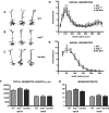
Dendrite arbors are normal in arg-/- and bs-dko mice at P21. A-C, Camera lucida drawings of representative wild-type (A), arg-/- (B), and bs-dko (C) layer 5 pyramidal neurons from P21 mice. Neurons are oriented as in Figure 3. D, E, Sholl analysis of apical (D) and basal (E) dendrites from wild-type, arg-/-, and bs-dko layer 5 pyramidal neurons. Each point represents mean ± SE. For all experiments in this figure, analysis was performed on the following: apical dendrites, wild type, n = 6 mice, 17 neurons; arg-/-, n = 5 mice, 16 neurons; bs-dko, n = 5 mice, 15 neurons; basal dendrites, wild type, n = 6 mice, 17 neurons; arg-/-, n = 6 mice, 17 neurons; bs-dko, n = 6 mice, 16 neurons. F, G, Mean total length (F) and branchpoint number (G) of apical (left) and basal (right) dendrite arbors from wild-type, arg-/-, and bs-dko layer 5 pyramidal neurons. Mean ± SE. ANOVA between all genotypes: apical length, p = 0.568; basal length, p > 0.999; apical branchpoints, p = 0.202; basal branchpoints, p = 0.436.
Figure 5.
Figure 5.
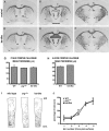
Cortical afferents appear normal in arg-/- and bs-dko mice. A-F, The major cortical afferent tracts were stained with anti-L1 antibodies in matched coronal sections from adult control (abl+/+arg+/-) (A-C) and bs-dko (D-F) mice. The corpus callosum (cc), internal capsule (ic), and dorsal hippocampal commissure (dhc) are indicated. G, H, Mean corpus callosum thickness from four to six matched coronal sections each from P18-P22 (G) and adult (H) control (abl+/+arg+/-), arg-/-, or bs-dko mice. Mean thickness is similar for all genotypes. P18-P22, n = 4 mice; adult, n = 4 mice. ANOVA between all genotypes in G, p = 0.88; Student's t test for bs-dko versus control in H, p = 0.16. I, J, Cortical axons were labeled by placement of a DiI crystal into the contralateral visual cortex. Representative tracings of axons in wild-type, arg-/-, and bs-dko cortex are shown in I. Each cortical section was overlaid with five equally sized bins that spanned area from the pial surface to the corpus callosum. The percentage of total axons in each bin is presented in J. Mean ± SE, n = 2 mice, four sections each per genotype. ANOVA between all genotypes for percentage axons in each bin: bin 1, p = 0.788; bin 2, p = 0.932; bin 3, p = 0.503; bin 4, p = 0.892; bin 5, p = 0.984.
Figure 6.
Figure 6.
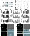
Arg promotes axon and dendrite branching in cultured cortical neurons. A, Diagram of Arg and Arg mutants used in these assays. The Arg N-terminal half contains SH3 (3), SH2 (2), and kinase domains. The Arg C-terminal half contains two F-actin-binding domains (F) and a microtubule-binding domain (MT). ArgKI has a point mutation rendering it kinase inactive. ArgΔC lacks the cytoskeleton-binding domains. ArgΔSH3 has a deletion of the SH3 domain, rendering it hyperactive for kinase activity. B, Camera lucida drawings of cortical neurons transfected with vector alone or with expression vectors for Arg and ArgKI made at 4 DIV. C-E, Quantitation of branchpoint number, total length, and branchpoints/length for neurites (C), dendrites (D), and axons (E) in neurons expressing vector alone or expression vectors for Arg, ArgKI, ArgΔC, and ArgΔSH3. All values represent mean ± SE. C, Vector, n = 202 neurons, eight transfections; Arg, n = 197 neurons, eight transfections; ArgKI, n = 187 neurons, six transfections; ArgΔC, n = 93 neurons, six transfections; ArgΔSH3, n = 53 neurons, four transfections. D, E, Vector, n = 126 neurons, five transfections; Arg, n = 123 neurons, five transfections; ArgKI, n = 123 neurons, four transfections; ArgΔC, n = 81 neurons, four transfections; ArgΔSH3, n = 45 neurons, three transfections. For neurites, ANOVA between all transfection conditions is as follows: branchpoints, p < 0.0001; length, p = 0.035; branchpoints/length, p < 0.0001. For dendrites, ANOVA between all transfection conditions: branchpoints, p < 0.0001; dendrite length, p = 0.0023; branchpoints/length, p < 0.0001. For axons, ANOVA between all transfection conditions is as follows: branchpoints, p < 0.0001; axon length, p = 0.0002; branchpoints/length, p < 0.0001. Posthoc Student-Newman-Keuls test for each Arg construct versus vector alone: *p < 0.05. E, F, Cultured cortical neurons immunostained with α-Arg (E) or α-Abl (F) antibodies at 5 or 6 DIV. Phase-contrast images are shown in the right panels. Axons are indicated by white arrowheads. Boxed regions are enlarged below each image. Endogenous Arg is concentrated in growth cones and at phase-dense spots along neurites. Endogenous Abl is present more homogeneously throughout neurites, with a slight concentration in the growth cone. Scale bars, 40 μm.
Figure 7.
Figure 7.
Arg is required for integrin-mediated neurite branching. Bar diagrams indicate mean branchpoint number (A, C, E) and total neurite arbor length (B, D, F). All values represent mean ± SE. A, B, Wild-type or arg-/- cortical neurons were plated on glass coverslips coated with polyornithine (ornithine) or polyornithine plus laminin-1 (laminin). Neurons were fixed at 24 h and traced with camera lucida software. Wild type on ornithine, n = 150 total neurons, five independent experiments; wild type on laminin, n = 136 neurons, five experiments; arg-/- on ornithine, n = 137 neurons, five experiments; arg-/- on laminin, n = 128 neurons, five experiments. C, D, Wild-type or arg-/- cortical neurons plated on ornithine or laminin in the presence of the Abl/Arg inhibitor STI571 (3.3 μm) or the β1/β3 integrin inhibitor echistatin (1 μm). Wild-type on ornithine plus echistatin, n = 61 neurons, three experiments; wild type on laminin plus echistatin, n = 59 neurons, three experiments, wild type on ornithine plus STI571, n = 61 neurons, three experiments; wild type on laminin plus STI571, n = 93 neurons, three experiments; arg-/- on ornithine plus echistatin, n = 20, one experiment; arg-/- on laminin plus echistatin, n = 59 neurons, three experiments; arg-/- on ornithine plus STI571, n = 57 neurons, three experiments; arg-/- on laminin plus STI571, n = 89 neurons, three experiments. For A-D, ANOVA between all plating conditions: branchpoints, p < 0.0001; length, p < 0.0001; post hoc Student-Newman-Keuls test for each condition versus wild type plated on ornithine, *p < 0.05. ANOVA between all plating conditions for each genotype: wild-type branchpoints, p < 0.0001; arg-/- branchpoints, p = 0.119; wild-type length, p < 0.0001; arg-/- length, p = 0.022. E, F, Wild type or arg-/- cortical neurons plated on Semaphorin7A (AP-Sema7A) or control protein (AP) substrates in the presence of STI571 (3.3 μm) or echistatin (1 μm). Wild type on AP, n = 70 neurons, four experiments; wild type on 20 nm Sema7A, n = 46 neurons, two experiments; wild type on 100 nm Sema7A, n = 72 neurons, three experiments; wild type on 100 nm Sema7A plus STI571, n = 27 neurons, one experiment; wild type on 100 nm Sema7A plus echistatin, n = 25 neurons, one experiment; arg-/- on AP, n = 74 neurons, three experiments; arg-/- on 20 nm Sema7A, n = 47 neurons, two experiments; arg-/- on 100 nm Sema7A, n = 72 neurons, three experiments; arg-/- on 100 nm Sema7A plus STI571, n = 25 neurons, one experiment; arg-/- on 100 nm Sema7A plus echistatin, n = 29 neurons, one experiment. For E and F, ANOVA between all plating conditions is as follows: branchpoints, p < 0.0001; length, p < 0.0001; post hoc Student-Newman-Keuls test for each condition versus wild type plated on 100 nm AP, *p < 0.05. ANOVA between all plating conditions for each genotype: wild-type branchpoints, p = 0.0004; arg-/- branchpoints, p = 0.015; wild-type length, p = 0.007; arg-/- length, p = 0.104. G, H, Wild type or arg-/- cortical neurons plated on ornithine (orni), laminin (lamin), or Netrin-1 (netrin) at the indicated concentrations. Neurons were fixed at 24 h. One experiment was performed for each condition. Wild-type neurons on ornithine, n = 20 neurons; wild type on laminin, n = 22 neurons; wild type on 5 μg/ml netrin, n = 25 neurons; wild type on 10 μg/ml netrin, n = 24 neurons; arg-/- neurons on ornithine, n = 24 neurons; arg-/- on laminin, n = 19 neurons; arg-/- on 5 μg/ml netrin, n = 24 neurons; arg-/- on 10 μg/ml netrin, n = 27 neurons. ANOVA between all plating conditions is as follows: branchpoints, p < 0.0001, length, p < 0.0001; post hoc Student-Newman-Keuls test for each condition versus wild type plated on ornithine, *p < 0.05. ANOVA between all plating conditions for each genotype: wild-type branchpoints, p = 0 < 0.0001; arg-/- branchpoints, p = 0.772; wild-type length, p < 0.0001; arg-/- length, p = 0.257.
Figure 8.
Figure 8.
arg-/- neurites have reduced dynamics and accumulate branches slower than wild-type neurites. A, Camera lucida drawings of representative arg-/- and wild-type neurons cultured on laminin-1-coated glass and imaged at 40 min intervals for 6 h at 1 DIV. Imaging times are indicated on the left. Branches that have been retracted before the subsequent imaging time are indicated with a red star. Branches that have been added since the previous imaging time are indicated with a green asterisk. Note that more stars and asterisks are associated with wild-type neurites than arg-/- neurites, indicating that their behavior is more dynamic than arg-/- neurites. B, C, Quantitation of dynamic behavior of wild-type and arg-/- neurites cultured on either polyornithine (ornithine) or polyornithine plus laminin-1 (laminin). Gray bars indicate the initial or final average number of branchpoints per neurite (B) or average neurite length (C) at the start of the imaging session. Green and red bars indicate the average cumulative number of branchpoints per neurite added or retracted (B) or cumulative neurite length extended or retracted (C) over the initial amount for the 6 h imaging session. Wild type on ornithine, n = 10 cells, 47 neurites; wild type on laminin, n = 15 cells, 63 neurites; arg-/- on ornithine, n = 9 cells, 43 neurites; arg-/- on laminin, n = 14 cells, 58 neurites. p values represent statistical significance by Student's t test of the difference between wild-type and arg-/- neurons cultured on laminin, *p = 0.08. D, E, Wild-type (n = 14) or arg-/- (n = 12) neurons cultured on laminin-1 were imaged at 7 and 17 h intervals from 18 to 96 h in vitro (1-4 DIV). The average number of branchpoints per cell (D) or total neurite length per cell (E) is plotted versus time in culture. Dashed lines are best-fit linear trendlines showing the decreased growth rate of arg-/- neurite arbors.

Similar articles

Cited by

References

    1. Anderson KL, Ferreira A (2004) alpha1 integrin activation: a link between beta-amyloid deposition and neuronal death in aging hippocampal neurons. J Neurosci Res 75: 688-697. - PubMed
    1. Bashaw GJ, Kidd T, Murray D, Pawson T, Goodman CS (2000) Repulsive axon guidance: abelson and enabled play opposing roles downstream of the roundabout receptor. Cell 101: 703-715. - PubMed
    1. Bastmeyer M, O'Leary DD (1996) Dynamics of target recognition by interstitial axon branching along developing cortical axons. J Neurosci 16: 1450-1459. - PMC - PubMed
    1. Beggs HE, Soriano P, Maness PF (1994) NCAM-dependent neurite outgrowth is inhibited in neurons from Fyn-minus mice. J Cell Biol 127: 825-833. - PMC - PubMed
    1. Bi X, Lynch G, Zhou J, Gall CM (2001) Polarized distribution of alpha5 integrin in dendrites of hippocampal and cortical neurons. J Comp Neurol 435: 184-193. - PubMed

Publication types