. 2005 Oct;54(10):1445-55.
doi: 10.1136/gut.2004.063164. Epub 2005 Jun 29.

Protein tyrosine phosphatase kappa and SHP-1 are involved in the regulation of cell-cell contacts at adherens junctions in the exocrine pancreas

Affiliations

Protein tyrosine phosphatase kappa and SHP-1 are involved in the regulation of cell-cell contacts at adherens junctions in the exocrine pancreas

J Schnekenburger et al. Gut. 2005 Oct.

Abstract

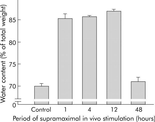
Background: We have previously shown that cell contacts between pancreatic acinar cells dissociate early in pancreatitis and that this is a prerequisite for the development of pancreatic oedema. Here we studied the underlying mechanism.

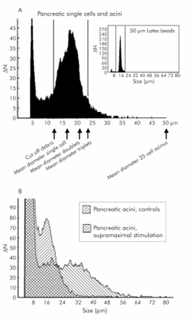
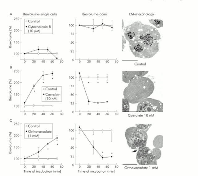
Methods: Employing experimental caerulein induced pancreatitis in vivo and isolated pancreatic acini ex vivo, in conjunction with protein chemistry, morphology, and electron microscopy, we determined whether cell contact regulation in the pancreas requires or involves: (1) changes in cadherin-catenin protein expression, (2) tyrosine phosphorylation of adhesion proteins, or (3) alterations in the actin cytoskeleton.

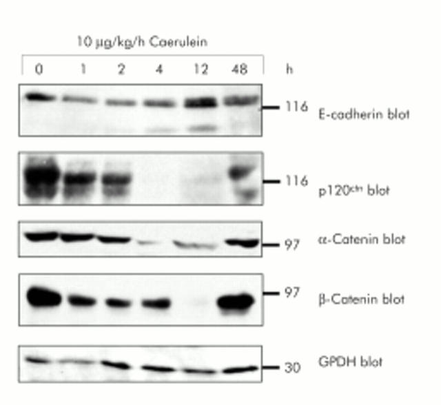
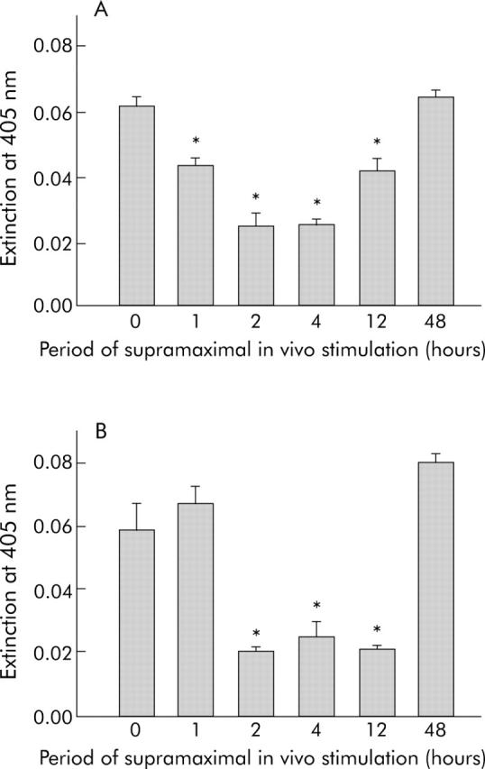
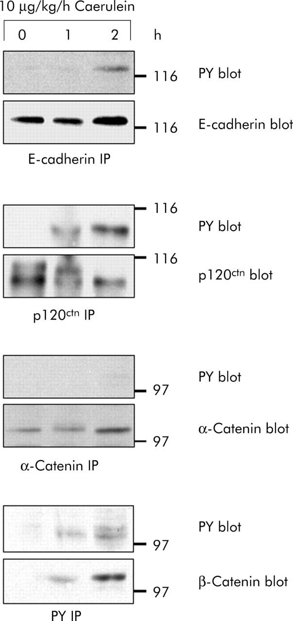
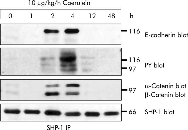
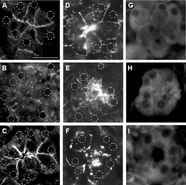
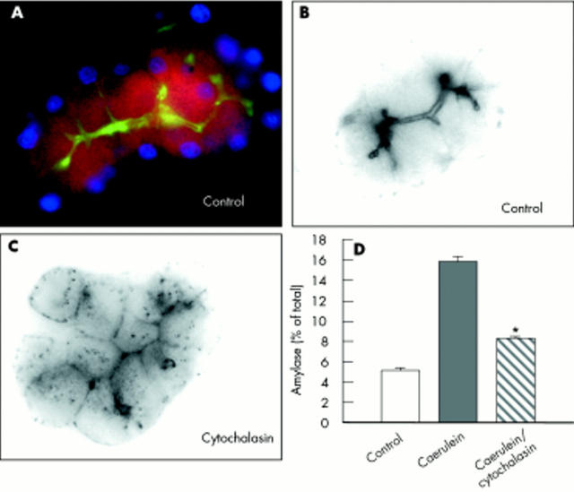
Results: During initial cell-cell contact dissociation at adherens junctions, expression of adhesion proteins remained stable. At time points of dissociated adherens junctions, the cadherin-catenin complex was found to be tyrosine phosphorylated and internalised. The receptor type protein tyrosine phosphatase (PTP)kappa was constitutively associated with the cadherin-catenin complex at intact cell contacts whereas following the dissociation of adherens junctions, the internalised components of the cadherin-catenin complex were tyrosine phosphorylated and associated with the cytosolic PTP SHP-1. In isolated acini, inhibition of endogenous protein tyrosine phosphatases alone was sufficient to induce dissociation of adherens junctions analogous to that found with supramaximal caerulein stimulation. Dissociation of actin microfilaments had no effect on adherens junction integrity.

Conclusions: These data identify tyrosine phosphorylation as the key regulator for cell contacts at adherens junctions and suggest a definitive role for the protein tyrosine phosphatases PTPkappa and SHP-1 in the regulation, maintenance, and restitution of cell adhesions in a complex epithelial organ such as the pancreas.


Figures

Figure 1
Figure 1
Expression of proteins of the cadherin/catenin complex during in vivo supramaximal secretagogue stimulation. Immunoblotting of E-cadherin, α-catenin, β-catenin, and p120ctn with specific antibodies in the same sodium dodecyl sulphate-polyacrylamide gel separated Triton X 100 lysates of rat pancreatic tissue. Animals were infused with either saline or caerulein (10 μg/kg/h) for the indicated time intervals. Lysates were standardised to 100 μg protein from control pancreas. Reblotting with specific GPDH antibody demonstrated loading of equal protein amounts.
Figure 2
Figure 2
Interstitial pancreatic fluid collection and dissociation of cell-cell contacts. As the formation of pancreatic oedema requires, and is ultrastructurally preceded by, the dissociation of cell contacts for fluid to enter the interstitial space, we determined the time course of oedema formation as an indicator of cell contact dissociation. The wet/dry weight ratio of pancreatic tissue was determined after desiccation and the results were expressed as pancreatic water content as a percentage of pancreatic weight. After 60 minutes of supramaximal caerulein infusion, entry of fluid into the pancreas was already complete and was only reabsorbed after 48 hours.
Figure 3
Figure 3
Enzymatic protein tyrosine phosphatase (PTP) (A) and protein tyrosine kinase (PTK) (B) activity in caerulein induced pancreatitis. To test whether PTK and PTP possess constitutive basal activity, we used pancreatic homogenates obtained at different time points during supramaximal caerulein stimulation to measure overall enzymatic activity, as indicated in materials and methods. PTP activity was already decreased one hours after the start of caerulein infusion (A) which was paralleled by an increase in PTK activity, mainly involving src kinases (B). Interestingly, after the decrease in PTP and PTK activity during the greatest disease severity (2 and 4 hours), the increase in PTP activity was faster than that of PTK activity. *Significantly different from controls at 0 h.
Figure 4
Figure 4
Time course of cadherin/catenin tyrosine phosphorylation in vivo. E-cadherin, α-catenin, β-catenin, and p120ctn were immunoprecipitated from pancreatic homogenates after supramaximal caerulein stimulation for the indicated time intervals. Precipitates were blotted with phosphotyrosine specific antibodies and with antibodies against E-cadherin, α-catenin, β-catenin, and p120ctn. Proteins were immunoprecipitated from 3 mg pancreatic protein.
Figure 5
Figure 5
In vivo association of protein tyrosine phosphatase (PTP)κ with proteins of the cadherin/catenin complex. Immunoprecipitation (IP) of PTPκ from pancreatic homogenates after supramaximal caerulein stimulation for the indicated time intervals. E-cadherin, α-catenin, and β-catenin were blotted with specific antibodies. PTPκ was detected by biotinylation of membrane bound proteins and staining with horseradish peroxidase conjugated streptavidin. Biotinylation was performed as PTPκ could not be reblotted when immunoprecipitated with the same antibody. Proteins were immunoprecipitated from 3 mg pancreatic protein.
Figure 6
Figure 6
In vivo association of protein tyrosine phosphatase (PTP) SHP-1 with proteins of the cadherin/catenin complex. Immunoprecipitation (IP) of PTP SHP-1 from pancreas homogenates after supramaximal caerulein stimulation for the indicated time intervals. Precipitates were blotted with antibodies against PTP SHP-1, E-cadherin, α-catenin, β-catenin, and antiphosphotyrosine. Proteins were immunoprecipitated from 3 mg pancreatic protein.
Figure 7
Figure 7
Localisation of protein tyrosine phosphatase (PTP)κ, PTP SHP-1, and E-cadherin during supramaximal caerulein stimulation in vivo. Sections from pancreatic tissue after in vivo treatment with supramaximal caerulein were labelled with monospecific antibodies directed against E-cadherin (A = 0 hours, B = one hour, C = 48 hours), PTPκ (D = 0 hours, E = four hours, F = 48 hours), or PTP SHP-1 (G = 0 hours, H = four hours, I = 48 hours) and fluorescent labelled secondary antibody for confocal microscopy, as described in materials and methods. Representative acini were digitally photographed at the same contrast and brightness setting and, with the exception of the SHP-1 label, the position of nuclei as determined by the DAPI stain was marked on the respective micrographs. Bar indicates 50 μm.
Figure 8
Figure 8
Dissociation of cell contacts in isolated pancreatic acini on supramaximal secretagogue stimulation. (A) Size frequency distribution of single cells in an acinar cell preparation. Single cells were prepared by a prolonged collagenase digestion. Average single cell size is calculated between the lower (11 μm) and upper (23 μm) diameter cut offs. Particles with diameters below 11 μm are cell debris; particles above 23 μm are cell aggregates of three or more cells (acini). Lower cut off, Ø = 11 μm (V = 697 μm3); average single cell diameter, Ø = 17 μm (2572 μm3), acini of 25 cells, Ø = 50 μm (65450 μm3). Insert: Size distribution of standard latex beads with an average diameter of 15 μm (Duke Scientific Corporation, Palo Alto, California, USA). (B) Overlay of size distribution of a culture of pancreatic acini before and 30 minutes after stimulation with a supramaximal concentration of caerulein (10 nM). There is a shift from complex acini (aggregates above the upper cut off of 23 μm) to single acinar cells (cells with diameters between 11 μm and 23 μm) caused by the caerulein induced disintegration of cell-cell contacts.
Figure 9
Figure 9
Dissociation of cell contacts in isolated pancreatic acini after protein tyrosine phosphatase (PTP) inhibition. The integrity of cell contacts in isolated pancreatic acini was evaluated as changes in the isovolume of the single cells fraction (left panels) or the intact acini fraction (centre panels), and the corresponding electron micrographs (EM) are shown in the right panels. Data are expressed as per cent of untreated controls for acini after treatment with cytochalasin B (10 μM, row A), supramaximal caerulein (10−8 M, row B), or 1 mM orthovanadate (row C) for up to 70 minutes. *Significant differences from respective control values (p<0.05). Bars indicate 20 μm.
Figure 10
Figure 10
Effect of cytochalasin on the cytoskeleton of pancreatic acini. To evaluate the effect of cytochalasin on microfilament disruption, untreated intact acini were labelled for G-actin (red) and F-actin (green) (A), which localised F-actin firmly to cell-cell contacts and the acinar lumen and G-actin to the cytosol (A, B). Incubation of acini with cytochalasin (10 μM, 40 minutes) caused not only rapid distribution of F-actin from cell contacts to the cytosol (C) but also greatly reduced stimulated pancreatic secretion (D).

Comment in

Similar articles

Cited by

References

    1. Lerch MM, Weidenbach H, Gress TM, et al. Effect of kinin inhibition in experimental acute pancreatitis. Am J Physiol 1995;269:G490–9. - PubMed
    1. Hinck L, Nathke IS, Papkoff J, et al. Dynamics of cadherin/catenin complex formation: novel protein interactions and pathways of complex assembly. J Cell Biol 1994;125:1327–40. - PMC - PubMed
    1. Ozawa M, Baribault H, Kemler R. The cytoplasmic domain of the cell adhesion molecule uvomorulin associates with three independent proteins structurally related in different species. Embo J 1989;8:1711–17. - PMC - PubMed
    1. Rimm DL, Koslov ER, Kebriaei P, et al. Alpha 1(E)-catenin is an actin-binding and -bundling protein mediating the attachment of F-actin to the membrane adhesion complex. Proc Natl Acad Sci U S A 1995;92:8813–17. - PMC - PubMed
    1. Yap AS, Brieher WM, Gumbiner BM. Molecular and functional analysis of cadherin-based adherens junctions. Annu Rev Cell Dev Biol 1997;13:119–46. - PubMed

Publication types

MeSH terms