

Assessment of the proliferative, apoptotic and cellular renovation indices of the human mammary epithelium during the follicular and luteal phases of the menstrual cycle
- PMID: 15987425
- PMCID: PMC1143573
- DOI: 10.1186/bcr994
Assessment of the proliferative, apoptotic and cellular renovation indices of the human mammary epithelium during the follicular and luteal phases of the menstrual cycle
Abstract
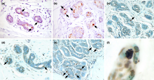
Introduction: During the menstrual cycle, the mammary gland goes through sequential waves of proliferation and apoptosis. In mammary epithelial cells, hormonal and non-hormonal factors regulate apoptosis. To determine the cyclical effects of gonadal steroids on breast homeostasis, we evaluated the apoptotic index (AI) determined by terminal deoxynucleotidyl transferase-mediated dUTP nick end labeling (TUNEL) staining in human mammary epithelial cells during the spontaneous menstrual cycle and correlated it with cellular proliferation as determined by the expression of Ki-67 during the same period.
Methods: Normal breast tissue samples were obtained from 42 randomly selected patients in the proliferative (n = 21) and luteal (n = 21) phases. Menstrual cycle phase characterization was based on the date of the last and subsequent menses, and on progesterone serum levels obtained at the time of biopsy.
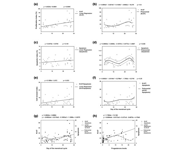
Results: The proliferation index (PI), defined as the number of Ki-67-positive nuclei per 1,000 epithelial cells, was significantly larger in the luteal phase (30.46) than in the follicular phase (13.45; P = 0.0033). The AI was defined as the number of TUNEL-positive cells per 1,000 epithelial cells. The average AI values in both phases of the menstrual cycle were not statistically significant (P = 0.21). However, the cell renewal index (CRI = PI/AI) was significantly higher in the luteal phase (P = 0.033). A significant cyclical variation of PI, AI and CRI was observed. PI and AI peaks occurred on about the 24th day of the menstrual cycle, whereas the CRI reached higher values on the 28th day.
Conclusions: We conclude that proliferative activity is dependent mainly on hormonal fluctuations, whereas apoptotic activity is probably regulated by hormonal and non-hormonal factors.
Figures
Similar articles
-
Effect of combined oral contraceptives on breast epithelial proliferation in young women.Breast J. 2008 Sep-Oct;14(5):450-5. doi: 10.1111/j.1524-4741.2008.00621.x. Epub 2008 Jul 24. Breast J. 2008. PMID: 18657146 Clinical Trial.
-
Cyclic changes in the mammary gland of cynomolgus macaques.Fertil Steril. 2004 Oct;82 Suppl 3:1160-70. doi: 10.1016/j.fertnstert.2004.04.035. Fertil Steril. 2004. PMID: 15474090
-
Morphometric analysis of fibroblasts of the mammary lobular stroma during the follicular and luteal phases of the menstrual cycle.Morphologie. 1998;82(257):7-10. Morphologie. 1998. PMID: 11928126
-
Role of progesterone in normal breast physiology.J Reprod Med. 1999 Feb;44(2 Suppl):172-9. J Reprod Med. 1999. PMID: 11392028 Review.
-
[Effect of menstrual cycle on the immune response to surgical stress].Minerva Ginecol. 1995 May;47(5):197-205. Minerva Ginecol. 1995. PMID: 7478085 Review. Italian.
Cited by
-
Examining the pathogenesis of breast cancer using a novel agent-based model of mammary ductal epithelium dynamics.PLoS One. 2013 May 21;8(5):e64091. doi: 10.1371/journal.pone.0064091. Print 2013. PLoS One. 2013. PMID: 23704974 Free PMC article.
-
Proliferative heterogeneity of murine epithelial cells in the adult mammary gland.Commun Biol. 2018 Aug 13;1:111. doi: 10.1038/s42003-018-0114-7. eCollection 2018. Commun Biol. 2018. PMID: 30271991 Free PMC article.
-
Neoadjuvant stereotactic body radiation therapy, capecitabine, and liver transplantation for unresectable hilar cholangiocarcinoma.Liver Transpl. 2014 Jan;20(1):81-8. doi: 10.1002/lt.23757. Epub 2013 Nov 21. Liver Transpl. 2014. PMID: 24115315 Free PMC article.
-
The relation between excess adiposity and breast cancer in women: Clinical implications and management.Crit Rev Oncol Hematol. 2024 Jan;193:104213. doi: 10.1016/j.critrevonc.2023.104213. Epub 2023 Nov 24. Crit Rev Oncol Hematol. 2024. PMID: 38008197 Free PMC article. Review.
-
Discordance in 21-gene recurrence scores between paired breast cancer samples is inversely associated with patient age.Breast Cancer Res. 2020 Aug 18;22(1):90. doi: 10.1186/s13058-020-01327-1. Breast Cancer Res. 2020. PMID: 32811558 Free PMC article.
References
-
- Greenlee RT, Murray T, Bolden S, Wingo PA. Cancer statistics, 2000. CA Cancer J Clin. 2000;50:7–33. - PubMed
-
- Wellings SR, Jensen HM, Marcum RG. An atlas of subgross pathology of the human breast with special reference to possible precancerous lesions. J Natl Cancer Inst. 1975;55:231–273. - PubMed
-
- Masters JR, Sangster K, Smith II. Hormonal sensitivity of human breast tumors in vitro: pentose-shunt activity. Cancer. 1977;39:1978–1980. - PubMed
-
- Meyer JS. Cell proliferation in normal human breast ducts, fibroadenomas, and other ductal hyperplasias measured by nuclear labeling with tritiated thymidine. Effects of menstrual phase, age, and oral contraceptive hormones. Hum Pathol. 1977;8:67–81. - PubMed
Publication types
MeSH terms
Substances
LinkOut - more resources
Full Text Sources
Research Materials
Miscellaneous
