. 2005 Aug;16(8):3642-58.
doi: 10.1091/mbc.e05-01-0076. Epub 2005 Jun 1.

EHD proteins associate with syndapin I and II and such interactions play a crucial role in endosomal recycling

Affiliations

EHD proteins associate with syndapin I and II and such interactions play a crucial role in endosomal recycling

Anne Braun et al. Mol Biol Cell. 2005 Aug.

Abstract

EHD proteins were shown to function in the exit of receptors and other membrane proteins from the endosomal recycling compartment. Here, we identify syndapins, accessory proteins in vesicle formation at the plasma membrane, as differential binding partners for EHD proteins. These complexes are formed by direct eps15-homology (EH) domain/asparagine proline phenylalanine (NPF) motif interactions. Heterologous and endogenous coimmunoprecipitations as well as reconstitutions of syndapin/EHD protein complexes at intracellular membranes of living cells demonstrate the in vivo relevance of the interaction. The combination of mutational analysis and coimmunoprecipitations performed under different nucleotide conditions strongly suggest that nucleotide binding by EHD proteins modulates the association with syndapins. Colocalization studies and subcellular fractionation experiments support a role for syndapin/EHD protein complexes in membrane trafficking. Specific interferences with syndapin-EHD protein interactions by either overexpression of the isolated EHD-binding interface of syndapin II or of the EHD1 EH domain inhibited the recycling of transferrin to the plasma membrane, suggesting that EH domain/NPF interactions are critical for EHD protein function in recycling. Consistently, both inhibitions were rescued by co-overexpression of the attacked protein component. Our data thus reveal that, in addition to a crucial role in endocytic internalization, syndapin protein complexes play an important role in endocytic receptor recycling.


Figures
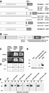
Figure 1.
Figure 1.
Syndapin I interacts specifically with EH domain-containing proteins of the EHD protein family. (A) Schematic representation of the syndapin isoforms and splice variants syndapin I, II-s, II-l, and III as well as of the different syndapin fragments used throughout this study (solid bars). (B) Schematic representation of the EH domain-containing protein EHD 3 and of two independent clones encoding for the C terminus isolated by Y2H screening with full-length syndapin I as a bait. (C and D) Activation of reporter genes assayed via growth on quadruple drop-out plates (C) and via β-galactosidase activity (D). (E) Affinity purifications of endogenous syndapin I from rat brain cytosol (RBC) with immobilized GST-fusion proteins of an EHD3 fragment and of full-length EHD3, verifying the Y2H results. (F) Affinity purifications of endogenous syndapin I from rat brain extracts with immobilized GST-fusion proteins of different EH domain-containing proteins reveal that syndapin I associates with EHD proteins but not with the EH domains of intersectin or Eps15. Equal amounts of fusion proteins and rat brain cytosol (1 mg) were used. Twenty micrograms of starting material was loaded for comparison. P, precipitates; S, supernatants.
Figure 2.
Figure 2.
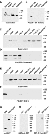
The interaction with EHD proteins is mediated by the NPF motif-encompassing region present in syndapin I and II. (A–F) Precipitates and supernatants from coprecipitation experiments with immobilized GST-fusion proteins of syndapins incubated with rat brain extracts (1 mg of protein each) were analyzed by immunoblotting with anti-EHD antibodies. (A and B) GST fusion proteins of syndapin I and of both syndapin II splice variants (Sdp II-s and Sdp II-l), but not of syndapin III or GST alone, efficiently coprecipitated endogenous EHD proteins. (C–F) Experiments with immobilized GST-fusion proteins of syndapin I (C and D) and syndapin II (E and F) reveal that the interaction of both syndapin I and II is mediated by the NPF motif-containing region (Sdp-NPF). (G) Alignment of the amino acid sequences of rat syndapin I (gi4324451, aa 336–386), rat syndapin II-s (gi6651165, aa 339–391), rat syndapin II-l (gi6651162, aa 339–432), and rat syndapin III (gi57471977, aa 336–367) by ClustalW (http://www.ebi.ac.uk/clustalW/). (H) Amino acid sequence identity between all four murine EHD family members as well as C. elegans Rme-1. (I–L) Coprecipitation analyses with extracts from HEK293 cells expressing GFP-fusion proteins of all four mouse full-length EHD proteins or GFP alone (I) and immobilized GST-fusion proteins of full-length syndapins. Whereas immobilized GST did not precipitate any GFP-fusion proteins (our unpublished data), the coprecipitates obtained with immobilized syndapin I (J), syndapin II-s (K), and syndapin II-l (L) revealed strong associations of all these syndapin variants with all EHD proteins except EHD2, as analyzed by anti-GFP immunoblotting.
Figure 3.
Figure 3.
The interaction between syndapins and EHD proteins is direct and depends on syndapin NPF motifs. (A–F) Immunoblot analyses of coprecipitations of FLAG-tagged wild-type and mutant syndapin I (A and B) and syndapin II-l (C–F) proteins overexpressed in HEK293 cells with immobilized GST-fusion proteins of the EH domain of EHD1 (A–D) and of full-length EHD1 (E and F). The coprecipitated material (B, D, and F) and the supernatants (A, C, and E) were analyzed by immunoblotting with anti-FLAG antibodies. (G–I) Blot overlay analysis of extracts from HEK293 cells overexpressing GFP and GFP-Sdp II-l. Overexpressed proteins were visualized by anti-GFP immunoblotting (G), with a GST-fusion protein of the EH domain of EHD1 (I) and GST (negative control) (H) as probes.
Figure 4.
Figure 4.
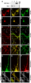
Syndapins and EHD proteins colocalize in neuronal and nonneuronal cells. (A) The new guinea pig anti-syndapin I antibodies specifically recognize syndapin I on Western blots of 25 μg (lane 1), 5 μg (lane 2) of rat brain extracts and 50 ng of MBP-syndapin I ΔSH3 (lane 3). (B–D) The affinity-purified anti-syndapin I antibodies (D) recognize FLAG-tagged mito-syndapin I expressed in COS-7 cells with the same specificity as monoclonal anti-FLAG antibodies (B), as seen by the complete overlap of the stainings in the merge (C). (E–M) Primary hippocampal neurons 2 DIV (E–G) and 24 DIV (H–M) were immunostained with anti-syndapin I antibodies (G, J, and M) and anti-EHD antibodies (E, H, and K). Colocalization is in yellow in merged images (F, I, and L). Examples of growth cones (E–G, arrow) and of sites within the periphery of the neuronal network that may represent synaptic contacts and also display an accumulation for both syndapin I and EHD proteins are marked (H—J, arrowheads). (N–O) GFP-EHD1 (N) and Xpress-syndapin II-l (P) expressed in primary hippocampal neurons also show a high degree of spatial overlap (O). (Q–S) FLAG-tagged EHD1 (Q, red in merge) and GFP-syndapin II-l (S, green in merge) coexpressed in HeLa cells exhibit a similarly high degree of colocalization, as observed by confocal microscopy. Insets represent magnifications of areas boxed. Bars, 10 μm.
Figure 5.
Figure 5.
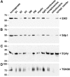
EHD and syndapin proteins are codistributed in rat brain fractions. Western blots of rat brain homogenate and indicated subcellular fractions probed with antibodies against EHD proteins (A), syndapin I (B), the synaptic vesicle marker synaptophysin (C), and the TGN marker protein TGN38 (D). S1, 1000 × g supernatant 1; S2, 12,000 × g supernatant 2; P2, 12,000 × g pellet 2 (crude membrane fraction). Myelin, light membranes, synaptosomes, and the fraction containing heavy membranes and mitochondria (mitochondria) were obtained by sucrose step gradient separation of P2. SM, 100,000 × g microsomal supernatant; PM, 100,000 × g microsomal pellet.
Figure 6.
Figure 6.
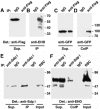
Syndapin I and II-l coimmunoprecipitate with EHD proteins. (A–D) Coimmunoprecipitation of GFP-syndapin II-l with FLAG-EHD1 coexpressed in HEK293 cells. Immunoblot analyses of immunoprecipitates (B and D) and supernatants (A and C). (E and F) Immunoblot analyses of supernatant, immunoprecipitated material and of the rat brain extract used for coimmunoprecipitations of endogenous EHD proteins along with endogenous syndapin I, which was immunoprecipitated by anti-syndapin I antibodies.
Figure 7.
Figure 7.
Syndapin II is recruited to mitochondria by a mitochondrially targeted EH domain of EHD1. (A–C) Mito-GFP-EH (A) is efficiently targeted to mitochondria of HeLa cells stained by Mito-Tracker (C). (D–F and J–L) Mitochondrially targeted EH domain (D and J) recruited both coexpressed full-length Xpress-syndapin II-l (F) and Xpress-syndapin II-l ΔSH3 (L). (G–I) In contrast, in cells cotransfected with the mito-GFP vector (G), no such recruitment was observable, but Xpress-syndapin II-l (I) shows diffuse localizations. B, E, H, and K are corresponding merged images. Bars, 20 μm.
Figure 8.
Figure 8.
Interference with the function of endogenous syndapin II by introduction of anti-syndapin immunoreagents leads to endocytosis impairments. Quantitation of endocytosis of FITC-transferrin in COS-7 cells incubated with BioPorter to introduce immunoreagents. Percentages of cells blocked (dark), reduced (gray), or wild-type (white) for endocytosis for weak (back row; gray values 60–119), medium (middle row; gray values 120–230), and strong content (front row; >90% of cytosol area in saturation [gray value 255]) of the respective immunoreagent in the cells evaluated. Note that the anti-syndapin immunoreagent #2521 directed against the C terminus of syndapin I, including the SH3 domain led to a dose-dependent impairment (381 cells), whereas BioPorter alone (322 cells) and the two different control immunoreagents (preimmune #2521 [555 cells] and fluorescently labeled IgG [670 cells]) did not.
Figure 9.
Figure 9.
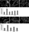
Interfering with EH domain/NPF motif interactions inhibits transferrin recycling and can be rescued by co-overexpression of the corresponding full-length proteins. (A–D) Interference with the syndapin/EHD1 interaction via GFP-syndapin II-l NPF overexpression and rescue by EHD1 co-overexpression in HeLa cells. (A–C) Pulse chase experiments with fluorescently labeled transferrin in cells overexpressing GFP-syndapin II-l NPF region (A), the corresponding triple NPF to NPV mutant (GFP-NPF***) (B) and a combination of GFP-syndapin II-l NPF and FLAG-EHD1 (C). (D) Quantitative data of scoring at least ∼100 transfected cells per coverslip on six to 12 coverslips of five to eight independent assays. Transfected cells scored, GFP-NPF, 1489; GFP, 1268; GFP-NPF***, 1064; GFP-NPF and EDH1, 688; EHD1, 646. Error bars represent SEM. ***p value < 0.0001 (Fisher's PLSD). (E-H) Interference with the syndapin/EHD1 interaction via the EH domain of EHD1 and rescue by syndapin II-l. (E–G) Pulse chase experiments with fluorescently labeled transferrin in cells overexpressing GFP-EHD1 EH domain (E), FLAG-syndapin II-l (F) and a combination of GFP-EHD1 EH domain and FLAG-syndapin II-l (G). (H) Quantitative determination as in D. Transfected cells scored, GFP-EH, 865; FLAG- and Xpress-syndapin II-l, 535; GFP-EH and FLAG-syndapin II-l, 504. Error bars represent SEM of at least ∼100 transfected cells on five to 12 coverslips of four to six independent assays. **p value = 0.0021. Cell borders of untransfected and transfected cells (marked by asterisks) are outlined. Insets in A–C and E–G are threefold enlargements of boxed areas in the corresponding images. Bars in C (for A–C) and in G (for E–G), 20 μm.
Figure 10.
Figure 10.
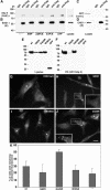
Nucleotide binding of EHD1 modulates interactions of the EH domain, which are crucial for receptor recycling. (A–D) Coimmunoprecipitation of GFP-syndapin II-l (A) but not GFP (C) with FLAG-EHD1 (B and D) from HEK293 cell lysates, which were preincubated for 10 min at room temperature without addition of nucleotides (—) or with addition of AMP, ADPβS, ATPγS, or ATP (5 mM final), by anti-FLAG antibodies covalently attached to protein G-Sepharose. The specific coimmunoprecipitation of GFP-SdpII-l was influenced by the preincubation with different nucleotides, as shown by anti-GFP immunoblotting (A). Quantitation of the bands of immunoprecipitated FLAG-EHD1 (with the use of the program Quantity One from Bio-Rad, Hercules, CA) confirmed the approximately equal efficiency of the immunoprecipitation. Quantitation of coimmunoprecipitated GFP-syndapin II-l and background subtraction yielded the following data: AMP, 22%; ADPβS, 50%; ATPγS, 105%; ATP, 130% of the value for no nucleotide addition (100%). (E and F) Affinity purification experiments of different GFP-EHD1 mutants expressed in HEK293 cells by immobilized GST-syndapin II-l. Analyses of lysates (E) and precipitated material (F) by anti-FLAG immunoblotting show that syndapin II-l coprecipitated wild-type EHD1 and EHD1 G429R but not the P-loop mutant G65R and the EH domain mutant W485A. (G–J) Analyses of fluorescently labeled transferrin after a chase of 20 min in untransfected HeLa cells (unmarked cells in G–J) and in cells overexpressing wild-type FLAG-EHD1 (G) and FLAG-EHD1 mutants (H, G65R; I, W485A; J, G429R). Transfected cells are marked by asterisks. Insets represent enlargements of boxed areas. Bar, 20 μm. (K) Quantitation of cells with remaining endosomal transferrin signal in percentage. Examined cells were from several independent transferrin recycling assays and analyzed by several independent investigators. Cells scored, untransfected cells, 350; wild-type EHD1 646; EHD1 G65R, 602; EHD1 G429R, 805; EHD1 W485A, 706. Error bars represent SEM.

Similar articles

Cited by

References

    1. Caplan, S., Naslavsky, N., Hartnell, L. M., Lodge, R., Polishchuk, R. S., Donaldson, J. G., and Bonifacino, J. S. (2002). A tubular EHD1-containing compartment involved in the recycling of major histocompatibility complex class I molecules to the plasma membrane. EMBO J. 21, 2557–2567. - PMC - PubMed
    1. Carroll, R. C., Beattie, E. C., Xia, H., Luscher, C., Altschuler, Y., Nicoll, R. A., Malenka, R. C., and von Zastrow, M. (1999). Dynamin-dependent endocytosis of ionotropic glutamate receptors. Proc. Natl. Acad. Sci. USA 96, 14112–14117. - PMC - PubMed
    1. Chen, W., Feng, Y., Chen, D., and Wandinger-Ness, A. (1998). Rab11 is required for trans-Golgi network-to-plasma membrane transport and a preferential target for GDP dissociation inhibitor. Mol. Biol. Cell. 9, 3241–3257. - PMC - PubMed
    1. de Beer, T., Hoofnagle, A. N., Enmon, J. L., Bowers, R. C., Yamabhai, M., Kay, B. K., and Overduin, M. (2000). Molecular mechanism of NPF recognition by EH domains. Nat. Struct. Biol. 7, 1018–1022. - PubMed
    1. de Renzis, S., Sonnichsen, B., and Zerial, M. (2002). Divalent Rab effectors regulate the sub-compartmental organization and sorting of early endosomes. Nat. Cell Biol. 4, 124–133. - PubMed

Publication types