. 2005 Jun;79(12):7792-802.
doi: 10.1128/JVI.79.12.7792-7802.2005.

Interaction between the human cytomegalovirus UL82 gene product (pp71) and hDaxx regulates immediate-early gene expression and viral replication

Affiliations

Interaction between the human cytomegalovirus UL82 gene product (pp71) and hDaxx regulates immediate-early gene expression and viral replication

Stacy R Cantrell et al. J Virol. 2005 Jun.

Abstract

The human cytomegalovirus UL82-encoded pp71 protein is required for efficient virus replication and immediate-early gene expression when cells are infected at a low multiplicity. Functions attributed to pp71 include the ability to enhance the infectivity of viral DNA, bind to and target hypophosphorylated Rb family member proteins for degradation, drive quiescent cells into the cell cycle, and bind to the cellular protein hDaxx. Using UL82 mutant viruses, we demonstrate that the LXCXD motif within pp71 is not necessary for efficient virus replication in fibroblasts, suggesting that pp71's ability to degrade hypophosphorylated Rb family members and induce quiescent cells into the cell cycle is not responsible for the growth defect associated with a UL82 deletion mutant. However, UL82 mutants that cannot bind to hDaxx are unable to induce immediate-early gene expression and are severely attenuated for viral replication. These results indicate that the interaction between the human cytomegalovirus UL82 gene product (pp71) and hDaxx regulates immediate-early gene expression and viral replication.


Figures
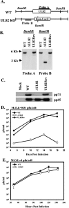
FIG. 1.
FIG. 1.
Generation of the UL82 deletion mutant BAC and recombinant virus. (A) Schematic representation of WT and UL82 deletion mutant genomes. (B) Southern blot analysis of WT pADCREGFP, pADΔUL82, and pADUL82Rev BACs digested with BamHI restriction enzyme and probed for either UL82 or the UL82 right flanking region. (C) Western blot analysis of pp71 expression at 72 h postinfection in HFF cells mock infected (M) or infected with WT, ADΔUL82, or ADUL82Rev virus. As a control, the expression of pp65 was also measured. (D and E) HFF cells were infected at a multiplicity of infection (M.O.I.) of 0.01 PFU/cell (D) or 4 PFU/cell (E) with WT ADCREGFP (•), ADΔUL82 (○), or ADUL82Rev (▾) virus. Cultures were harvested at the indicated times postinfection, and infectious virus was quantified by plaque assay n UL82-complementing cells. Error bars indicate standard deviations.
FIG. 2.
FIG. 2.
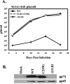
Growth kinetics of ADUL82-C219G mutant virus. (A) HFF cells were infected (multiplicity of infection [M.O.I.] of 0.01 PFU/cell) with WT ADCREGFP (•), ADUL82-C219G (○), or ADΔUL82 (▾) virus. Cultures were harvested at the indicated times postinfection, and infectious virus was quantified by plaque assay on UL82-complementing cells. Error bars indicate standard deviations. (B) Western blot analysis of pp71 expression at 72 h postinfection in HFF cells mock infected or infected with WT, ADUL82-C219G, or ADΔUL82 virus at a multiplicity of 2 PFU/cell. As a control, the expression of pp65 was also measured.
FIG. 3.
FIG. 3.
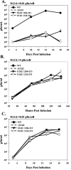
Growth kinetics of wild-type and UL82-hDaxx binding mutants. (A and B) HFF cells were infected with (A) 0.01 PFU/cell or (B) 4 PFU/cell of WT (•), ADΔUL82 (○), ADUL82Δ206-213 (▾), or ADUL82Δ324-331 (▿) virus. Cultures were harvested at the indicated times postinfection, and infectious virus was quantified by plaque assay on UL82 complementing cells. (C) UL82-complementing cells were infected (0.01 PFU/cell) with WT (•), ADΔUL82 (○), ADUL82Δ206-213 (▾), or ADUL82Δ324-331 (▿) virus. Cultures were harvested at the indicated times postinfection, and infectious virus was quantified by plaque assay on UL82-complementing cells. M.O.I., multiplicity of infection. Error bars indicate standard deviations.
FIG. 4.
FIG. 4.
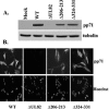
Expression of pp71 from UL82 mutants. (A) HFF cells were infected with WT, ADΔUL82, ADUL82Δ206-213, or ADUL82Δ324-331 virus at a multiplicity of 2 PFU/cell. Lysates were prepared at 72 h postinfection and assayed for pp71 expression by Western blotting. Antitubulin was included as an internal loading control. (B) HFF cells were infected at a multiplicity of 1.0 PFU/cell with WT, ADΔUL82, ADUL82Δ206-213, or ADUL82Δ324-331 virus. Cells were fixed at 72 h postinfection and immunostained with monoclonal antibody against pp71. Cell nuclei were detected with Hoechst stain.
FIG. 5.
FIG. 5.
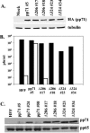
Cell lines expressing pp71-hDaxx mutant proteins fail to complement the UL82 deletion mutant growth defect. (A) Cell lysates from stable HFF cell lines expressing WT pp71 or pp71 which is unable to interact with hDaxx were examined for pp71 expression by Western blot analysis. Two clones of each pp71-hDaxx binding mutant and the pp71-complementing cells (pp71 no. 5) were examined. Antitubulin was included as an internal loading control. (B) HFF cells or cells expressing the various pp71 mutant proteins were infected (0.01 PFU/cell) with WT (black bars) or ADΔUL82 (open bars) virus. Cultures were harvested and infectious virus was quantified by plaque assay on UL82-complementing cells. (C) HFF cells or cells expressing the WT or various pp71 mutant proteins were infected (4 PFU/cell) with ADΔUL82 virus. At 96 h postinfection, virus was collected and purified via ultracentrifugation. Virus particles were lysed and examined for tegument incorporation of pp71 by Western blot analysis. As a control, the tegument protein pp65 was also examined.
FIG. 6.
FIG. 6.
pp71 interaction with hDaxx during virus infections. (A) HFF cells were infected with WT virus at a multiplicity of 3 PFU/cell. Lysates were prepared at the indicated times postinfection (p.i.) and assayed for hDaxx expression by Western blotting. (B) HFF cells were infected with WT virus at a multiplicity of 2 PFU/cell. Lysates were prepared at the indicated times postinfection and incubated with antibody against hDaxx. Immune complexes were collected, separated by SDS-PAGE, transferred to membranes, and probed for pp71 via Western blotting. Lysates were also examined for pp71 expression by Western blot analysis. IP, immunoprecipitation. (C) HFF cells were infected with WT, ADΔUL82, ADUL82Δ206-213, or ADUL82Δ324-331 virus (1 PFU/cell). Cell lysates were prepared at 72 h postinfection and incubated with hDaxx antibody. Immune complexes were collected, separated by SDS-PAGE, transferred to membranes, and probed for pp71 via Western blotting. Cell lysates were also examined for pp71 expression by Western blot analysis. (D) HFF cells were infected at a multiplicity of 10 PFU/cell with WT, ADΔUL82, ADUL82Δ206-213, or ADUL82Δ324-331 virus in the presence of 100 μg/ml cycloheximide. Cells were fixed at 2 h postinfection and immunostained with monoclonal antibody against pp71 (green) and polyclonal antibody against PML (red) to detect ND10 domains.
FIG. 7.
FIG. 7.
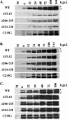
IE gene expression following infection with UL82 mutant viruses. HFF cells were infected at a multiplicity of 0.1 PFU/cell (A) or 2.0 PFU/cell (C) with WT, ADΔUL82, ADUL82Δ206-213, ADUL82Δ324-331, or ADUL82-C219G virus. Cell lysates were prepared at the indicated times postinfection (p.i.) and assayed for IE1 (◊) and IE2 (*) expression by Western blotting. (B) UL82-complementing cells were infected (0.1 PFU/cell) with WT, ADΔUL82, ADUL82Δ206-213, ADUL82Δ324-331, or ADUL82-C219G virus. Cell lysates were prepared at the indicated times postinfection and examined for IE1 (◊) and IE2 (*) expression by Western blotting.
FIG. 8.
FIG. 8.
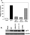
Effect of pp71 mutant proteins on viral DNA infectivity. (A) HFF cells were transfected with WT ADCREGFP viral DNA (1 μg) and 5 μg pCGN vector (open bar), pCGNpp71 (black bar), pCGNpp71Δ206-213 (diagonally striped bar), pCGNpp71Δ324-331 (vertically striped bar), or pCGNpp71-C219G (crosshatched bar). Cells were plated in six-well dishes and overlaid with agarose. Plaques were fixed, stained, and counted at 15 days posttransfection. (B) Cell lysates from one well of each transfection described in A was harvested at 36 h posttransfection and assayed for pp71 expression by Western blot analysis. Antitubulin was included as an internal loading control.

Similar articles

Cited by

References

    1. Baldick, C. J., Jr., A. Marchini, C. E. Patterson, and T. Shenk. 1997. Human cytomegalovirus tegument protein pp71 (ppUL82) enhances the infectivity of viral DNA and accelerates the infectious cycle. J. Virol. 71:4400-4408. - PMC - PubMed
    1. Borst, E. M., G. Hahn, U. H. Koszinowski, and M. Messerle. 1999. Cloning of the human cytomegalovirus (HCMV) genome as an infectious bacterial artificial chromosome in Escherichia coli: a new approach for construction of HCMV mutants. J. Virol. 73:8320-8329. - PMC - PubMed
    1. Bradford, M. M. 1976. A rapid and sensitive method for the quantitation of microgram quantities of protein utilizing the principle of protein-dye binding. Anal. Biochem. 72:248-254. - PubMed
    1. Bresnahan, W. A., I. Boldogh, T. Ma, T. Albrecht, and E. A. Thompson. 1996. Cyclin E/Cdk2 activity is controlled by different mechanisms in the G0 and G1 phases of the cell cycle. Cell Growth Differ. 7:1283-1290. - PubMed
    1. Bresnahan, W. A., G. E. Hultman, and T. Shenk. 2000. Replication of wild-type and mutant human cytomegalovirus in life-extended human diploid fibroblasts. J. Virol. 74:10816-10818. - PMC - PubMed

Publication types

MeSH terms

LinkOut - more resources