Male sterility in mice lacking retinoic acid receptor alpha involves specific abnormalities in spermiogenesis
- PMID: 15901285
- PMCID: PMC3785313
- DOI: 10.1111/j.1432-0436.2005.00018.x
Male sterility in mice lacking retinoic acid receptor alpha involves specific abnormalities in spermiogenesis
Abstract
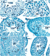
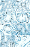
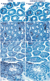
The severe degeneration of the germinal epithelium and subsequent male sterility observed in mice null for the retinoic acid receptor alpha (RARalpha) gene suggested its critical role in spermatogenesis, although the etiology and progression of these abnormalities remain to be determined. Previous studies have revealed that elongated spermatids in RARalpha(-/-) testes were improperly aligned at the tubular lumen and did not undergo spermiation at stage VIII(*). We now report a distinctive failure of step 8-9 spermatids to orient properly with regard to the basal aspect of Sertoli cells, resulting in stage VIII(*)-IX(*) tubules with randomly oriented spermatids. By in situ terminal deoxynucleotidyltransferase-mediated deoxy-UTP nick end labeling (TUNEL), we noted that elongating spermatids frequently underwent apoptosis. Immunohistochemical analysis revealed that while activated caspase-3, the primary effector caspase in the apoptotic cell death machinery, was detected in the nuclei of primary spermatocytes in the first wave of spermatogenesis and occasionally in spermatogonia of both normal and mutant testes, it was not involved in the death of elongating spermatids in RARalpha(-/-) testes. Thus, sterility in RARalpha(-/-) males was associated with specific defects in spermiogenesis, which may correlate with a failure in both spermatid release and spermatid orientation to the basal aspect of Sertoli cells at stage VIII(*) in young adult RARalpha(-/-) testis. Further, the resulting apoptosis in elongating spermatids appears to involve pathways other than that mediated by activated caspase-3.
Figures


Similar articles
-
Potential functions of retinoic acid receptor A in Sertoli cells and germ cells during spermatogenesis.Ann N Y Acad Sci. 2007 Dec;1120:114-30. doi: 10.1196/annals.1411.008. Epub 2007 Sep 28. Ann N Y Acad Sci. 2007. PMID: 17905941
-
Loss of TSLC1 causes male infertility due to a defect at the spermatid stage of spermatogenesis.Mol Cell Biol. 2006 May;26(9):3595-609. doi: 10.1128/MCB.26.9.3595-3609.2006. Mol Cell Biol. 2006. PMID: 16611999 Free PMC article.
-
Seipin deficiency increases chromocenter fragmentation and disrupts acrosome formation leading to male infertility.Cell Death Dis. 2015 Jul 16;6(7):e1817. doi: 10.1038/cddis.2015.188. Cell Death Dis. 2015. PMID: 26181198 Free PMC article.
-
Retinoid signaling during spermatogenesis as revealed by genetic and metabolic manipulations of retinoic acid receptor alpha.Soc Reprod Fertil Suppl. 2007;63:11-23. Soc Reprod Fertil Suppl. 2007. PMID: 17566257 Free PMC article. Review.
-
Role of retinoid signaling in the regulation of spermatogenesis.Cytogenet Genome Res. 2004;105(2-4):189-202. doi: 10.1159/000078189. Cytogenet Genome Res. 2004. PMID: 15237207 Free PMC article. Review.
Cited by
-
New approaches to male non-hormonal contraception.Contraception. 2013 Mar;87(3):296-9. doi: 10.1016/j.contraception.2012.08.016. Epub 2012 Sep 17. Contraception. 2013. PMID: 22995542 Free PMC article. Review.
-
Periodic production of retinoic acid by meiotic and somatic cells coordinates four transitions in mouse spermatogenesis.Proc Natl Acad Sci U S A. 2017 Nov 21;114(47):E10132-E10141. doi: 10.1073/pnas.1710837114. Epub 2017 Nov 6. Proc Natl Acad Sci U S A. 2017. PMID: 29109271 Free PMC article.
-
Function of Retinoic Acid in Development of Male and Female Gametes.Nutrients. 2022 Mar 18;14(6):1293. doi: 10.3390/nu14061293. Nutrients. 2022. PMID: 35334951 Free PMC article. Review.
-
The future of male contraception: a fertile ground.Transl Androl Urol. 2018 May;7(Suppl 2):S220-S235. doi: 10.21037/tau.2018.03.23. Transl Androl Urol. 2018. PMID: 29928620 Free PMC article. Review.
-
Spermatogenesis: The Commitment to Meiosis.Physiol Rev. 2016 Jan;96(1):1-17. doi: 10.1152/physrev.00013.2015. Physiol Rev. 2016. PMID: 26537427 Free PMC article. Review.
References
-
- Akmal KM, Dufour JM, Kim KH. Retinoic acid receptor alpha gene expression in the rat testis: potential role during the prophase of meiosis and in the transition from round to elongating spermatids. Biol Reprod. 1997;56:549–556. - PubMed
-
- Akmal KM, Dufour JM, Vo M, Higginson S, Kim KH. Ligand-dependent regulation of retinoic acid receptor alpha in rat testis: in vivo response to depletion and repletion of vitamin A. Endocrinology. 1998;139:1239–1248. - PubMed
-
- Alnemri ES, Livingston DJ, Nicholson DW, Salvesen G, Thornberry NA, Wong WW, Yuan J. Human ICE/CED-3 protease nomenclature. Cell. 1996;87:171. - PubMed
-
- Beumer TL, Roepers-Gajadien HL, Gademan LS, Rutgers DH, de Rooij DG. P21(Cip1/WAF1) expression in the mouse testis before and after X irradiation. Mol Reprod Dev. 1997;47:240–247. - PubMed
-
- Billig H, Furuta I, Rivier C, Tapanainen J, Parvinen M, Hsueh AJ. Apoptosis in testis germ cells: developmental changes in gonadotropin dependence and localization to selective tubule stages. Endocrinology. 1995;136:5–12. - PubMed
Publication types
MeSH terms
Substances
Grants and funding
LinkOut - more resources
Full Text Sources
Other Literature Sources
Medical
Molecular Biology Databases
Research Materials

