. 2005 Jun;79(11):7182-94.
doi: 10.1128/JVI.79.11.7182-7194.2005.

Elimination of ie1 significantly attenuates murine cytomegalovirus virulence but does not alter replicative capacity in cell culture

Affiliations

Elimination of ie1 significantly attenuates murine cytomegalovirus virulence but does not alter replicative capacity in cell culture

Peter Ghazal et al. J Virol. 2005 Jun.

Abstract

The major immediate-early (MIE) genes of cytomegaloviruses (CMV) are broadly thought to be decisive regulators of lytic replication and reactivation from latency. To directly assess the role of the MIE protein IE1 during the infection of murine CMV (MCMV), we constructed an MCMV with exon 4 of the ie1 gene deleted. We found that, independent of the multiplicity of infection, the resulting recombinant virus, MCMVdie1, which fails to express the IE1 protein, was fully competent for early gene expression and replicated in different cultured cell types with identical kinetics to those of parental or revertant virus. Immunofluorescence microscopy studies revealed that MCMVdie1 was greatly impaired in its capacity to disrupt promyelocytic leukemia bodies in NIH 3T3 cells early after infection, a process that has been proposed to increase viral transcription efficiency. We examined MCMVdie1 in the murine model using both immunocompetent BALB/c and severe combined immunodeficient (SCID) mice. When MCMVdie1 was inoculated into these two types of mice, significantly lower viral titers were detected in infected organs than in those of the wild-type virus-infected animals. Moreover, the ie1-deficient MCMV exhibited a markedly reduced virulence. While all animals infected with 5 x 10(4) PFU of parental virus died by 30 days postinfection, SCID mice infected with a similar dose of MCMVdie1 did not succumb before 60 days postinfection. The in vivo defective growth phenotype of MCMVdie1 was abrogated upon rescue of ie1. These results demonstrate the significance of the ie1 gene for promoting an acute MCMV infection and virulence yet indicate that MCMV is able to grow in vivo, although impaired, in the absence of the ie1 gene.


Figures

FIG. 1.
FIG. 1.
Construction of an MCMV BAC genome lacking exon 4 of the ie1 gene. (A) Schematic representation of the parental pSM3fr, pSM3frdie1, and the revertant pSM3frdie1-rev BAC. The HindIII map of the MCMV genome is shown at the top. The expanded map of the HindIII K and HindIII L fragments represents the MCMV MIE gene region. Coding exons are shown as black rectangles, and the first noncoding exon of the ie1/ie3 transcription unit is shown as an white rectangle. The gray box represents the MCMV enhancer ie1/ie3 promoter. The structures of the ie1 and ie3 transcripts are indicated below line 1 (of the expanded map). Starting with the parental BAC pSM3fr (line 1), the mutant BAC pSM3frdie1 (line 2) and the revertant BAC pSM3frdie1-rev (line 3) were generated by successive rounds of homologous recombination in E. coli as described in Materials and Methods. The deletion of the fourth exon of the ie1 gene is marked by the delta (Δ). The sizes of the natural HindIII K and L fragments in pSM3fr and pSM3frdie1-rev and that of the new HindIII fragment in pSM3frdie1 are indicated. (B) Genomic structure of pSM3fr, pSM3frdie1, and pSM3frdie1-rev. Ethidium bromide-stained 0.7% agarose gel of HindIII-digested BACs pSM3fr (lane 1), pSM3frdie1 (lane 2), and pSM3frie1-rev (lane 3). Size markers are indicated in the left margin. The names of the MCMV HindIII fragments (16) are shown in the right margin.
FIG. 2.
FIG. 2.
Absence of IE1 protein in cells infected with MCMVdie1. NIH 3T3 cells were either mock infected (M) or infected with the parental MCMV (lanes 1), MCMVdie1 (lanes 2), or MCMVrev (lanes 3) at an MOI of 5 PFU/cell in the absence or presence of cycloheximide (CHX) as indicated in Materials and Methods. CHX-treated samples were subsequently exposed to CHX for the 3 h after the adsorption period, at which time the CHX was removed and actinomycin D was added for 4 h before the cells were harvested at 7 hpi. At the time points after infection indicated (7 h for the CHX samples), cells were lysed, subjected to sodium dodecyl sulfate-polyacrylamide gel electrophoresis analysis on 7.5% polyacrylamide gels and Western blots performed with anti-IE1 (IE1)- or anti-E1 (E1)-specific monoclonal antibodies or an IE3-specific antiserum (IE3).
FIG. 3.
FIG. 3.
Growth of MCMVdie1 in NIH 3T3 cells. NIH 3T3 cells were infected at an MOI of 0.01 (A) or 2 (B) PFU/cell with the parental MCMV, MCMVdie1, or MCMVrev. Culture supernatants were collected at the indicated times after infection (days postinfection [dpi]) and titrated by standard plaque assays on NIH 3T3 cells. Each data point represents the average and standard deviation of results from three separate cultures.
FIG. 4.
FIG. 4.
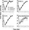
Multistep growth curves of MCMVdie1 on different cell types. Mouse embryonic fibroblasts, SVEC4 cells, C127I cells, and peritoneal macrophages were infected with 0.05 PFU/cell, except for macrophages, which were infected at an MOI of 0.5 PFU/cell, of parental MCMV, MCMVdie1, or MCMVrev. Culture supernatants were collected at the designated times postinfection (days postinfection [dpi]) and titrated by plaque assays on NIH 3T3 cells. In the case of peritoneal macrophages, intracellular viral titers were determined. Each data point represents the average and standard deviation of results from three separate cultures.
FIG. 5.
FIG. 5.
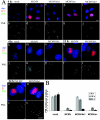
MCMVdie1 fails to disrupt PML bodies in NIH 3T3 cells at immediate-early times after infection. NIH 3T3 cells were mock infected or infected with MCMV, MCMVdie1, MCMVrev, or MCMVdie3 at an MOI of 0.3. At the different times postinfection indicated, cells were fixed with paraformaldehyde and double labeling was performed for MCMV IE1 or E1 and PML as described in Materials and Methods. DNA was counterstained with DAPI. (A) Shown are photomicrographs from the different cultures. Panels a to p, 4 hpi; panels q to t, 15 hpi; panels u to x, 24 hpi. IE1 is shown in red (a, c, e, g, i), E1 in red (k, m, o, q, s, u, w), PML in green and black/white, and DAPI in blue. Bars, 10 μm. (B) The number of PML bodies in mock-infected cells and cells infected with MCMV, MCMVdie1, or MCMVrev were counted at the various times after infection indicated. For mock-infected samples, 100 cells were considered in the quantitation. For infected samples, only infected cells (E1 positive) were included in the quantitation: at 4 hpi, 100 cells per sample; at 15 hpi, >50 cells per sample; at 24 hpi, 14 to 29 cells per sample.
FIG. 6.
FIG. 6.
Excision of BAC vector sequences from the MCMVdie1 genome. (A) PCRs were carried out using genomic DNAs isolated from a full-length MCMV BAC (lanes 1 and 5) or from cells infected with the viral stocks of MCMV (lanes 2 and 6), MCMVdie1 (lanes 3 and 7), and MCMVrev (lanes 4 and 8) as templates. Two distinct primer sets were used to analyze the appropriate excision of the BAC vector sequences (see Materials and Methods for details). The first primer set includes a primer that binds to the BAC sequence and a primer that anneals with a sequence located within the EcoRI g fragment (lanes 1 to 4), and the second primer set contains two primers that anneal with MCMV sequences flanking the excision site (lanes 5 to 8). Amplified products were separated on a 1% agarose gel and visualized by ethidium bromide staining. Products obtained in the PCR are marked by arrows indicating their sizes. The positions of the size markers are shown on the right. (B) Schematic representation of the viral genomes before and after excision of the BAC sequences. The probe detects a fragment of 12.1 kbp (before excision of the BAC sequences) and 3.7 kbp (after excision of the BAC sequences) in MluI-digested viral DNA. Nucleotide positions refer to the MCMV sequence (53). (C) Ethidium bromide-stained 0.7% agarose gel of MluI-digested viral DNAs from recombinant viral stocks (left panel) and corresponding Southern blot analysis (right panel). The sizes of the molecular size markers and detected DNA fragments are indicated in the left and right margins, respectively. Lanes 1 and 5, full-length MCMV BAC; lanes 2 and 6, MCMV; lanes 3 and 7, MCMVdie1; lanes 4 and 8, MCMVrev. The blot was overexposed to clearly show the absence of signals corresponding to BAC sequences in the viral stock DNAs.
FIG. 7.
FIG. 7.
Growth of MCMVdie1 in different organs of BALB/c mice. Groups of BALB/c mice (four to six per group) were inoculated intraperitoneally with 1 × 10⁶ PFU of tissue culture-propagated parental MCMV (gray circles), MCMVdie1 (white circles), or MCMVrev (black circles). On days 3, 7, and 14 after infection, mice were sacrificed and the indicated organs removed, weighed, and sonicated as a 10% (wt/vol) tissue homogenate in DMEM. Viral titers of the resulting homogenates were determined by standard plaque assays on NIH 3T3 cells. Shown are the titers corresponding to the different organs for each individual mouse within a group. The dashed line shows the detection limit. Horizontal bars, median values; Saliv., salivary.
FIG. 8.
FIG. 8.
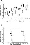
Growth and virulence of MCMVdie1 in SCID mice. (A) Groups of six SCID mice (CB17) were inoculated intraperitoneally with 1 × 10⁶ PFU of tissue culture-propagated parental MCMV (gray circles), MCMVdie1 (white circles), or MCMVrev (black circles). On days 7 and 14 after infection, mice were sacrificed and the indicated organs removed, weighed, and sonicated as a 10% (wt/vol) tissue homogenate in DMEM. Viral titers of the resulting homogenates were determined by standard plaque assays on NIH 3T3 cells. Shown are the titers corresponding to the different organs for each individual mouse within a group. All values were among the limits of detection of the assay. Horizontal bars, median values. Data shown correspond to results from one of two independently performed experiments in which similar results were obtained. (B) Groups of six SCID mice (CB17) were infected intraperitoneally with 5 × 10⁴ PFU of either parental MCMV (gray circles), MCMVdie1 (white circles), or MCMVrev (black circles) and observed daily for mortality.

Similar articles

Cited by

References

    1. Ahn, J. H., and G. S. Hayward. 1997. The major immediate-early proteins IE1 and IE2 of human cytomegalovirus colocalize with and disrupt PML-associated nuclear bodies at very early times in infected permissive cells. J. Virol. 71:4599-4613. - PMC - PubMed
    1. Ahn, J. H., and G. S. Hayward. 2000. Disruption of PML-associated nuclear bodies by IE1 correlates with efficient early stages of viral gene expression and DNA replication in human cytomegalovirus infection. Virology 274:39-55. - PubMed
    1. Angulo, A., P. Ghazal, and M. Messerle. 2000. The major immediate-early gene ie3 of mouse cytomegalovirus is essential for viral growth. J. Virol. 74:11129-11136. - PMC - PubMed
    1. Angulo, A., M. Messerle, U. H. Koszinowski, and P. Ghazal. 1998. Enhancer requirement for murine cytomegalovirus growth and genetic complementation by the human cytomegalovirus enhancer. J. Virol. 72:8502-8509. - PMC - PubMed
    1. Balthesen, M., L. Dreher, P. Lucin, and M. J. Reddehase. 1994. The establishment of cytomegalovirus latency in organs is not linked to local virus production during primary infection. J. Gen. Virol. 75:2329-2336. - PubMed

Publication types

MeSH terms

LinkOut - more resources