. 2005 Sep 1;390(Pt 2):529-40.
doi: 10.1042/BJ20041711.

The semi-phosphorylative Entner-Doudoroff pathway in hyperthermophilic archaea: a re-evaluation

Affiliations

The semi-phosphorylative Entner-Doudoroff pathway in hyperthermophilic archaea: a re-evaluation

Hatim Ahmed et al. Biochem J. .

Abstract

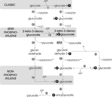
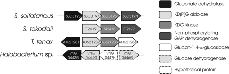
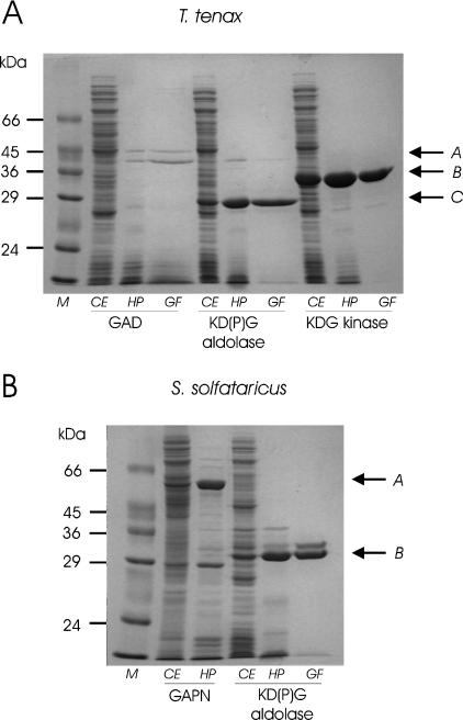
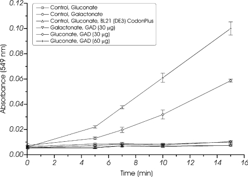
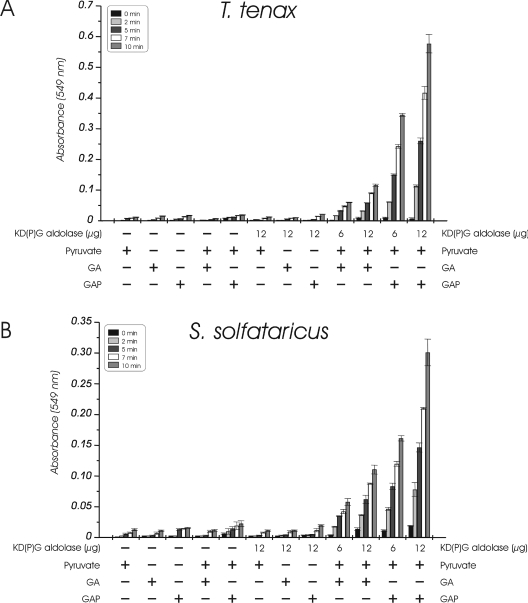
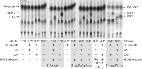
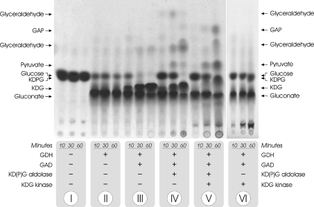
Biochemical studies have suggested that, in hyperthermophilic archaea, the metabolic conversion of glucose via the ED (Entner-Doudoroff) pathway generally proceeds via a non-phosphorylative variant. A key enzyme of the non-phosphorylating ED pathway of Sulfolobus solfataricus, KDG (2-keto-3-deoxygluconate) aldolase, has been cloned and characterized previously. In the present study, a comparative genomics analysis is described that reveals conserved ED gene clusters in both Thermoproteus tenax and S. solfataricus. The corresponding ED proteins from both archaea have been expressed in Escherichia coli and their specificity has been identified, revealing: (i) a novel type of gluconate dehydratase (gad gene), (ii) a bifunctional 2-keto-3-deoxy-(6-phospho)-gluconate aldolase (kdgA gene), (iii) a 2-keto-3-deoxygluconate kinase (kdgK gene) and, in S. solfataricus, (iv) a GAPN (non-phosphorylating glyceraldehyde-3-phosphate dehydrogenase; gapN gene). Extensive in vivo and in vitro enzymatic analyses indicate the operation of both the semi-phosphorylative and the non-phosphorylative ED pathway in T. tenax and S. solfataricus. The existence of this branched ED pathway is yet another example of the versatility and flexibility of the central carbohydrate metabolic pathways in the archaeal domain.


Figures

Figure 1
Figure 1. Glucose catabolism via the different ED pathways
Overview of the classical and modifications of the ED pathway, each with the characteristic phosphorylation level indicated. Non-phosphorylated intermediates are depicted on the left, and phosphorylated intermediates on the right. The key phosphorylation reactions for the different ED versions are highlighted in grey boxes (glucokinase/hexokinase for the classical ED, KDG kinase for the semi-phosphorylative ED and glycerate kinase for the non-phosphorylative ED). Key to enzymes: 1, glucokinase/hexokinase; 2, glucose-6-phosphate dehydrogenase; 3, 6-phosphogluconate dehydratase; 4, KDPG aldolase; 5, GAPDH; 6, 3-phosphoglycerate kinase; 7, phosphoglycerate mutase; 8, enolase; 9, pyruvate kinase; 10, GAPN/GAP oxidoreductase; 11, GDH; 12, GAD; 13, KDG kinase; 14, KD(P)G aldolase; 15, aldehyde dehydrogenase/aldehyde oxidoreductase; and 16, glycerate kinase.
Figure 2
Figure 2. ED gene clusters in archaea identified by conserved genome context analysis
Schematic representation of the conserved gene clusters in archaeal genomes, comprising key genes of the semi-phosphorylative ED pathway. The genes are indicated by their systematic gene name; except for T. tenax, the accession numbers are displayed and orthologous genes are shaded in the same grey scale. Genes are not drawn to scale.
Figure 3
Figure 3. Recombinant production and purification of T. tenax and S. solfataricus ED proteins
(A) SDS/PAGE of recombinant expression and purification of T. tenax GAD, KD(P)G aldolase and KDG kinase. Arrows indicate the purified recombinant GAD (A), KD(P)G aldolase (C) and KDG kinase (B). (B) SDS/PAGE of recombinant expression and purification of S. solfataricus KD(P)G aldolase and GAPN. Arrows indicate the purified recombinant GAPN (A) and KD(P)G aldolase (B). Lanes containing crude cell extracts (CE), soluble fractions after heat precipitation (HP) and gel filtration (GF) were loaded with 20, 10 and 5 μg of protein respectively. Lane M corresponds to the protein marker, Dalton Mark VII-L (Sigma).
Figure 4
Figure 4. GAD activity of T. tenax
GAD activity (protein fraction after gel filtration) was monitored at 70 °C using the discontinuous TBA assay. Activity on gluconate (10 mM; 30 and 60 μg of protein) as well as galactonate (10 mM; 30 μg of protein) and controls without enzyme (10 mM gluconate and galactonate) or substrate (results not shown) and heat-precipitated extract of BL21 (DE3) CodonPlus with pET-15b without insert are shown. All experiments were performed in triplicate and the S.D. is given. GAD activity was only observed in the presence of gluconate and the observed activity is proportional to the amount of enzyme.
Figure 5
Figure 5. KD(P)G aldolase activity of T. tenax (A) and S. solfataricus (B)
The formation of KDG and KDPG from pyruvate (5 mM) and GA or GAP (2 mM) respectively was monitored at 70 °C using the discontinuous TBA assay. The dependence on the amount of protein (6 and 12 μg of protein, fraction after gel filtration) and controls with one (GA, GAP or pyruvate) or both (pyruvate and GA or GAP respectively) substrates without enzyme and with one substrate (GA, GAP or pyruvate respectively) in the presence of enzyme are shown. For each probe, three independent measurements were performed and the experimental error is given. In the presence of KD(P)G aldolase, the formation of KDG from GA and pyruvate as well as KDPG from GAP and pyruvate was observed. The activity with non-phosphorylated and phosphorylated substrates is proportional to the amount of enzyme used in the assay.
Figure 6
Figure 6. Detection of 14C-labelled KDG and KDPG through TLC and autoradiography
The KD(P)G aldolases of T. tenax and S. solfataricus (fraction after heat precipitation) were incubated at 70 °C in the presence of labelled pyruvate ([2-14C]pyruvate) and either GA or GAP. Control samples containing different combinations of KD(P)G aldolase and substrates are shown as indicated. In addition, control samples of the expression host BL21 (DE3) CodonPlus with pET-15b without insert after heat precipitation, indicated by ‘+’, and formation of 14C-labelled KDG and KDPG by the KDPG aldolase (EDA) of T. maritima (fraction after heat precipitation) [34] are shown. The formation of 14C-labelled KDG and KDPG was monitored via TLC and visualized using autoradiography. As for the KDPG aldolase of T. maritima, the formation of both KDG (from GA and pyruvate) and KDPG (from GAP and pyruvate) is observed in the presence of the KD(P)G aldolase of T. tenax or S. solfataricus.
Figure 7
Figure 7. KDG kinase activity of T. tenax
The KDG kinase activity was determined in a continuous assay at 70 °C by monitoring the formation of GAP after KDPG cleavage via KD(P)G aldolase and GAPN of T. tenax. The rate dependence on the KDG concentration, determined by the TBA assay, is shown. The enzyme follows Michaelis–Menten kinetics for KDG. The inset shows the linear transformation according to Hanes. For the assay, it was ensured that the amount of auxiliary enzymes is not rate-limiting and the measured enzyme activity was directly proportional to the amount of enzyme added to the assay (results not shown). Three independent assays were performed for each substrate concentration and the S.D. is given. The rate-dependent formation of GAP was only monitored in the presence of KDG, ATP, Mg2+, auxiliary enzymes and the KDG kinase of T. tenax.
Figure 8
Figure 8. Reconstruction of the ED pathway in vitro
[U-14C]glucose was incubated in the presence of different combinations of ED enzymes from T. tenax [GDH, GAD, KD(P)G aldolase and KDG kinase; protein fractions after heat precipitation] as indicated (10, 30 and 60 min at 70 °C respectively), and the labelled intermediates were separated by TLC and visualized using autoradiography. The labelling pattern in the presence of KDG kinase and KD(P)G aldolase (V) indicates the co-existence of both the semi-phosphorylative and non-phosphorylative ED modifications in T. tenax.

Similar articles

Cited by

References

    1. Ronimus R. S., Morgan H. W. Distribution and phylogenies of enzymes of the Embden-Meyerhof-Parnas pathway from archaea and hyperthermophilic bacteria support a gluconeogenic origin of metabolism. Archaea. 2002;1:199–221. - PMC - PubMed
    1. Verhees C. H., Kengen S. W. M., Tuininga J. E., Schut G. J., Adams M. W. W., de Vos W. M., van der Oost J. The unique features of glycolytic pathways in Archaea. Biochem. J. 2003;375:231–246. - PMC - PubMed
    1. Verhees C. H., Kengen S. W. M., Tuininga J. E., Schut G. J., Adams M. W. W., de Vos W. M., van der Oost J. The unique features of glycolytic pathways in Archaea. Biochem. J. 2004;377:819–822. - PMC - PubMed
    1. Siebers B., Tjaden B., Michalke K., Dörr C., Ahmed H., Zaparty M., Gordon P., Sensen C. W., Zibat A., Klenk H., et al. Reconstruction of the central carbohydrate metabolism of Thermoproteus tenax by use of genomic and biochemical data. J. Bacteriol. 2004;186:2179–2194. - PMC - PubMed
    1. De Rosa M., Gambacorta A., Nicolaus B., Giardina P., Poerio E., Buonocore V. Glucose metabolism in the extreme thermoacidophilic archaebacterium Sulfolobus solfataricus. Biochem. J. 1984;224:407–414. - PMC - PubMed

Publication types

MeSH terms

Substances