Endosomal proteolysis of the Ebola virus glycoprotein is necessary for infection
- PMID: 15831716
- PMCID: PMC4797943
- DOI: 10.1126/science.1110656
Endosomal proteolysis of the Ebola virus glycoprotein is necessary for infection
Abstract
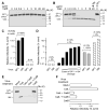
Ebola virus (EboV) causes rapidly fatal hemorrhagic fever in humans and there is currently no effective treatment. We found that the infection of African green monkey kidney (Vero) cells by vesicular stomatitis viruses bearing the EboV glycoprotein (GP) requires the activity of endosomal cysteine proteases. Using selective protease inhibitors and protease-deficient cell lines, we identified an essential role for cathepsin B (CatB) and an accessory role for cathepsin L (CatL) in EboV GP-dependent entry. Biochemical studies demonstrate that CatB and CatL mediate entry by carrying out proteolysis of the EboV GP subunit GP1 and support a multistep mechanism that explains the relative contributions of these enzymes to infection. CatB and CatB/CatL inhibitors diminish the multiplication of infectious EboV-Zaire in cultured cells and may merit investigation as anti-EboV drugs.
Figures


Similar articles
-
Role of endosomal cathepsins in entry mediated by the Ebola virus glycoprotein.J Virol. 2006 Apr;80(8):4174-8. doi: 10.1128/JVI.80.8.4174-4178.2006. J Virol. 2006. PMID: 16571833 Free PMC article.
-
Novel proteolytic activation of Ebolavirus glycoprotein GP by TMPRSS2 and cathepsin L at an uncharted position can compensate for furin cleavage.Virus Res. 2024 Sep;347:199430. doi: 10.1016/j.virusres.2024.199430. Epub 2024 Jul 8. Virus Res. 2024. PMID: 38964470 Free PMC article.
-
Inhibitors of signal peptide peptidase and subtilisin/kexin-isozyme 1 inhibit Ebola virus glycoprotein-driven cell entry by interfering with activity and cellular localization of endosomal cathepsins.PLoS One. 2019 Apr 11;14(4):e0214968. doi: 10.1371/journal.pone.0214968. eCollection 2019. PLoS One. 2019. PMID: 30973897 Free PMC article.
-
[Ebola and Marburg viruses: the humans strike back].Med Sci (Paris). 2006 Apr;22(4):405-10. doi: 10.1051/medsci/2006224405. Med Sci (Paris). 2006. PMID: 16597410 Review. French.
-
Molecular Mechanism of Externalization of Phosphatidylserine on the Surface of Ebola Virus Particles.DNA Cell Biol. 2019 Feb;38(2):115-120. doi: 10.1089/dna.2018.4485. Epub 2019 Jan 7. DNA Cell Biol. 2019. PMID: 30615471 Review.
Cited by
-
Multimerization of Ebola GPΔmucin on protein nanoparticle vaccines has minimal effect on elicitation of neutralizing antibodies.Front Immunol. 2022 Aug 24;13:942897. doi: 10.3389/fimmu.2022.942897. eCollection 2022. Front Immunol. 2022. PMID: 36091016 Free PMC article.
-
Niemann-Pick C1 (NPC1)/NPC1-like1 chimeras define sequences critical for NPC1's function as a flovirus entry receptor.Viruses. 2012 Oct 25;4(11):2471-84. doi: 10.3390/v4112471. Viruses. 2012. PMID: 23202491 Free PMC article.
-
Diminished reovirus capsid stability alters disease pathogenesis and littermate transmission.PLoS Pathog. 2015 Mar 4;11(3):e1004693. doi: 10.1371/journal.ppat.1004693. eCollection 2015 Mar. PLoS Pathog. 2015. PMID: 25738608 Free PMC article.
-
Ready, set, fuse! The coronavirus spike protein and acquisition of fusion competence.Viruses. 2012 Apr;4(4):557-80. doi: 10.3390/v4040557. Epub 2012 Apr 12. Viruses. 2012. PMID: 22590686 Free PMC article. Review.
-
Haploid Genetic Screen Reveals a Profound and Direct Dependence on Cholesterol for Hantavirus Membrane Fusion.mBio. 2015 Jun 30;6(4):e00801. doi: 10.1128/mBio.00801-15. mBio. 2015. PMID: 26126854 Free PMC article.
References
Publication types
MeSH terms
Substances
Grants and funding
LinkOut - more resources
Full Text Sources
Other Literature Sources
Medical
Molecular Biology Databases

