Comparative Study
. 2005 Apr 15;19(8):933-41.
doi: 10.1101/gad.333205. Epub 2005 Apr 1.

The nucleolus as a stress sensor: JNK2 inactivates the transcription factor TIF-IA and down-regulates rRNA synthesis

Affiliations
Comparative Study

The nucleolus as a stress sensor: JNK2 inactivates the transcription factor TIF-IA and down-regulates rRNA synthesis

Christine Mayer et al. Genes Dev. .

Abstract

Cells respond to a variety of extracellular and intracellular forms of stress by down-regulating rRNA synthesis. We have investigated the mechanism underlying stress-dependent inhibition of RNA polymerase I (Pol I) transcription and show that the Pol I-specific transcription factor TIF-IA is inactivated upon stress. Inactivation is due to phosphorylation of TIF-IA by c-Jun N-terminal kinase (JNK) at a single threonine residue (Thr 200). Phosphorylation at Thr 200 impairs the interaction of TIF-IA with Pol I and the TBP-containing factor TIF-IB/SL1, thereby abrogating initiation complex formation. Moreover, TIF-IA is translocated from the nucleolus into the nucleoplasm. Substitution of Thr 200 by valine as well as knock-out of Jnk2 prevent inactivation and translocation of TIF-IA, leading to stress-resistance of Pol I transcription. Our data identify TIF-IA as a downstream target of the JNK pathway and suggest a critical role of JNK2 to protect rRNA synthesis against the harmful consequences of cellular stress.


Figures
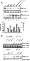
Figure 1.
Figure 1.
Stress-induced activation of JNK inhibits Pol I transcription. (A) Cellular stress induces JNK activity and impairs rRNA synthesis. RNA from HEK293T cells that were mock-treated or treated with H2O2 (200 μM, 60 min), BSO (1 mM, 16 h), anisomycin (10 μM, 60 min), SB203580 (2 μM, 20 min), or SP600125 (5 μM, 30 min) followed by anisomycin (10 μM, 60 min) was analyzed on Northern blots for 45S pre-rRNA (upper panel) and JNK2 activity and levels (lower two panels). To measure JNK2 activity, HEK293T cells were transfected with pcDNA3.1HA-JNK2 and subjected to the indicated stress stimuli. HA-JNK2 was immunopurified from 2 × 10⁶ cells and tested for kinase activity using purified GST-tagged c-Jun (amino acids 1-166). Western blots of cellular lysates were probed using antibodies against overexpressed HA-JNK2. A bar diagram shows quantification of levels of pre-rRNA (normalized to actin-mRNA signals) and phospho-c-Jun (normalized to HA-JNK western blot units). Error bars denote the standard deviation derived from three independent experiments. (B) Stress does not impair pre-rRNA synthesis in Jnk2 single and Jnk1,2 double-knockout fibroblasts. 45S pre-rRNA was monitored on Northern blots using 3 μg RNA from parental and Jnk1,2-/- MEFs (top panel) or immortalized Jnk1-/- and Jnk2-/- NIH3T3 cells (bottom panel) treated with H2O2 (200 μM, 60 min), BSO (1 mM, 16 h), or anisomycin (10 μM, 60 min). In the bottom panel, the filter was reprobed to detect actin-mRNA.
Figure 2.
Figure 2.
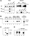
Stress-induced inhibition of Pol I transcription is due to inactivation of TIF-IA by JNK-mediated phosphorylation. (A) TIF-IA complements transcriptional activity of nuclear extracts from H2O2- and anisomycin-treated cells. Transcription reactions containing 30 μg nuclear extract protein from untreated (-), anisomycin-treated (NEaniso), or H2O2-treated (NEH2O2) FM3A (left) or HEK293T cells (right) were supplemented with fractions containing partially purified TIF-IA, TIF-IB/SL1, Pol I, or recombinant UBF, as indicated. (B) TIF-IA from H2O2- or anisomycin-treated cells is transcriptionally inactive. Forty and 80 ng of Flag-TIF-IA immunopurified from HEK293T cells that were either untreated or treated with anisomycin + SB203580, anisomycin + SP600125, or H2O2 were assayed for their capability to complement transcriptional activity of nuclear extracts from density-arrested cells. A silver-stained SDS-polyacrylamide gel with 200 ng of TIF-IA added to the transcription reactions is shown on the left. (C) In vitro phosphorylation of TIF-IA. GST-tagged TIF-IA purified from Eschericheria coli was phosphorylated with immunopurified HA-tagged JNK2 or p38. (D) Phosphorylation by JNK2 inactivates TIF-IA. (Lanes 1,2) A reconstituted TIF-IA-responsive transcription system consisting of partially purified Pol I, TIF-IB/SL1 and recombinant UBF (Schnapp and Grummt 1996) was used to assay the activity of TIF-IA. In lanes 3-5, TIF-IA was preincubated for 30 min in the presence of ATP with blocked control beads (lane 3), bead-bound HA-JNK2 (lane 4), or HA-p38 (lane 5) before adding to the transcription reactions.
Figure 3.
Figure 3.
The phosphorylation pattern of TIF-IA is altered upon cellular stress. (A) Two-dimensional tryptic phosphopeptide maps of TIF-IA labelled in vivo. U2OS cells stably overexpressing Flag-tagged TIF-IA were metabolically labeled for 2 h with [32P]orthophosphate in the absence or presence of H2O2 (200 μM, 60 min) or anisomycin (10 μM, 60 min), or in the presence of constitutively active (CA) MEKK-1. Immunopurified TIF-IA was subjected to two-dimensional tryptic phosphopeptide mapping. (B) Phosphoamino acid analysis of peptide a of TIF-IA from mock- and anisomycin-treated U2OS cells. A phylogenetic comparison of the amino acid sequence of the phosphopetide a is given on the left. Thr 200 is shown in bold, the conserved JNK target site TP is shaded gray. (C) Two-dimensional tryptic phosphopeptide maps of TIF-IA phosphorylated by JNK2 in vitro. GST-tagged TIF-IA (wild-type and T200V mutant) purified from E. coli was incubated with immunopurified HA-tagged JNK2 and (γ-32P)-ATP for 30 min and subjected to two-dimensional tryptic phosphopeptide mapping.
Figure 4.
Figure 4.
Phosphorylation of Thr 200 inactivates TIF-IA. (A) Transcriptional activity of TIF-IA mutants in vivo. HEK293T cells were cotransfected with 8 μg of a Pol I reporter plasmid and 0.5 or 1.5 μg expression vector encoding wild-type or mutant TIF-IA (pcDNA3.1-TIF-IA or TIF-IAT200V/E). Transcripts from the reporter were analyzed on Northern blots. Quantitation of transcripts by PhosphorImager analysis is shown below.(B) Substitution of Thr 200 by valine renders TIF-IA resistant towards oxidative stress. Cells were cotransfected with 8 μg Pol I reporter plasmid and 0.5 or 1.5 μg of pcDNA3.1-TIF-IA or TIF-IAT200V. After 40 h, cells were incubated for 60 min with 200 μM H2O2 and transcripts from the reporter plasmid were analyzed on Northern blots. Quantitation of transcripts by PhosphorImager analysis is shown below.
Figure 5.
Figure 5.
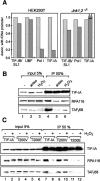
Cellular stress impairs the interaction of TIF-IA with TIF-IB/SL1 and Pol I. (A) Anisomycin treatment prevents the association of Pol I and TIF-IA with the rDNA promoter. Cross-linked chromatin from HEK293T cells (left) or Jnk1,2-/- MEFs (right) was immunoprecipitated with antibodies against TIF-IB/SL1 (α-TAFI110), UBF, Pol I (α-RPA116), and TIF-IA, and precipitated rDNA was monitored by real-time PCR with primers that amplify the 5′-terminal region of human rDNA. The results of three independent experiments are plotted. Light-gray and dark-gray columns represent the relative association of the analyzed proteins (calculated as bound normalized to input) from mock- and anisomycin-treated cells with the rDNA promoter, respectively. (B) Stress inhibits the interaction between TIF-IA and Pol I/TIF-IB. Lysates from mock-, H2O2-, or anisomycin-treated HEK293T cells expressing Flag-tagged TIF-IA were incubated with α-Flag antibodies, and immunoprecipitated TIF-IA, Pol I, and TIF-IB/SL1 were visualized on Western blots using α-Flag, α-RPA116, and α-TAFI68 antibodies, respectively. (C) Substitution of Thr 200 by glutamic acid interferes with the interaction of TIF-IA with Pol I and TIF-IB/SL1. Flag-tagged TIF-IA or the indicated point mutants were overexpressed in HEK293T cells and immunoprecipitated using α-Flag antibodies. Cells were either treated with H2O2 (200 μM) for 60 min or were left untreated. Coprecipitated Pol I and TIF-IB/SL1 were monitored on Western blots using α-RPA116 and α-TAFI68 antibodies, respectively.
Figure 6.
Figure 6.
Anisomycin treatment leads to accumulation of TIF-IA in the nucleoplasm. Cellular TIF-IA and UBF were visualized in parental or Jnk1,2-/- MEFs by indirect immunostaining. A merged image is shown on the right. Where indicated, MEFs were treated with anisomycin (10 μM) for 60 min.

Similar articles

Cited by

References

    1. Barr R.K. and Bogoyevitch, M.A. 2001. The c-jun N-terminal kinase family of mitogen-activated protein kinases (JNK MAPKs). Int. J. Biochem. Cell Biol. 33: 1047-1063. - PubMed
    1. Bodem J., Dobreva, G., Hoffmann-Rohrer, U., Iben, S., Zentgraf, H., Delius, H., Vingron, M., and Grummt, I. 2000. TIF-IA, the factor mediating growth-dependent control of ribosomal RNA synthesis, is the mammalian homolog of yeast Rrn3p. EMBO Rep. 1: 171-175. - PMC - PubMed
    1. Boyd K.E., Wells, J., Gutman, J., Bartley, S.M., and Farnham, P.J. 1998. c-Myc target gene specificity is determined by a post-DNA binding mechanism. Proc. Natl. Acad. Sci. 95: 13887-13892. - PMC - PubMed
    1. Boyle W.J., van der Geer, P., and Hunter, T. 1991. Phosphopeptide mapping and phosphoamino acid analysis by two-dimensional separation on thin-layer cellulose plates. Meth. Enzymol. 201: 110-149. - PubMed
    1. Cavanaugh A.H., Hirschler-Laszkiewicz, I., Hu, Q., Dundr, M., Smink, T., Misteli, T., and Rothblum, L.I. 2002. Rrn3 phosphorylation is a regulatory checkpoint for ribosome biogenesis. J. Biol. Chem. 277: 27423-27432. - PubMed

Publication types

MeSH terms