Comparative Study
. 2005 Mar 30;25(13):3259-69.
doi: 10.1523/JNEUROSCI.5168-04.2005.

Adherens junctions in myelinating Schwann cells stabilize Schmidt-Lanterman incisures via recruitment of p120 catenin to E-cadherin

Affiliations
Comparative Study

Adherens junctions in myelinating Schwann cells stabilize Schmidt-Lanterman incisures via recruitment of p120 catenin to E-cadherin

Nicolas Tricaud et al. J Neurosci. .

Abstract

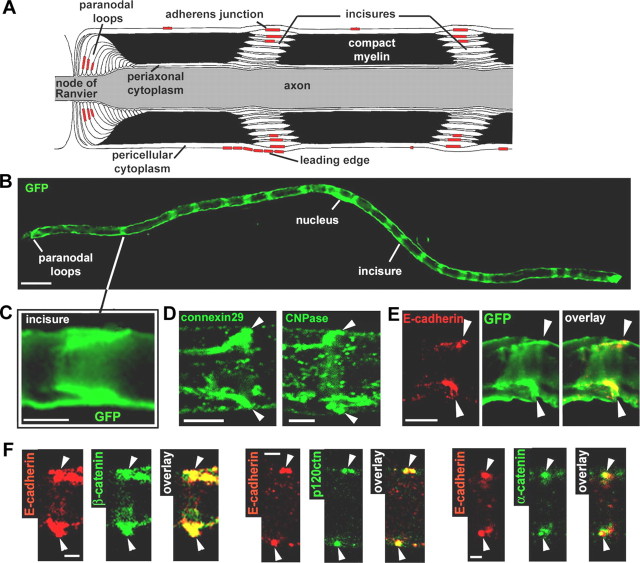
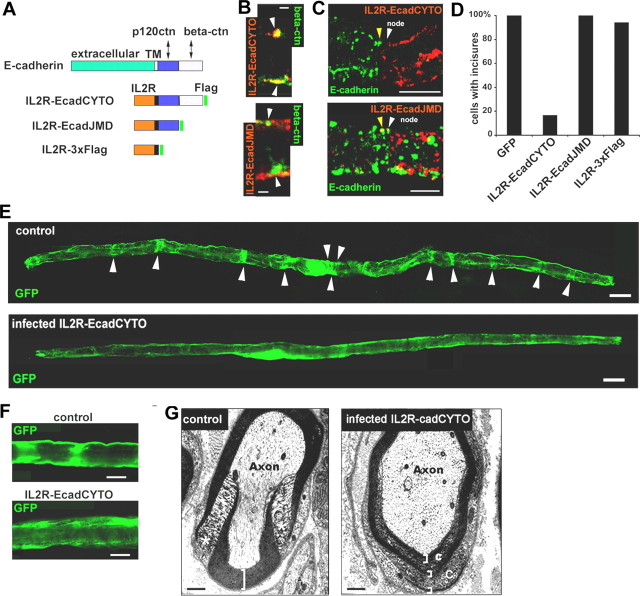
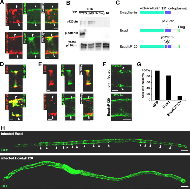
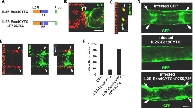
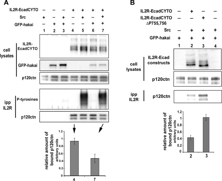
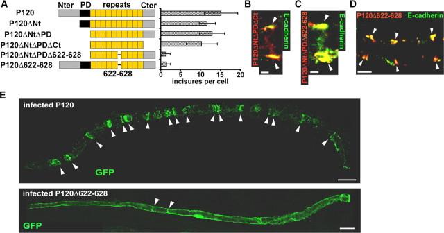
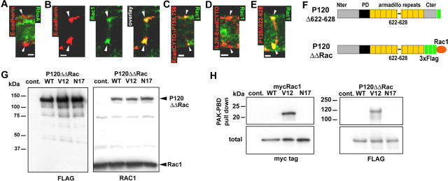
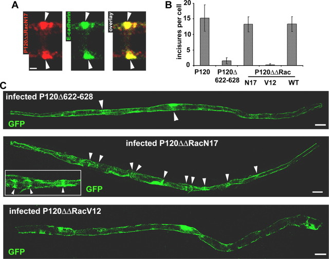
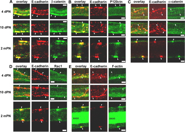
Schwann cell myelin contains highly compacted layers of membrane as well as noncompacted regions with a visible cytoplasm. One of these cytoplasmic compartments is the Schmidt-Lanterman incisure, which spirals through the compacted layers and is believed to help sustain the growth and function of compact myelin. Incisures contain adherens junctions (AJs), the key components of which are E-cadherin, its cytoplasmic partners called catenins, and F-actin. To explore in vivo the role of cadherin and catenins in incisures, E-cadherin mutant proteins that completely replace endogenous cadherin have been delivered to the cells using adenovirus. When the introduced cadherin lacked its extracellular domain, association of p120 catenin (p120ctn) with the cadherin did not occur, and incisures disappeared. Remarkably, the additional replacement of two phosphorylatable tyrosines by phenylalanine in the cytoplasmic tail of the mutant cadherin restored both p120ctn binding and incisure architecture, indicating that p120ctn recruitment is critical for incisures maintenance and might be regulated by phosphorylations. In addition, the ability of the p120ctn/cadherin complex to support incisures was blocked by mutation of the Rho GTPase regulatory region of the p120ctn, and downregulation of Rac1 activity at the junction reversed this inhibition. Because Rho GTPases regulate the state of the actin filaments, these findings suggest that one role of p120ctn in incisures is to organize the cytoskeleton at the AJ. Finally, developmental studies of Schwann cells demonstrated that p120ctn recruitment from the cytoplasm to the AJ occurs before the appearance of Rac1 GTPase and F-actin at the junction.


Figures

Figure 1.
Figure 1.
AJ and architecture of the myelinated Schwann cell. A, Longitudinal view of a myelinated Schwann cell showing the different compartments and the respective location of the AJs. B, Myelinated Schwann cell infected with an adenovirus expressing GFP and showing repeated bands of cytoplasm corresponding to the location of incisures. Scale bar, 50 μm. C, GFP-filled incisure showing the pair of angled cytoplasmic regions representing one incisure as depicted in A. Scale bar, 10 μm. D, Immunostaining of connexin29 (left) and CNPase (right) showing their enrichment in an incisure (arrowheads). Scale bar, 5 μm. E, Pair of E-cadherin clusters (red) that localize at the outer edge of a GFP-filled incisure (arrowheads). Scale bar, 10 μm. F, Coimmunostaining of E-cadherin (red) and β-catenin, p120ctn, and α-catenin (green) on sciatic nerve samples at 2 months postnatal (arrowheads indicate pairs of E-cadherin clusters). Scale bar, 2.5 μm.
Figure 2.
Figure 2.
Full-length E-cadherin is necessary for maintenance of the cell architecture. A, IL2R/E-cadherin constructs used in this study. The distal domain of E-cadherin cytoplasmic tail (light blue) interacts with β-catenin, whereas the juxtamembrane domain (dark blue) interacts with p120ctn. All constructs are tagged with 3xFlag tag (green). IL2R, Extracellular and transmembrane domains of IL2R. B, Top, IL2R-EcadCYTO construct (red) expressed in a myelinated Schwann cell colocalizes with β-catenin (green) in clusters (arrowheads) at the periphery of the cell. Bottom, IL2R-EcadJMD construct (red) does not colocalize with β-catenin (green) in clusters (arrowheads). Scale bars, 2.5 μm. C, Top, Node of Ranvier having on one side a cell expressing IL2R-EcadCYTO (white arrowhead) but no E-cadherin and on the other side a noninfected cell expressing E-cadherin (yellow arrowhead). Bottom, At a node, a cell (white arrowhead) expressing IL2R-EcadJMD (red) also displays E-cadherin (green) as does a noninfected cell (yellow arrowhead). Scale bars, 10 μm. D, Percentage of cells displaying incisures was determined after infection with viruses expressing different constructs. The presence of incisures was assessed by the presence of transverse bands labeled with GFP or with connexin 29/CNPase immunostaining. Only 16% of the cells infected with IL2R-EcadCYTO maintained one or more incisure. Wounded or dying cells were not counted. Number of cells scored: GFP, 15; IL2R-EcadCYTO, 12; IL2R-EcadJMD, 7; IL2R-3xFlag, 17. E, Myelinated Schwann cells infected with control GFP adenovirus (top) or IL2R-EcadCYTO adenovirus (bottom) as observed by the expression of GFP in the cytoplasm. Scale bars, 20 μm. F, Detail of the GFP distribution in cells infected with GFP virus (top) or IL2R-EcadCYTO virus (bottom). Scale bars, 10 μm. G, Electron micrographs of transversal sections of an IL2R-CcadCYTO-infected nerve (right) and a noninfected nerve (left). Asterisks indicate a normal incisure, brackets indicate the compact myelin, and “C” indicates the cytoplasm inside the compact myelin. Scale bars, 500 nm.
Figure 3.
Figure 3.
Absence of interaction between p120ctn and E-cadherin perturbs the incisures. A, IL2R-EcadCYTO (red; top) and IL2R-EcadJMD (red; bottom) does not colocalize with p120ctn (green). Scale bars, 2.5 μm. B, CHO cells were infected with virus expressing IL2R-EcadCYTO, IL2R-EcadJMD, or IL2R-3xFlag or not infected (NI). The expressed proteins were immunoprecipitated with an antibody against IL2R, and the precipitates were subjected to Western blotting for detection of p120ctn and β-catenin. The same amount of cell lysate was subjected to Western blotting to show that a similar amount of p120ctn was available for coimmunoprecipitation. ipp, Immunoprecipitated proteins. C, Ecad is a full-length E-cadherin construct that is Flag tagged. EcadΔP120 is a full-length human E-cadherin construct Flag tagged and mutated so as to be unable to bind p120ctn. D, Ecad (red; top) and EcadΔP120 (red; bottom) colocalize with β-catenin (green). Scale bars, 2.5 μm. E, Ecad (red; top) but not EcadΔP120 (red; bottom) colocalizes with p120ctn (green). Scale bars, 2.5 μm. F, In a noninfected cell (top), p120ctn (green) forms a pair of clusters (arrowheads) at the periphery of the cell. In an EcadΔP120-infected cell (bottom), p120ctn has a diffuse distribution. Scale bars, 5 μm. G, Percentage of cells displaying incisures determined after infection with viruses expressing GFP alone, Ecad, or EcadΔP120. Thirteen percent of the cells infected with EcadΔP120 and 83% of the cells infected with Ecad virus maintained one or more incisure. Number of cell scored: GFP, 15; Ecad, 22; EcadΔP120, 15. H, GFP distribution in myelinated Schwann cells infected with Ecad (top) or EcadΔP120 (bottom) adenoviruses. Scale bars, 20 μm.
Figure 4.
Figure 4.
Removal of the phosphorylation sites in the juxtamembrane region of IL2R-EcadCYTO restores its interaction with p120ctn and the Schwann cell structure. A, IL2R-EcadCYTOΔP755,756 is a IL2R-EcadCYTO construct mutated on the two phosphorylatable tyrosines, 755 and 756 (YY to FF), in the E-cadherin juxtamembrane domain. B, At a node, a cell infected with the IL2R-EcadCYTOΔP755,756 adenovirus (white arrowhead) expresses GFP (green) but very little E-cadherin (red) compared with the noninfected cell (yellow arrowhead). Scale bar, 10 μm. C, IL2R-EcadCYTOΔP755,756 (red) colocalizes with p120ctn (green). Scale bar, 2.5 μm. D, Detail of the GFP distribution in a myelinated Schwann cell expressing GFP alone (top), IL2R-EcadCYTO (middle), and IL2R-EcadCYTOΔP755,756 (bottom). Arrows indicate incisures. Scale bar, 10 μm. E, IL2R-EcadCYTOΔP755,756 (red) forms clusters in the outer region of an incisure (arrowheads) filled with CNPase (green). Scale bar, 5 μm. F, Percentage of cells displaying incisures determined expression of GFP, IL2R-EcadCYTO, or IL2R-EcadCYTOΔP755,756. Incisures are maintained by 84.7% of the cells expressing IL2R-EcadCYTOΔP755,756. Number of cells scored: GFP, 15; IL2R-EcadCYTO, 12; IL2R-EcadCYTOΔP755,756, 13.
Figure 5.
Figure 5.
p120ctn interaction with IL2R-EcadCYTO is regulated by phosphorylations of the E-cadherin intracellular domain. A, IL2R-EcadCYTO was expressed in 293 cells with or without v-src or GFP-hakai. The cells were solubilized, and equal amounts of solubilized proteins (cell lysates) were immunoprecipitated with an anti-IL2R antibody. The immunoprecitated proteins (ipp) were probed with an anti-phosphotyrosines antibody (P-tyrosines) and an anti-p120ctn antibody. The amount of coimmunoprecipitated p120ctn was quantified by densitometry, normalized to the amount of p120ctn present in the cell lysate (relative amount of bound p120ctn) and the value obtained for lane 4 (IL2R-EcadCYTO alone) and lane 7 (IL2R-EcadCYTO plus v-src and GFP-hakai) plotted in a graph. The amount of bound p120ctn drop of 50% (results from 3 independent experiments). B, IL2R-EcadCYTO (lane2) and IL2R-EcadCYTOΔP755,756 (lane 3) were expressed in 293 cells with v-src and GFP-hakai. The cells were solubilized, and an equal amount of solubilized proteins (cell lysates) was immunoprecipitated with IL2R antibody. The amount of coimmunoprecipitated p120ctn was detected, quantified by densitometry, normalized to the amount of p120ctn present in the cell lysate (relative amount of bound p120ctn), and the value obtained for lanes 2 and 3 plotted in a graph. The amount of bound p120ctn is 2.4 times higher for IL2R-EcadCYTOΔP755,756 than for IL2R-EcadCYTO (results from 3 independent experiments). Error bars represent SD.
Figure 6.
Figure 6.
Cadherin-bound p120ctn requires amino acids 622-628 to maintain incisures. A, Left, A series of mutants of p120ctn produced by deleting the N-terminal domain (Nter; P120ΔNt), plus the phosphorylation domain, P120ΔNtΔPD, plus the C-terminal domain (Cter; P120ΔNtΔPDΔCt), and plus amino acids 622-628 in the sixth armadillo repeat (P120ΔNtΔPDΔ622-628). In addition, only the 622-628 region was deleted in full-length p120ctn (P120Δ622-628). All of these constructs were labeled at their C terminus with a Flag tag. Right, Each construct was expressed in myelinated Schwann cells, and the number of incisures displayed by each cell (assessed via GFP labeling or CNPase/connexin29 staining) was determined. Number of cells scored: P120, 27; P120ΔNt, 14; P120ΔNtΔPD, 17; P120ΔNtΔPDΔCt, 13; P120ΔNtΔPDΔ622-628, 5; P120Δ622-628, 22. B, P120ΔNtΔPDΔCt (red) colocalizes with E-cadherin (green). Scale bar, 2.5 μm. C, P120ΔNtΔPDΔ622-628 (red) colocalizes with E-cadherin (green). Scale bar, 2.5 μm. D, Schwann cell expressing P120Δ622-628 (red) displays pairs of E-cadherin clusters (green). Scale bar, 5 μm. E, A Schwann cell expressing P120 (top) displays 17 incisures (arrowheads). A cell expressing P120Δ622-628 (bottom) displays only two GFP-filled incisures (arrowheads). Scale bars, 20 μm.
Figure 7.
Figure 7.
Rac1 colocalization with AJ correlates with the recruitment of p120ctn to cadherin. A, RhoA (green) does not colocalize with E-cadherin (red) at incisures (arrowheads). Scalebar, 2.5 μm. B, Rac1 (green) partially colocalizes with E-cadherin (red). Scalebar, 2.5 μm. C, IL2R-EcadCYTOΔP755,756 (red) partially colocalizes with Rac1 (green). Scalebar, 2.5 μm. D, IL2R-EcadCYTO (red) does not colocalize with Rac1 (green). Scalebar, 2.5 μm. E, P120Δ622-628 construct (red) colocalizes with Rac1 (green). Scalebar, 2.5 μm. F, The C terminal (Cter) of P120Δ622-628 was fused to a 3xFlag tag and Rac1 wild-type, dominant-active mutant (V12) and dominant-negative mutant (N17) to obtain P120ΔΔRacWT, V12, and N17. G, P120ΔΔRac constructs were expressed in CHO cells and immunoblotted with mouse monoclonal anti-Flag (left) or anti-Rac1 (right) antibodies. Note that anti-Rac1 antibody recognizes both Rac1 and P120ΔΔRac. H, myc-Rac1 (left) and P120ΔΔRac (right) constructs expressed in CHO were subjected to PAK-PBD pull down, and the precipitated proteins were detected by Western blots hybridized with anti-myc and anti-Flag antibodies. A sample of the total amount of protein subjected to precipitation was also processed (total) to show that the same amount of protein was used for each construct. Nter, N terminal; Cont., control.
Figure 8.
Figure 8.
Fusion of dominant-negative Rac1 with P120Δ622-628 restores p120ctn ability to maintain the incisures. A, P120ΔΔRacN17 (red) expressed in Schwann cell colocalizes with E-cadherin (green). Scale bar, 2.5 μm. B, Each construct was introduced into myelinated Schwann cells, and the number of incisures displayed by each cell (assessed via GFP labeling or CNPase/connexin29 staining) was determined. Number of cells scored: P120, 27; P120Δ622-628, 22; P120ΔΔRacN17, 17; P120ΔΔRacV12, 24; P120ΔΔRacWT, 11. C, A Schwann cell expressing P120Δ622-628 (top) shows two GFP-filled incisures (arrowheads); a cell expressing P120ΔΔRacN17 (middle) shows 10 incisures (arrowheads), and some of them are shown in detail (inclusion on the right side); a cell expressing P120ΔΔRacV12 (bottom) displays no incisure. Scale bars, 20 μm. Error bars represent SD.
Figure 9.
Figure 9.
Sequential appearance of AJ components during myelination. A-E, Coimmunostaining of E-cadherin (red) and β-catenin (A), p120ctn (B), α-catenin (C), Rac1 (D), or F-actin (E) on sciatic nerve samples at 4 d postnatal, 10 d postnatal, and 2 months postnatal. Arrowheads indicate cadherin clusters. The staining of F-actin with phalloidin was intense inside the axon at 2 mPN. Scale bars, 2.5 μm.

Similar articles

Cited by

References

    1. Anastasiadis PZ, Moon SY, Thoreson MA, Mariner DJ, Crawford HC, Zheng Y, Reynolds AB (2000) Inhibition of RhoA by p120 catenin. Nat Cell Biol 2: 637-644. - PubMed
    1. Aono S, Nakagawa S, Reynolds AB, Takeichi M (1999) p120(ctn) acts as an inhibitory regulator of cadherin function in colon carcinoma cells. J Cell Biol 145: 551-562. - PMC - PubMed
    1. Arroyo EJ, Scherer SS (2000) On the molecular architecture of myelinated fibers. Histochem Cell Biol 113: 1-18. - PubMed
    1. Benard V, Bohl BP, Bokoch GM (1999) Characterization of rac and cdc42 activation in chemoattractant-stimulated human neutrophils using a novel assay for active GTPases. J Biol Chem 274: 13198-13204. - PubMed
    1. Chen X, Kojima S, Borisy GG, Green KJ (2003) p120 catenin associates with kinesin and facilitates the transport of cadherin-catenin complexes to intercellular junctions. J Cell Biol 163: 547-557. - PMC - PubMed

Publication types

MeSH terms

LinkOut - more resources