. 2005 Apr;25(8):3348-56.
doi: 10.1128/MCB.25.8.3348-3356.2005.

Posttranscriptional downregulation of c-IAP2 by the ubiquitin protein ligase c-IAP1 in vivo

Affiliations

Posttranscriptional downregulation of c-IAP2 by the ubiquitin protein ligase c-IAP1 in vivo

Dietrich B Conze et al. Mol Cell Biol. 2005 Apr.

Abstract

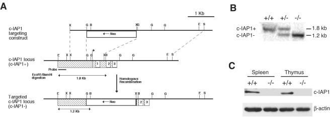
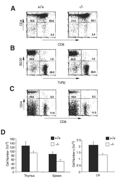
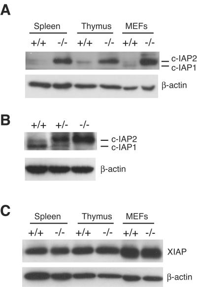
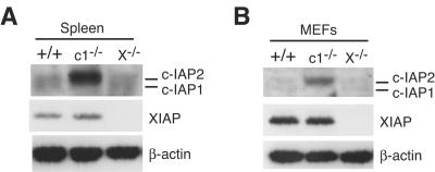
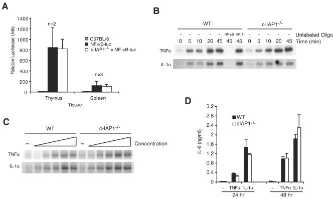
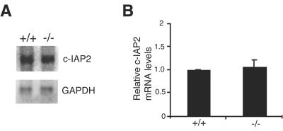
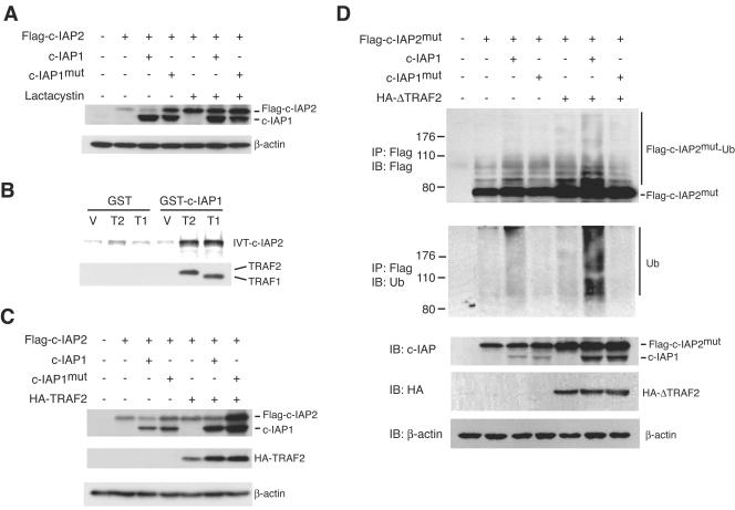
Inhibitor of apoptosis proteins (IAPs) c-IAP1 and c-IAP2 were identified as part of the tumor necrosis factor receptor 2 (TNFR2) signaling complex and have been implicated as intermediaries in tumor necrosis factor alpha signaling. Like all RING domain-containing IAPs, c-IAP1 and c-IAP2 have ubiquitin protein ligase (E3) activity. To explore the function of c-IAP1 in a physiologic setting, c-IAP1-deficient mice were generated by homologous gene recombination. These animals are viable and have no obvious sensitization to proapoptotic stimuli. Cells from c-IAP1(-/-) mice do, however, express markedly elevated levels of c-IAP2 protein in the absence of increased c-IAP2 mRNA. In contrast to reports implicating c-IAPs in the activation of NF-kappaB, resting and cytokine-induced NF-kappaB activation was not impaired in c-IAP1-deficient cells. Transient transfection studies with wild-type and E3-defective c-IAP1 revealed that c-IAP2 is a direct target for c-IAP1-mediated ubiquitination and subsequent degradation, which are potentiated by the adaptor function of TRAF2. Thus, the c-IAPs represent a pair of TNFR-associated ubiquitin protein ligases in which one regulates the expression of the other by a posttranscriptional and E3-dependent mechanism.


Figures

FIG. 1.
FIG. 1.
Generation of c-IAP1-deficient mice. (A) Strategy for c-IAP1 gene disruption by homologous recombination. The hatched box represents exon 1, the numbered boxes represent the three baculovirus IAP repeats, and the arrow indicates the transcription initiation start codon. Insertion of the neomycin gene (Neo) by homologous recombination replaced the transcription initiation start codon and the first BIR domain. Restriction sites are indicated as follows: E, EcoRI; X, XbaI; G, BglII; B, BamH1; S, SalI. (B) Genomic DNA from wild-type (+/+), c-IAP1 heterozygous (+/−), and c-IAP1 homozygous (−/−) deficient mice was digested with EcoRI and BamHI and probed with the radiolabeled probe indicated in panel A. The upper band (1.8 kb) corresponds to the wild-type c-IAP1 allele (c-IAP1+), and the lower band (1.2 kb) corresponds to the mutant c-IAP1 allele (c-IAP1−). (C) Immunoblot analysis of c-IAP1 expression in spleen and thymus whole-cell lysates from +/+ and −/− mice. Expression of c-IAP1 was determined using a polyclonal antibody that recognizes the C terminus of c-IAP1. β-actin expression was used as a loading control.
FIG. 2.
FIG. 2.
T- and B-cell development in wild-type and c-IAP1−/− mice. (A) Thymocytes from wild-type (+/+) and c-IAP1−/− (−/−) mice were isolated, stained with anti-CD4 and anti-CD8 monoclonal antibodies, and analyzed by flow cytometry. (B and C) Splenocytes from +/+ and −/− mice were isolated and stained with anti-TCRβ and anti-CD45R/B220 monoclonal antibodies (B) or anti-CD4 and anti-CD8 monoclonal antibodies (C) and analyzed by flow cytometry. Numbers displayed in the histograms in panels A, B, and C represent the percentages of cells in each quadrant. (D) Cell suspensions were made from thymus (n = 6), spleen (n = 7), and lymph nodes (LN; n = 5) from +/+ and −/− mice. Viable cell recovery was determined by microscopy and trypan blue exclusion. The numbers displayed for lymph nodes reflect the number of cells per lymph node. Error bars represent standard errors of the means.
FIG. 3.
FIG. 3.
Dramatic upregulation of c-IAP2 but not XIAP in c-IAP1−/− mice. (A) Immunoblot analysis of c-IAP expression in spleen, thymus, and murine embryonic fibroblasts (MEFs) using an anti-c-IAP antiserum. (B) Immunoblot analysis of c-IAP expression in splenocytes harvested from +/+, c-IAP1 heterozygous (+/−), and −/− mice. (C) Immunoblot analysis of XIAP expression in spleen, thymus, and MEF lysates from +/+ and −/− mice. β-actin expression was used as a loading control.
FIG. 4.
FIG. 4.
Elevated c-IAP2 in c-IAP1-deficient cells but not XIAP-deficient cells. Immunoblot analysis of c-IAP expression in spleen (A) and MEF (B) lysates from wild-type (+/+), c-IAP1−/− (c1−/−), and XIAP−/− (X−/−) mice using anti-c-IAP antiserum. β-actin expression was used as a loading control.
FIG. 5.
FIG. 5.
Neither c-IAP1 deficiency nor upregulation of c-IAP2 affects NF-κB-mediated transcription. (A) Thymocytes and splenocytes harvested from C57BL/6, NF-κB-luciferase (NF-κB-luc), and c-IAP1−/− × NF-κB-luciferase (c-IAP1−/− × NF-κB-luc) reporter transgenic mice were lysed and assayed for luciferase activity. Values presented are the averages for the indicated numbers (n) of mice. Error bars represent standard deviations. (B) Kinetic analysis of NF-κB DNA binding in stimulated wild-type (WT) and c-IAP1−/− MEFs. Nuclear extracts from unstimulated MEFs or MEFs stimulated with TNF-α (33 ng/ml) or IL-1α (5 ng/ml) for the indicated periods of time were incubated with 32P-end-labeled double-stranded oligonucleotide containing an NF-κB consensus binding site. WT extracts stimulated with TNF-α or IL-1α for 45 min were also incubated with a 32P-end-labeled NF-κB double-stranded oligonucleotide in the absence (−) or presence of a 100-fold excess of unlabeled NF-κB or AP-1 oligonucleotides. (C) Dose-response analysis of NF-κB DNA binding in WT and c-IAP1−/− MEFs by EMSA. Nuclear extracts from unstimulated MEFs or MEFs stimulated with increasing concentrations of TNF-α (0.206, 4.12, 82.5, 1,650, and 33,000 pg/ml) or IL-1α (0.031, 0.625, 12.5, 250, and 5,000 pg/ml) for 25 min were incubated with 32P-end-labeled double-stranded oligonucleotide containing an NF-κB consensus binding site. (D) IL-6 production from WT and c-IAP1−/− MEFs. WT and c-IAP1−/− MEFs were incubated in medium alone or stimulated with TNF-α (33 ng/ml) or IL-1α (5 ng/ml) for 24 and 48 h. The IL-6 concentrations in the supernatants were determined by ELISA. Triplicate samples were assayed, and the error bars represent the standard errors of the means.
FIG. 6.
FIG. 6.
c-IAP1 deficiency does not affect c-IAP2 gene expression. (A) c-IAP2 mRNA expression in splenocytes harvested from wild-type (+/+) and c-IAP1−/− (−/−) mice was determined by Northern blot analysis. Expression of GAPDH was used as a loading control. (B) c-IAP2 mRNA expression in splenocytes harvested from +/+ and −/− mice was determined by real-time PCR. Values for c-IAP2 mRNA expression in −/− splenocytes (n = 3) are expressed relative to the levels of c-IAP2 mRNA in +/+ splenocytes (n = 3). Error bars represent standard errors of the means.
FIG. 7.
FIG. 7.
c-IAP1 ubiquitinates and downregulates c-IAP2 in an E3-dependent manner. (A) Immunoblot analysis of 293 cells transiently transfected with Flag-tagged-c-IAP2 in combination with vector (−), c-IAP1, or c-IAP1mut. Cells were transfected for 17 h and then treated with or without lactacystin (25 μM) for an additional 7 h. The amounts of Flag-c-IAP2, c-IAP1, and c-IAP1mut were determined with an anti-c-IAP antiserum. β-actin expression was used as a loading control. (B) c-IAP1 binds c-IAP2 only in the presence of TRAF1 or TRAF2. Bacterially expressed and purified GST- or GST-c-IAP1-coated beads were incubated with the lysate of bacteria transformed with vector (V) or His-tagged TRAF1 (189-416) (T1) or His-tagged TRAF2 (272-501) (T2) cDNAs. The beads were washed and incubated with in vitro-translated and 35S-labeled c-IAP2. After extensive washing, the bound material was eluted and resolved by SDS-PAGE. The presence of His-TRAF1 (189-416) and His-TRAF2 (272-501) in the protein complexes was determined by immunoblotting. (C) 293 cells were transiently transfected with Flag-tagged-c-IAP2 (Flag-c-IAP2) in combination with vector (−), c-IAP1, c-IAP1mut, or HA-tagged TRAF2 (HA-TRAF2). The amounts of Flag-c-IAP2, c-IAP1, c-IAP1mut, and HA-TRAF2 were determined by immunoblot analysis. Flag-c-IAP2, c-IAP1, and c-IAP1mut expression was determined using anti-c-IAP antiserum. (D) 293 cells were transfected with Flag-tagged-c-IAP2mut alone or in combination with the indicated cDNAs. After overnight culture, the cells were treated with lactacystin for an additional 7 h and then harvested and lysed. Flag-c-IAP2mut was immunoprecipitated (IP) and immunoblotted (IB) with anti-Flag to detect the native molecule as well as higher-molecular-weight species representing ubiquitinated c-IAP2mut (Flag-c-IAP2mut-Ub). The membrane was stripped and reblotted with anti-ubiquitin polyclonal antibody to directly detect the presence of ubiquitinated Flag-c-IAP2mut. Expression of Flag-c-IAP2mut, c-IAP1, c-IAP1mut, and HA-ΔTRAF2 in cell lysates was detected by immunoblotting.

Similar articles

Cited by

References

    1. Abbondanzo, S. J., I. Gadi, and C. L. Stewart. 1993. Derivation of embryonic stem cell lines. Methods Enzymol. 225:803-823. - PubMed
    1. Akira, S., and T. Kishimoto. 1992. IL-6 and NF-IL6 in acute-phase response and viral infection. Immunol. Rev. 127:25-50. - PubMed
    1. Borden, K. L. 2000. RING domains: master builders of molecular scaffolds. J. Mol. Biol. 295:1103-1112. - PubMed
    1. Burstein, E., L. Ganesh, R. D. Dick, B. van De Sluis, J. C. Wilkinson, L. W. Klomp, C. Wijmenga, G. J. Brewer, G. J. Nabel, and C. S. Duckett. 2004. A novel role for XIAP in copper homeostasis through regulation of MURR1. EMBO J. 23:244-254. - PMC - PubMed
    1. Chu, Z. L., T. A. McKinsey, L. Liu, J. J. Gentry, M. H. Malim, and D. W. Ballard. 1997. Suppression of tumor necrosis factor-induced cell death by inhibitor of apoptosis c-IAP2 is under NF-kappaB control. Proc. Natl. Acad. Sci. USA 94:10057-10062. - PMC - PubMed

MeSH terms