Comparative Study
. 2005 Mar 23;25(12):3199-208.
doi: 10.1523/JNEUROSCI.4201-04.2005.

An essential Drosophila glutamate receptor subunit that functions in both central neuropil and neuromuscular junction

Affiliations
Comparative Study

An essential Drosophila glutamate receptor subunit that functions in both central neuropil and neuromuscular junction

David E Featherstone et al. J Neurosci. .

Abstract

A Drosophila forward genetic screen for mutants with defective synaptic development identified bad reception (brec). Homozygous brec mutants are embryonic lethal, paralyzed, and show no detectable synaptic transmission at the glutamatergic neuromuscular junction (NMJ). Genetic mapping, complementation tests, and genomic sequencing show that brec mutations disrupt a previously uncharacterized ionotropic glutamate receptor subunit, named here "GluRIID." GluRIID is expressed in the postsynaptic domain of the NMJ, as well as widely throughout the synaptic neuropil of the CNS. In the NMJ of null brec mutants, all known glutamate receptor subunits are undetectable by immunocytochemistry, and all functional glutamate receptors are eliminated. Thus, we conclude that GluRIID is essential for the assembly and/or stabilization of glutamate receptors in the NMJ. In null brec mutant embryos, the frequency of periodic excitatory currents in motor neurons is significantly reduced, demonstrating that CNS motor pattern activity is regulated by GluRIID. Although synaptic development and molecular differentiation appear otherwise unperturbed in null mutants, viable hypomorphic brec mutants display dramatically undergrown NMJs by the end of larval development, suggesting that GluRIID-dependent central pattern activity regulates peripheral synaptic growth. These studies reveal GluRIID as a newly identified glutamate receptor subunit that is essential for glutamate receptor assembly/stabilization in the peripheral NMJ and required for properly patterned motor output in the CNS.


Figures

Figure 1.
Figure 1.
brec mutations disrupt the CG18039 gene encoding an ionotropic glutamate receptor subunit GluRIID. Gray boxes represent predicted exons in CG18039, a putative ionotropic glutamate receptor subunit most similar in sequence to mammalian kainite receptor subunits. The entire 3.8 kb genomic region was sequenced in brec1 and brec2 mutants, as well as the control genotypes rucuca and Oregon R (WT). Partial sequences were obtained for brecP5 and its parental strain. In brec1 mutants, there is a 50 bp deletion in the fourth exon. In brec2 mutants, there is a 7 bp duplication in the 11th exon. brecP5 mutants containan I-element-like insert (P5 insert) ∼2 kb long between exons 6 and 7 and a predicted splice-site alteration at the end of exon 7 (asterisk).
Figure 4.
Figure 4.
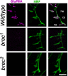
Homozygous brec mutant embryonic NMJs have no detectable glutamate receptors. Confocal images show intersegmental nerve branch b, visualized using anti-HRP antibody (neuronal membrane epitope; green), innervating several ventral longitudinal muscles. At WT NMJs (top row), glutamate receptors visualized using an anti-GluRIIA antibody (magenta) form distinct clusters opposite presynaptic terminals. In homozygous brec mutants (rows 2, 3), there are no detectable glutamate receptor clusters. Similar results were obtained using antibodies against the GluRIIB and GluRIIC subunits (see Fig. S1, available at www.jneurosci.org as supplemental material). Scale bar, 5 μm.
Figure 2.
Figure 2.
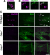
GluRIID is expressed in the embryonic NMJ. A, High-magnification confocal image of typical GluRIID immunoreactivity in a wild-type embryonic NMJ. Note that GluRIID immunoreactivity appears as small (∼300-nm-diameter) clusters. Scale bar, 1 μm. B, As in A but showing both GluRIID and GluRIIA immunoreactivity. Note that only some GluRIID clusters are colabeled with GluRIIA. C, Confocal images of glutamatergic NMJs on embryonic ventral muscles 7, 6, 13, 12, and 15, double stained using antibodies for GluRIID (magenta) and DLG (green). GluRIID immunoreactivity (but not DLG immunoreactivity) is eliminated in homozygous brec1 and brec2 mutants. Scale bar, 10 μm.
Figure 3.
Figure 3.
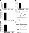
Homozygous null brec mutant embryonic NMJs have no functional glutamate receptors. A, B, brec mutants show no spontaneous synaptic currents (A) of any size (B). C, Evoked synaptic transmission is eliminated in brec mutant NMJs. D, Representative evoked synaptic currents; the robust EJCs observed at WT NMJs (top traces) are completely eliminated in the brec mutants (bottom traces). E, Pressure ejection of 1 mm glutamate directly onto the postsynaptic muscle elicits no response in the brec mutants. F, Representative glutamate-gated currents are large and reproducible in WT muscle (top traces) but completely absent in brec mutant muscles (bottom traces). Arrows mark the time of glutamate pressure ejection. Note also the spontaneous synaptic currents visible in the WT traces but absent in the recordings from the mutants.
Figure 7.
Figure 7.
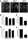
NMJ presynaptic morphology is relatively normal in brec null mutant embryos but underdeveloped in hypomorphic brec mutant third-instar larvae. A, Confocal images of embryonic NMJ terminals visualized using antibodies against the synaptic vesicle marker CSP, from WT (top) and homozygous brec mutants (middle, bottom). B, C, Quantification of embryonic NMJ area (B) and number of presynaptic boutons (C). D, E, Quantification of larval NMJ area (D) and number of presynaptic boutons (E). P5, brecP5.
Figure 5.
Figure 5.
GluRIID is necessary for efficient expression of transgenic GluRIIA. In heterozygous brec mutants (top row), postsynaptically expressed transgenic myc-tagged GluRIIA (visualized using an anti-myc antibody; magenta) clusters predominantly at postsynaptic sites, as shown by the colocalization (white) with the postsynaptic marker DLG (green). In homozygous brec mutants (bottom row), overexpression of myc-tagged glutamate receptor protein does not rescue mutant lethality, and synaptic GluRIIA localization remains greatly reduced compared with controls.
Figure 6.
Figure 6.
Hypomorphic brec mutants have greatly reduced NMJ glutamate receptor expression and synaptic activity. A, Confocal images of third-instar larval NMJs on adjacent muscles 6 and 7, from heterozygous brecP5 mutant larvae (brecP5/TM6; controls) and homozygous brecP5 larvae. The presynaptic terminal is visualized using the neuronal membrane marker anti-HRP (green). Glutamate receptors, visualized using anti-GluRIIA antibodies (magenta), are reduced to barely detectable levels at the NMJ in homozygous brecP5 mutants. B, Sample voltage-clamp recordings from muscle 6, showing frequent spontaneous EJCs (downward deflections) in wild-type larvae (top trace), which are essentially abolished in homozygous brecP5 larvae (bottom trace). C, Cumulative frequency histogram of sEJC amplitudes from WT and homozygous brecP5 mutant larvae, quantifying a significant decrease in sEJC amplitudes measured in mutants. D, Quantification of sEJC frequency in WT and homozygous brecP5 mutant larvae, showing greatly decreased frequency in mutants. P5, brecP5; K-S test, Kolmogorov-Smirnov test.
Figure 8.
Figure 8.
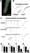
GluRIID is localized to the CNS neuropil and modulates excitatory neuronal activity. A, Confocal image showing an embryonic ventral ganglion stained with GluRIID antibodies (green) and anti-DLG antibodies (magenta). Note the GluRIID immunoreactivity throughout the synaptic neuropil. B, Representative acetylcholine-gated currents recorded from WT and homozygous brec2 mutant neurons. C, Voltage-clamp recordings from WT, heterozygous brec2/TM6 controls, and homozygous brec2 mutant motor neurons, showing endogenous SRCs. Events outlined by boxes are shown at an expanded time scale to the right. D-G, Quantification of ACh-gated current amplitudes (D), SRC amplitudes (E), percentage of neurons displaying SRCs (F), and SRC frequency in the neurons that display SRCs (G) in control (WT and heterozygous brec2/TM6 mutants) and homozygous brec2 mutant embryos.

Similar articles

Cited by

References

    1. Baines RA, Bate M (1998) Electrophysiological development of central neurons in the Drosophila embryo. J Neurosci 18: 4673-4683. - PMC - PubMed
    1. Baines RA, Robinson SG, Fujioka M, Jaynes JB, Bate M (1999) Postsynaptic expression of tetanus toxin light chain blocks synaptogenesis in Drosophila Curr Biol 9: 1267-1270. - PMC - PubMed
    1. Baines RA, Uhler JP, Thompson A, Sweeney ST, Bate M (2001) Altered electrical properties in Drosophila neurons developing without synaptic transmission. J Neurosci 21: 1523-1531. - PMC - PubMed
    1. Barry MF, Ziff EB (2002) Receptor trafficking and the plasticity of excitatory synapses. Curr Opin Neurobiol 12: 279-286. - PubMed
    1. Bate M, Broadie K (1995) Wiring by fly: the neuromuscular system of the Drosophila embryo. Neuron 15: 513-525. - PubMed

Publication types

MeSH terms