Primary human lymphocytes transduced with NY-ESO-1 antigen-specific TCR genes recognize and kill diverse human tumor cell lines
- PMID: 15778407
- PMCID: PMC2174604
- DOI: 10.4049/jimmunol.174.7.4415
Primary human lymphocytes transduced with NY-ESO-1 antigen-specific TCR genes recognize and kill diverse human tumor cell lines
Abstract
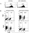
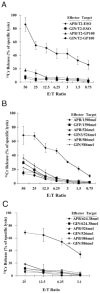
cDNAs encoding TCR alpha- and beta-chains specific for HLA-A2-restricted cancer-testis Ag NY-ESO-1 were cloned using a 5'RACE method from RNA isolated from a CTL generated by in vitro stimulation of PBMC with modified NY-ESO-1-specific peptide (p157-165, 9V). Functionality of the cloned TCR was confirmed by RNA electroporation of primary PBL. cDNA for these alpha- and beta-chains were used to construct a murine stem cell virus-based retroviral vector, and high titer packaging cell lines were generated. Gene transfer efficiency in primary T lymphocytes of up to 60% was obtained without selection using a method of precoating retroviral vectors onto culture plates. Both CD4(+) and CD8(+) T cells could be transduced at the same efficiency. High avidity Ag recognition was demonstrated by coculture of transduced lymphocytes with target cells pulsed with low levels of peptide (<20 pM). TCR-transduced CD4 T cells, when cocultured with NY-ESO-1 peptide pulsed T2 cells, could produce IFN-gamma, GM-CSF, IL-4, and IL-10, suggesting CD8-independent, HLA-A2-restricted TCR activation. The transduced lymphocytes could efficiently recognize and kill HLA-A2- and NY-ESO-1-positive melanoma cell lines in a 4-h (51)Cr release assay. Finally, transduced T cells could efficiently recognize NY-ESO-1-positive nonmelanoma tumor cell lines. These results strongly support the idea that redirection of normal T cell specificity by TCR gene transfer can have potential applications in tumor adoptive immunotherapy.
Figures


Similar articles
-
High efficiency TCR gene transfer into primary human lymphocytes affords avid recognition of melanoma tumor antigen glycoprotein 100 and does not alter the recognition of autologous melanoma antigens.J Immunol. 2003 Sep 15;171(6):3287-95. doi: 10.4049/jimmunol.171.6.3287. J Immunol. 2003. PMID: 12960359 Free PMC article.
-
Allorestricted T lymphocytes with a high avidity T-cell receptor towards NY-ESO-1 have potent anti-tumor activity.Int J Cancer. 2009 Aug 1;125(3):649-55. doi: 10.1002/ijc.24414. Int J Cancer. 2009. PMID: 19444908
-
A rare population of tumor antigen-specific CD4+CD8+ double-positive αβ T lymphocytes uniquely provide CD8-independent TCR genes for engineering therapeutic T cells.J Immunother Cancer. 2019 Jan 9;7(1):7. doi: 10.1186/s40425-018-0467-y. J Immunother Cancer. 2019. PMID: 30626427 Free PMC article.
-
WT1-specific T cell receptor gene therapy: improving TCR function in transduced T cells.Blood Cells Mol Dis. 2008 Jan-Feb;40(1):113-6. doi: 10.1016/j.bcmd.2007.06.018. Epub 2007 Sep 12. Blood Cells Mol Dis. 2008. PMID: 17855129 Review.
-
Molecular immunology lessons from therapeutic T-cell receptor gene transfer.Immunology. 2010 Feb;129(2):170-7. doi: 10.1111/j.1365-2567.2009.03227.x. Immunology. 2010. PMID: 20561357 Free PMC article. Review.
Cited by
-
Transfer of mRNA Encoding Invariant NKT Cell Receptors Imparts Glycolipid Specific Responses to T Cells and γδT Cells.PLoS One. 2015 Jun 29;10(6):e0131477. doi: 10.1371/journal.pone.0131477. eCollection 2015. PLoS One. 2015. PMID: 26121617 Free PMC article.
-
Impact of Cysteine Residues on MHC Binding Predictions and Recognition by Tumor-Reactive T Cells.J Immunol. 2020 Jul 15;205(2):539-549. doi: 10.4049/jimmunol.1901173. Epub 2020 Jun 22. J Immunol. 2020. PMID: 32571843 Free PMC article.
-
Enhanced receptor expression and in vitro effector function of a murine-human hybrid MART-1-reactive T cell receptor following a rapid expansion.Cancer Immunol Immunother. 2010 Oct;59(10):1551-60. doi: 10.1007/s00262-010-0882-5. Epub 2010 Jul 14. Cancer Immunol Immunother. 2010. PMID: 20628878 Free PMC article.
-
Gene editing: Towards the third generation of adoptive T-cell transfer therapies.Immunooncol Technol. 2019 Jun 14;1:19-26. doi: 10.1016/j.iotech.2019.06.001. eCollection 2019 Jul. Immunooncol Technol. 2019. PMID: 35755321 Free PMC article. Review.
-
Treating cancer with genetically engineered T cells.Trends Biotechnol. 2011 Nov;29(11):550-7. doi: 10.1016/j.tibtech.2011.04.009. Epub 2011 Jun 12. Trends Biotechnol. 2011. PMID: 21663987 Free PMC article. Review.
References
-
- Scanlan MJ, Gure AO, Jungbluth AA, Old LJ, Chen YT. Cancer/testis antigens: an expanding family of targets for cancer immunotherapy. Immunol Rev. 2002;188:22. - PubMed
-
- Scanlan MJ, Simpson AJ, Old LJ. The cancer/testis genes: review, standardization, and commentary. Cancer Immun. 2004;4:1. - PubMed
-
- Lethe B, Lucas S, Michaux L, De Smet C, Godelaine D, Serrano A, De Plaen E, Boon T. LAGE-1, a new gene with tumor specificity. Int J Cancer. 1998;76:903. - PubMed
-
- Alpen B, Gure AO, Scanlan MJ, Old LJ, Chen YT. A new member of the NY-ESO-1 gene family is ubiquitously expressed in somatic tissues and evolutionarily conserved. Gene. 2002;297:141. - PubMed
MeSH terms
Substances
Grants and funding
LinkOut - more resources
Full Text Sources
Other Literature Sources
Research Materials

