Inhibitors of type II NADH:menaquinone oxidoreductase represent a class of antitubercular drugs
- PMID: 15767566
- PMCID: PMC555520
- DOI: 10.1073/pnas.0500469102
Inhibitors of type II NADH:menaquinone oxidoreductase represent a class of antitubercular drugs
Abstract
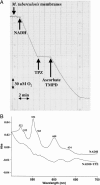
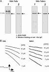
Mycobacterium tuberculosis (Mtb) is an obligate aerobe that is capable of long-term persistence under conditions of low oxygen tension. Analysis of the Mtb genome predicts the existence of a branched aerobic respiratory chain terminating in a cytochrome bd system and a cytochrome aa(3) system. Both chains can be initiated with type II NADH:menaquinone oxidoreductase. We present a detailed biochemical characterization of the aerobic respiratory chains from Mtb and show that phenothiazine analogs specifically inhibit NADH:menaquinone oxidoreductase activity. The emergence of drug-resistant strains of Mtb has prompted a search for antimycobacterial agents. Several phenothiazines analogs are highly tuberculocidal in vitro, suppress Mtb growth in a mouse model of acute infection, and represent lead compounds that may give rise to a class of selective antibiotics.
Figures


Similar articles
-
Type II NADH: menaquinone oxidoreductase of Mycobacterium tuberculosis.Infect Disord Drug Targets. 2007 Jun;7(2):169-81. doi: 10.2174/187152607781001781. Infect Disord Drug Targets. 2007. PMID: 17970227 Review.
-
Steady-state kinetics and inhibitory action of antitubercular phenothiazines on mycobacterium tuberculosis type-II NADH-menaquinone oxidoreductase (NDH-2).J Biol Chem. 2006 Apr 28;281(17):11456-63. doi: 10.1074/jbc.M508844200. Epub 2006 Feb 9. J Biol Chem. 2006. PMID: 16469750
-
Antitubercular pharmacodynamics of phenothiazines.J Antimicrob Chemother. 2013 Apr;68(4):869-80. doi: 10.1093/jac/dks483. Epub 2012 Dec 9. J Antimicrob Chemother. 2013. PMID: 23228936 Free PMC article.
-
Development of ssDNA aptamers as potent inhibitors of Mycobacterium tuberculosis acetohydroxyacid synthase.Biochim Biophys Acta. 2015 Oct;1854(10 Pt A):1338-50. doi: 10.1016/j.bbapap.2015.05.003. Epub 2015 May 16. Biochim Biophys Acta. 2015. PMID: 25988243
-
Type-II NADH Dehydrogenase (NDH-2): a promising therapeutic target for antitubercular and antibacterial drug discovery.Expert Opin Ther Targets. 2017 Jun;21(6):559-570. doi: 10.1080/14728222.2017.1327577. Epub 2017 May 15. Expert Opin Ther Targets. 2017. PMID: 28472892 Review.
Cited by
-
Mycobacterium tuberculosis nuoG is a virulence gene that inhibits apoptosis of infected host cells.PLoS Pathog. 2007 Jul;3(7):e110. doi: 10.1371/journal.ppat.0030110. PLoS Pathog. 2007. PMID: 17658950 Free PMC article.
-
Transcriptional Approach for Decoding the Mechanism of rpoC Compensatory Mutations for the Fitness Cost in Rifampicin-Resistant Mycobacterium tuberculosis.Front Microbiol. 2018 Nov 30;9:2895. doi: 10.3389/fmicb.2018.02895. eCollection 2018. Front Microbiol. 2018. PMID: 30555440 Free PMC article.
-
The F1Fo-ATP synthase of Mycobacterium smegmatis is essential for growth.J Bacteriol. 2005 Jul;187(14):5023-8. doi: 10.1128/JB.187.14.5023-5028.2005. J Bacteriol. 2005. PMID: 15995221 Free PMC article.
-
Isoniazid Bactericidal Activity Involves Electron Transport Chain Perturbation.Antimicrob Agents Chemother. 2019 Feb 26;63(3):e01841-18. doi: 10.1128/AAC.01841-18. Print 2019 Mar. Antimicrob Agents Chemother. 2019. PMID: 30642937 Free PMC article.
-
Vitamin K2 in electron transport system: are enzymes involved in vitamin K2 biosynthesis promising drug targets?Molecules. 2010 Mar 10;15(3):1531-53. doi: 10.3390/molecules15031531. Molecules. 2010. PMID: 20335999 Free PMC article. Review.
References
-
- Maher, D. & Raviglionem M. C. (1999) in Tuberculosis and Nontuberculous Mycobacterial Infections, ed. Schlossberg, D. (Saunders, Philadelphia), 4th Ed., pp. 104–115.
-
- Wayne, L. G. (1994) Eur. J. Clin. Microbiol. Infect. Dis. 13, 908–914. - PubMed
-
- Cole, S. T., Brosch, R., Parkhill, J., Garnier, T., Churcher, C., Harris, D., Gordon, S. V., Eiglmeier, K., Gas, S., Barry, C. E., III, et al. (1998) Nature 393, 537–544. - PubMed
-
- Ferguson-Miller, S. & Babcock, G. T. (1996) Chem. Rev. 96, 2889–2907. - PubMed
Publication types
MeSH terms
Substances
Grants and funding
LinkOut - more resources
Full Text Sources
Other Literature Sources
Molecular Biology Databases

