. 2005 Apr;79(7):4415-24.
doi: 10.1128/JVI.79.7.4415-4424.2005.

Epstein-Barr virus transforming protein LMP1 plays a critical role in virus production

Affiliations

Epstein-Barr virus transforming protein LMP1 plays a critical role in virus production

Nazmul Ahsan et al. J Virol. 2005 Apr.

Abstract

The Epstein-Barr virus (EBV) latent membrane protein 1 (LMP1), which is critical for EBV-induced B-cell transformation, is also abundantly expressed during the lytic cycle of viral replication. However, the biological significance of this strong LMP1 induction remains unknown. We engineered a bacterial artificial chromosome clone containing the entire genome of Akata strain EBV to specifically disrupt the LMP1 gene. Akata cell clones harboring the episomes of LMP1-deleted EBV were established, and the effect of LMP1 loss on virus production was investigated. We found that the degree of viral DNA amplification and the expression levels of viral late gene products were unaffected by LMP1 loss, demonstrating that the LMP1-deleted EBV entered the lytic replication cycle as efficiently as the wild-type counterpart. This was confirmed by our electron microscopic observation that nucleocapsid formation inside nuclei occurred even in the absence of LMP1. By contrast, loss of LMP1 severely impaired virus release into culture supernatants, resulting in poor infection efficiency. The expression of truncated LMP1 in Akata cells harboring LMP1-deleted EBV rescued the virus release into the culture supernatant and the infectivity, and full-length LMP1 partially rescued the infectivity. These results indicate that inducible expression of LMP1 during the viral lytic cycle plays a critical role in virus production.


Figures

FIG. 1.
FIG. 1.
Construction of d.LMP1-EBV. (A) Schematic maps of AK-BAC-GFP (18) and d.LMP1-EBV. The locations of the BamHI fragments of Akata strain EBV are shown on top. Triple stop codons (indicated by crosses) were inserted after codon 9 of the LMP1 gene of AK-BAC-GFP via GET recombination (29, 31). A zeocin resistance gene (Zeor), which served as a marker gene in GET recombination, was subsequently removed by in vitro Cre recombinase treatment. The positions of wild-type and 5171 loxP sites (mloxP) are indicated by open arrowheads. The chloramphenicol resistance gene (Cmr), neomycin resistance gene (Neor), kanamycin resistance gene (Kmr), right origin of lytic replication (oriLyt), and internal repeat 4 (IR4) are also indicated. (B) Partial sequence of the PCR product that was used for GET recombination to generate d.LMP1-EBV. The coding sequence of LMP1 is in the complementary strand of this sequence, and an arrow indicates its translation start site. The sequences homologous to the LMP1 gene are in bold. The positions of the primer sequences (NA03 and NA04) are underlined, the artificial stop codons (which appear in the complementary strand) are in italic, and the mutated loxP sites (23) are indicated by parentheses. Mutated loxP sites were used to avoid unwanted recombination, as AK-BAC-GFP had one wild-type loxP site located upstream of the BAC vector sequence (18). Stars indicate base substitution mutations created in 5171 loxP sites.
FIG. 2.
FIG. 2.
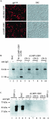
(A) Expression of viral glycoprotein gp110 after anti-IgG treatment in cells harboring AK-BAC-GFP and in cell clone 28-1 harboring d.LMP1-EBV episomes. The corresponding differential interference contrast (DIC) images are also shown. (B) Viral DNA amplification of d.LMP1-EBV in anti-IgG-treated Akata cells. Genomic DNAs of Akata cells harboring AK-BAC-GFP (BAC, lanes 1 and 2), cell clone 28 harboring both wild-type EBV and d.LMP1-EBV (W+d.LMP1-EBV, lanes 3 and 4), and cell clones harboring only d.LMP1-EBV (lanes 5through 10) were prepared before (−) and after (+) anti-IgG treatment. BamHI-digested genomic DNA was analyzed by Southern blotting with the BamHI X fragment as a probe. The sizes of the BamHI fragments are 8.6 kb for BAC-inserted EBV genomes and 2.1 kb for the wild-type EBV genome (18). Note that viral DNA amplification levels of d.LMP1-EBV (lanes 5 through 10) are comparable to that of AK-BAC-GFP (lanes 1 and 2). (C) Detection of LMP1 protein in Akata cell harboring various EBV episomes. Whole-cell extracts were prepared before (−) and after (+) anti-IgG treatment and subjected to Western blotting. The results for Akata cells harboring wild-type EBV episomes (EBV+, lanes 1 and 2), Akata cells harboring AK-BAC-GFP (BAC, lanes 3 and 4), Akata cells harboring d.LMP1-EBV (lanes 5 through 10), and EBV-negative Akata cells (EBV−, lane 11) are shown. The positions of protein standards are indicated on the left.
FIG. 3.
FIG. 3.
Electron microscopic observations of nucleocapsid formation inside cell nuclei after anti-IgG treatment. Representative images of cells harboring AK-BAC-GFP episomes (with the intact LMP1 gene) (A) and cell clone 28-1 harboring d.LMP1-EBV episomes (B) are shown. Abundant nucleocapsids (arrowheads) are visible inside nuclei (N). The nuclear membranes (NM) are indicated. Bars, 100 nm.
FIG. 4.
FIG. 4.
Impaired infection efficiency of the LMP1-deleted virus. (A) EBV-negative Akata cells were treated with the culture supernatant of anti-IgG-treated AK-BAC-GFP cells (top) or with the culture supernatant of anti-IgG-treated cell clone 28-1 harboring d.LMP1-EBV (bottom). GFP-positive cells represent infected cells. Note the marked reduction in the frequency of GFP-positive cells in the recipient cells that were infected with the culture supernatant of LMP1-deleted virus. The phase contrast images are also shown. (B) Expression of viral glycoprotein gp350/220 after anti-IgG treatment in cells harboring AK-BAC-GFP (top) and in cell clone 28-1 harboring d.LMP1-EBV (bottom). (C) Effect of transient expression of truncated LMP1 in Akata cells harboring d.LMP1-EBV (clone 28-1). Cells were transiently transfected with either pSG5-ΔXLMP1 or the pSG5 vector control, and transfected cells were subsequently treated with anti-IgG. EBV-negative Akata cells were then infected with the resultant culture supernatants. Note that the transient expression of truncated LMP1 resulted in increased infection efficiency (top) compared to the case when no LMP1 protein was supplied (bottom). The corresponding phase contrast images are also shown.
FIG. 5.
FIG. 5.
(A) Rescue experiments via constitutively expressed truncated LMP1 in Akata cells harboring d.LMP1-EBV (clone 28-1). The results with a lymphoblastoid cell line established with EGFP-EBV (27) (lane 1) and five independent cell clones obtained by stable transfection of truncated LMP1 (lanes 2 through 6) are shown. Note that four out of the five cell clones examined expressed truncated LMP1 (45 kDa). The positions of protein standards are indicated on the left. (B) Constitutive expression of truncated LMP1 rescued the infection efficiency of the culture supernatants of d.LMP1-EBV cells. EBV-negative Akata cells were infected with the culture supernatant of anti-IgG-treated AK-BAC-GFP cells (top) or anti-IgG-treated clone 3 (lane 3 in A, bottom). (C) Expression of truncated LMP1 increased the amounts of virus that were released into the culture supernatant. Culture supernatants of various anti-IgG-treated Akata cell clones were ultracentrifuged, and DNAs were extracted from the pelleted viruses. EcoRI-digested DNAs were subjected to Southern analysis with the EcoRI-K fragment as a probe. The results for d.LMP1-EBV cells (lane 1), d.LMP1-EBV cells with constitutive expression of truncated LMP1 (clone 3) (lane 2), and a cell clone harboring AK-BAC-GFP (lane 3) are shown. Note that cells expressing truncated LMP1 exhibit a more intense band of viral DNA (lane 2) than the cells lacking LMP1 expression (lane 1).
FIG. 6.
FIG. 6.
(A) Rescue experiments via constitutively expressed full-length LMP1 in Akata cells harboring d.LMP1-EBV (clone 28-1). The results of a lymphoblastoid cell line established with EGFP-EBV (27) (lane 1) and five independent cell clones obtained by stable transfection of full-length LMP1 (lanes 2 through 6) are shown. Note that the expression levels of full-length LMP1 (which is highest in clone 27) are far less than that of a lymphoblastoid cell line. The positions of protein standards are indicated on the left. (B) Constitutive expression of full-length LMP1 partially rescued the infectivity of the culture supernatant of d.LMP1-EBV cells. EBV-negative Akata cells were infected with the culture supernatant of anti-IgG-treated d.LMP1-EBV cells (clone 28-1) (top) or anti-IgG-treated cell clone 27 (lane 4 in A, bottom).

Similar articles

Cited by

References

    1. Adler, B., E. Schaadt, B. Kempkes, U. Zimber-Strobl, B. Baier, and G. W. Bornkamm. 2002. Control of Epstein-Barr virus reactivation by activated CD40 and viral latent membrane protein 1. Proc. Natl. Acad. Sci. USA 99:437-442. - PMC - PubMed
    1. Baer, R., A. T. Bankier, M. D. Biggin, P. L. Deininger, P. J. Farrell, T. J. Gibson, G. Hatfull, G. S. Hudson, S. C. Satchwell, C. Seguin, et al. 1984. DNA sequence and expression of the B95-8 Epstein-Barr virus genome. Nature 310:207-211. - PubMed
    1. Baichwal, V. R., and B. Sugden. 1989. The multiple membrane-spanning segments of the BNLF-1 oncogene from Epstein-Barr virus are required for transformation. Oncogene 4:67-74. - PubMed
    1. Baichwal, V. R., and B. Sugden. 1987. Posttranslational processing of an Epstein-Barr virus-encoded membrane protein expressed in cells transformed by Epstein-Barr virus. J. Virol. 61:866-875. - PMC - PubMed
    1. Boos, H., R. Berger, C. Kuklik-Roos, T. Iftner, and N. Mueller-Lantzsch. 1987. Enhancement of Epstein-Barr virus membrane protein (LMP) expression by serum, TPA, or n-butyrate in latently infected Raji cells. Virology 159:161-165. - PubMed

Publication types

Substances