. 2005 Jul 15;389(Pt 2):333-41.
doi: 10.1042/BJ20050244.

Binding of microtubule-associated protein 1B to LIS1 affects the interaction between dynein and LIS1

Affiliations

Binding of microtubule-associated protein 1B to LIS1 affects the interaction between dynein and LIS1

Eva M Jiménez-Mateos et al. Biochem J. .

Abstract

For neuronal migration to occur, the cell must undergo morphological changes that require modifications of the cytoskeleton. Several different MAPs (microtubule-associated proteins) or actin-binding proteins are proposed to be involved in the migration of neurons. Therefore we have specifically analysed how two members of the MAP family, MAP1B and LIS1 (lissencephaly-related protein 1), interact with one another and participate in neuronal migration. Our results indicate that, in hippocampal neurons, MAP1B and LIS1 co-localize, associate and interact with each another. The interaction between these two MAPs is regulated by the phosphorylation of MAP1B. Furthermore, this interaction interferes with the association between LIS1 and the microtubule-dependent molecular motor, dynein. Clearly, the differential binding of these cytoskeletal proteins could regulate the functions attributed to the LIS1-dynein complex, including those related to extension of the neural processes necessary for neuronal migration.


Figures
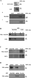
Figure 1
Figure 1. MAP1B interacts with LIS1
(A) Proteins from brain extracts were pulled down using GST–LIS1. (a) GST and GST–LIS1 were expressed and characterized (see the Materials and methods section). (b) MAP1B is specifically pulled-down with GST–LIS1; (c) the control of LIS1 is shown. The electrophoretic mobilities of the 200, 70, 45 and 30 kDa markers are indicated. (B) MAP1B and LIS1 proteins co-immunoprecipitate. Upper panel: immunoprecipitation experiments performed with antibodies raised against MAP1B (IPP MAP1B) or LIS1 (IPP LIS1), and proteins were visualized by Western blot (WB) with anti-MAP1B antibody. Lower panel: a similar experiment was conducted but the proteins were visualized with an anti-LIS1 antibody. The total protein (T) before immunoprecipitation and the immunoprecipitated (P) and non-immunoprecipitated (S) proteins are indicated. (C) To analyse the specificity of the interaction described in (A, B), a pellet fraction obtained after immunoprecipitation with MAP1B and LIS1 antibodies was revealed using antibodies against MAP2 and DCX. (D) The interaction between LIS1 and MAP1B is also specific when compared with mDab, a protein involved in the reelin signalling pathway. Molecular mass (kDa) is indicated on the right.
Figure 2
Figure 2. Cross-linking of MAP1B with LIS1
Protein from a mouse brain extract was incubated for 30 min at room temperature in the presence or absence of formaldehyde (FA). After this incubation, the protein was separated by gel electrophoresis and characterized by Western blotting (WB) using an antibody raised against MAP1B or LIS1. Additionally, the amount of actin in each sample was determined by dot blot (DB). The electrophoretic mobilities for MAP1B and LIS1 as well as the 200 and 40 kDa markers are indicated.
Figure 3
Figure 3. The interaction between MAP1B and LIS1 is regulated by MAP1B phosphorylation
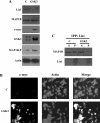
(A) Immunoprecipitation of brain extracts with LIS1 was performed using specific antibodies and the phosphorylated MAP1B was analysed. The MAP1B recovered in the pellet fraction was rich in mode II-phosphorylated MAP1B, whereas mode I-phosphorylated MAP1B was recovered mostly in the supernatant fraction. (B) Double immunofluorescence of hippocampal neurons in culture, showing the distribution of LIS1 and mode I-phosphorylated MAP1B or mode II-phosphorylated MAP1B. (C) Immunoprecipitation with A6 (an antibody that recognizes MAP1B irrespective of its level of phosphorylation). The precipitated proteins were fractionated by gel electrophoresis and visualized with an antibody raised against LIS1. The immunoprecipitated proteins from cultured neurons incubated in the presence of lithium (a specific inhibitor of mode I MAP1B phophorylation) or in the presence of OA (which prevents the dephosphorylation of MAP1B phosphorylated by mode I) are shown, together with those from untreated cells (control, C).
Figure 4
Figure 4. GSK3 regulates the interaction of MAP1B with LIS1
(A) N2A neuroblastoma cells were transfected with DNA encoding GSK3β bearing a c-Myc peptide tag. The amounts of LIS1, MAP1B, c-Myc, total GSK3, mode I-phosphorylated MAP1B (MAP1B-P) and actin were measured in untransfected (C) and GSK3-transfected cells. (B) The proportion of transfected cells was measured using the reaction with an antibody raised against the c-Myc peptide. (C) A protein extract from untransfected cells (C) or transfected (GSK3) cells was immunoprecipitated (IPP) with an antibody raised against LIS1, and the presence of MAP1B or LIS1 in the immunoprecipitated (P) or soluble (S) fraction was determined by Western blotting.
Figure 5
Figure 5. Dynein does not bind to MAP1B
(A) Protein from mouse brain extracts was immunoprecipitated (IP) with antibodies raised against LIC dynein or MAP1B, and then visualized with an anti-LIC dynein antibody. (B) Double immunofluorescence analysed by confocal microscopy, showing the distribution of dynein, of mode II-phosphorylated MAP1B and the merged image.
Figure 6
Figure 6. The absence of MAP1B does not affect the overall levels of dynein or LIS1 proteins, but results in an increase in LIS1–dynein interaction
(A) Western-blot analyses of brain extracts derived from control (WT) and MAP1B-deficient (KO) mice are shown. Actin was tested as a loading control. (B, a) These experiments are similar to the pull-down experiments indicated in Figure 1(A) using GST–LIS1 recombinant protein mixed with brain extracts from control (WT) and from MAP1B-deficient (KO) animals. The first panel shows that MAP1B was recovered from the WT brain extracts after pull down with GST–LIS1, whereas no MAP1B was recovered from KO mice brain extracts. An increase in the amount of dynein subunits recovered after GST–LIS1 pull down was observed when the proteins were mixed with KO mice brain extracts. No MAP2 was recovered after pull down. Tubulin was used as an internal control to compare the protein concentration in the two brain extracts. (B, b) Quantification of the proteins recovered after pull-down experiments. (C, a) Co-immunoprecipitation experiments show that dynein LICs bind to LIS1 in the presence (WT) or absence (KO) of MAP1B. In the absence of MAP1B, a decrease of protein 1 was detected as well as an increase of protein 2, corresponding to the dynein LICs. (C, b) Quantification of the results by densitometry analysis.
Figure 7
Figure 7. Association of LIS1 with neuronal microtubules in the presence or absence of MAP1B
Microtubule protein was polymerized in vitro and the association of LIS1 with the polymers was examined. The first panel shows that MAP1B was present in the microtubule fraction derived from control animals (WT) but was absent from those from mutant animals (KO). Furthermore, LIS1 was enriched in the pellet fraction after microtubule polymerization. The level of assembled tubulin is also shown. A decrease (30%) in the amount of LIS1 bound to microtubules was found in the KO-derived polymer compared with that found in microtubules from WT mice.
Figure 8
Figure 8. Golgi localization in neurons with or without MAP1B
Hippocampal neurons at culture stage 2 (see [5]) from WT (A) or mutant MAP1B (B) mice were incubated with the antibody LL-FITC to localize the Golgi complex (see the Material and methods section). In neurons from WT mice, it is localized to a specific site close to the nucleus, whereas it appears to be dispersed in neurons lacking MAP1B. Scale bars, 50 μm.
Figure 9
Figure 9. Microtubule network in growth cones in the presence or absence of MAP1B
(A) Hippocampal neurons from WT or KO mice were cultured, and the presence of microtubules in their growth cones was studied by immunofluorescence analysis using an antibody raised against Tyr-tubulin. A difference in the amounts of peripheral microtubules was found. Scale bars, 5 μm. The outline of the growth cones is indicated in each case by white dots. (B) Same as (A), but the growth cones were analysed by immunofluorescence using phalloidin to identify their shape.

Similar articles

Cited by

References

    1. Tucker R. P., Garner C. C., Matus A. In situ localization of microtubule-associated protein mRNA in the developing and adult rat brain. Neuron. 1989;2:1245–1256. - PubMed
    1. Diaz-Nido J., Avila J. Characterization of proteins immunologically related to brain microtubule-associated protein MAP-1B in non-neural cells. J. Cell Sci. 1989;92:607–620. - PubMed
    1. Schoenfeld T. A., McKerracher L., Obar R., Vallee R. B. MAP 1A and MAP 1B are structurally related microtubule associated proteins with distinct developmental patterns in the CNS. J. Neurosci. 1989;9:1712–1730. - PMC - PubMed
    1. Roos J., Hummel T., Ng N., Klambt C., Davis G. W. Drosophila Futsch regulates synaptic microtubule organization and is necessary for synaptic growth. Neuron. 2000;26:371–382. - PubMed
    1. Gonzalez-Billault C., Demandt E., Wandosell F., Torres M., Bonaldo P., Stoykova A., Chowdhury K., Gruss P., Avila J., Sanchez M. P. Perinatal lethality of microtubule-associated protein 1B-deficient mice expressing alternative isoforms of the protein at low levels. Mol. Cell. Neurosci. 2000;16:408–421. - PubMed

Publication types

Substances