. 2005 Apr;3(4):e96.
doi: 10.1371/journal.pbio.0030096. Epub 2005 Mar 15.

Two distinct E3 ubiquitin ligases have complementary functions in the regulation of delta and serrate signaling in Drosophila

Affiliations

Two distinct E3 ubiquitin ligases have complementary functions in the regulation of delta and serrate signaling in Drosophila

Roland Le Borgne et al. PLoS Biol. 2005 Apr.

Abstract

Signaling by the Notch ligands Delta (Dl) and Serrate (Ser) regulates a wide variety of essential cell-fate decisions during animal development. Two distinct E3 ubiquitin ligases, Neuralized (Neur) and Mind bomb (Mib), have been shown to regulate Dl signaling in Drosophila melanogaster and Danio rerio, respectively. While the neur and mib genes are evolutionarily conserved, their respective roles in the context of a single organism have not yet been examined. We show here that the Drosophila mind bomb (D-mib) gene regulates a subset of Notch signaling events, including wing margin specification, leg segmentation, and vein determination, that are distinct from those events requiring neur activity. D-mib also modulates lateral inhibition, a neur- and Dl-dependent signaling event, suggesting that D-mib regulates Dl signaling. During wing development, expression of D-mib in dorsal cells appears to be necessary and sufficient for wing margin specification, indicating that D-mib also regulates Ser signaling. Moreover, the activity of the D-mib gene is required for the endocytosis of Ser in wing imaginal disc cells. Finally, ectopic expression of neur in D-mib mutant larvae rescues the wing D-mib phenotype, indicating that Neur can compensate for the lack of D-mib activity. We conclude that D-mib and Neur are two structurally distinct proteins that have similar molecular activities but distinct developmental functions in Drosophila.


Figures

Figure 1
Figure 1. Molecular and Genetic Characterization of D-mib Mutations
(A) Molecular map of the D-mib locus showing the position of the P-element inserted into the 5′ untranslated region (allele D-mib1) and the 13.6 kb deletion that removes the D-mib and the RpS31 genes (allele D-mib2). Transcribed regions are indicated with arrows, and exons are indicated with boxes. Open reading frames are shown in black. (B) Domain composition of D-mib and D. rerio Mib. Both proteins show identical domain organization. D-mib has an N-terminal ZZ zinc finger flanked on either side by a Mib/HERC2 (M-H) domain, followed by two Mib repeats, six ankyrin repeats, two atypical RING domains, and a C-terminal protypical RING that has been associated with catalytic E3 ubiquitin ligase activity. The D-mib3 mutant allele is predicted to produce a truncated protein devoid of E3 ubiquitin ligase activity whereas the D-mib4 protein carries a mutation at a conserved position in the second Mib repeat. (C and C′) Western blot analysis of D-mib (C). The endogenous D-mib protein (predicted size: 130 kDa) was detected in S2 cells (lane 2) and in imaginal discs from wild-type larvae (lane 3) but was not detectable in homozygous D-mib1 (lane 4) and D-mib1/D-mib3 (lane 5) third instar larvae. The D-mib protein produced in transfected S2 cells from the cDNA used in this study (lane 1) runs exactly as endogenous D-mib (lane 2). Panel C′ shows a Red Ponceau staining of the gel with the same protein samples as in panel C. (D–H) Wings from wild-type (D), D-mib1 (E), SerRX82/Serrev6.1 (F), D-mib2/D-mib4 (G), and UAS-D-mib2/+; D-mib1/D-mib2 flies (H). D-mib (E) and Ser (F) mutant flies showed similar wing loss phenotypes. The D-mib mutant phenotype could be almost fully rescued by a leaky UAS-D-mib transgene (H). (D′) and (G′) show high magnification views of (D) and (G), respectively, to show that D-mib2/D-mib4 mutant flies (G′) exhibited ectopic sensilla (arrowheads) along vein L3. (I–N) Nota (I–K) and legs (L–N) from wild-type (I and L), D-mib1 (J and M), and SerRX82/Serrev6.1 (K and N) flies. D-mib mutant flies showed a weak neurogenic phenotype (J) that was not observed in Ser mutant flies (K). Ectopic sensory organs in D-mib mutant flies developed from ectopic sensory organ precursor cells (not shown). D-mib (M) and Ser (N) mutant legs also showed distinct growth and/or elongation defects. Arrows in (J) show ectopic macrochaetes. Arrows in (L–N) indicate the joints. Ti, tibia; t1 to t5, tarsal segments 1 to 5.
Figure 2
Figure 2. The D-mib and neur Genes Have Distinct Functions during Wing Development
(A–E) Wing imaginal discs (B–E) from wild-type (B and D), D-mib1 (C), and D-mib1/D-mib2 (E) third instar larvae stained for Cut (B and C) and wg-lacZ (D and E). D-mib mutant discs showed a dramatically reduced size of the wing pouch (see diagram in [A] showing the different regions of the wing imaginal disc; V, ventral; D, dorsal), as well as a complete loss of Cut and wg-lacZ (red arrows in [B–E]) expression at the wing margin. Expression of wg-lacZ in the hinge region (arrowheads in [D] and [E]) and the accumulation of Cut in sensory cells (small arrows in [B] and [C]) and muscle precursor cells (large arrowheads in [B] and [C]) appeared to be largely unaffected). (F and F′) Expression of Cut (red) at the wing margin was not affected by the complete loss of neur activity in neur1F65 mutant clones (indicated by the loss of the nuclear green fluorescent protein [GFP] marker, in green). Bar is 50 μm in (B–E) and 20 μm in (F and F′).
Figure 3
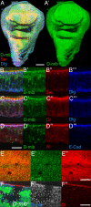
Figure 3. D-mib Co-Localizes with Dl and Ser at the Apical Cell Cortex
(A and A′) D-mib (green) is detected in all cells of the wing imaginal disc. In (A), Ser is in red and Discs-large (Dlg) is in blue. (B–D′′′) D-mib (green in B, B′, C, C′, D, and D′) co-localized with Ser (red in [B and B′′]), Dl (red in [C and C′′]), N (red in [D and D′′]), and E-Cadherin (E-Cad; blue in [D and D′′′]) and was found apical to Discs-large (Dlg; blue in [B, B′′′, C, and C′′′]) in notum cells located at the edges of the wing discs. (E–E′′) D-mib (green in [E and E′]) co-localized with Dl (red in [E and E′′]) at the apical cortex of wing pouch cells. (F–F′′) D-mib staining at the apical cortex (blue in [F and F′]) was not detected in D-mib2 mutant clone (marked by loss of nuclear GFP staining; green in [F]). Loss of D-mib activity has no detectable effect on the apical accumulation of Dl (red in [F and F′′]). Bar is 50 μm for (A and A′) and 10 μm for (B–F′′).
Figure 6
Figure 6. D-mib Is Required in Dorsal Cells for Margin Expression of Cut
Large dorsal clones of D-mib2 mutant cells (marked by the loss of nuclear GFP, in green) resulted in a complete loss of Cut (red) expression (A and B). This indicates that D-mib is required for Ser signaling by dorsal cells. In contrast, ventral clones did not prevent the expression of Cut (C and D), implying that D-mib is not strictly required for Dl signaling. Note that mutant ventral cells abutting wild-type dorsal cells expressed Cut (arrow in [D]), indicating that D-mib is not required for N signal transduction. Low-magnification views of the wing portion of the discs are shown in (A) and (C). (B) and (D) show high-magnification views of the areas boxed in (A) and (C), respectively.
Figure 4
Figure 4. D-mib Is Required to Down-Regulate Ser at the Apical Cortex
(A–F′) Distribution of Dl (green) and Ser (red) in the notum region of wild-type (A–C′) and D-mib1 mutant (D–F′) wing imaginal discs. The boxed areas in (A) and (D) are shown at higher magnification in (B–F′). The specific loss of Ser accumulation into intracellular vesicles (compare [E′] with [B′]) correlated with the elevated levels of Ser seen at the apical cortex of D-mib mutant cells (compare [E] with [B]). (G–J′) Ser (red in [H and H′]) accumulated at the apical cortex (H) as well as in intracellular dots (H′) in D-mib2 mutant cells (marked by the loss of nuclear GFP; green in [G]). Cut is shown in blue (G). The distribution of Dl (red in [J and J′]) was not affected by the loss of D-mib activity. Low-magnification views of the wing portion of the discs are shown in (G) and (I). (H and H′) and (J and J′) show high magnification views of the areas boxed in (G) and (I), respectively. Clone boundaries are outlined in (H and H′) and (J and J′). Bar is 40 μm for (A, D, G), 5 μm for (B–C′ and E–F′), and 10 μm for (H–J′).
Figure 5
Figure 5. D-mib Is Required for Ser Endocytosis
Localization of the anti-Ser (red) and anti-Dl (green) antibodies that have been internalized by wild-type (A–C′′) and D-mib1 mutant (D–F′′) cells in the notum region of wing discs. (A–A′′) and (D–D′′) show apical sections and (B–B′′) and (E–E′′) show basal sections. (C–C′′) and (F–F′′) show confocal z-sections. The z-section axes are shown with a double-headed arrow in (A) and (D). Internalized anti-Ser and anti-Dl antibodies co-localized in wild-type cells. In contrast, high levels of anti-Ser antibodies were detected at the cell surface of D-mib mutant epithelial cells whereas anti-Dl antibodies were efficiently internalized. Bar is 10 μm for all panels.
Figure 7
Figure 7. Expression of D-mib in Dorsal Cells Is Sufficient to Rescue the D-mib Mutant Phenotype
(A) Expression of D-mib (green) in dorsal cells, using Ser-GAL4, rescued the growth of the wing pouch and margin Cut (red) expression in D-mib2/D-mib3 mutant discs. (B–D′′′) Ser-GAL4-driven expression of YFP::D-mib (green) rescued the D-mib2/D-mib3 phenotype and strongly reduced the level of Dl (blue in [B, B′, C, C′′, D, and D′′]) and Ser (red in [B, B′′, C, C′′′, D, and D′′′]) in dorsal cells. (C–D′′′) are high-magnification views (apical [C–C′′′] and basal [D–D′′′]) of the disc shown in (B–B′′). YFP::D-mib co-localized with Dl and Ser at the apical cortex in cells expressing only low levels of YFP::D-mib. Bar is 50 μm for (A–B′′) and 10 μm for (C–D′′′).
Figure 8
Figure 8. Expression of Neur in Dorsal Cells Is Sufficient to Rescue the D-mib Mutant Phenotype
D-mib2/D-mib3 mutant discs expressing GFP (A) (GFP staining not shown), Ser (B), Ncdc10 (C), or Neur (D) under the control of Ser-GAL4 were stained for Cut (red). Expression of Ser in dorsal cells did not rescue the D-mib2/D-mib3 wing pouch mutant phenotype (compare [B] with [A]), consistent with D-mib being required for Ser signaling. By contrast, expression of Ncdc10, an activated version of N, led to the deregulated growth of the dorsal compartment and the expression of Cut in most dorsal cells (C), indicating that activated N acts downstream of D-mib. Expression of Neur in dorsal cells was sufficient to compensate for the loss of D-mib activity (D). Bar is 40 μm for all panels.

Similar articles

Cited by

References

    1. Lai EC. Notch signaling: Control of cell communication and cell fate. Development. 2004;131:965–973. - PubMed
    1. Schweisguth F. Notch signaling activity. Curr Biol. 2004;14:R129–R138. - PubMed
    1. Fleming RJ. Structural conservation of Notch receptors and ligands. Semin Cell Dev Biol. 1998;9:599–607. - PubMed
    1. Lehmann R, Jimenez F, Dietrich U, Campos-Ortega JA. On the phenotype and development of mutants of early neurogenesis in Drosophila melanogaster . Rouxs Arch Dev Biol. 1983;192:62–74. - PubMed
    1. Wiellette EL, McGinnis W. Hox genes differentially regulate Serrate to generate segment-specific structures. Development. 1999;126:1985–1995. - PubMed

Publication types

Associated data