Review
. 2005 Mar;69(1):101-23.
doi: 10.1128/MMBR.69.1.101-123.2005.

Initiation of protein synthesis in bacteria

Affiliations
Review

Initiation of protein synthesis in bacteria

Brian Søgaard Laursen et al. Microbiol Mol Biol Rev. 2005 Mar.

Abstract

Valuable information on translation initiation is available from biochemical data and recently solved structures. We present a detailed description of current knowledge about the structure, function, and interactions of the individual components involved in bacterial translation initiation. The first section describes the ribosomal features relevant to the initiation process. Subsequent sections describe the structure, function, and interactions of the mRNA, the initiator tRNA, and the initiation factors IF1, IF2, and IF3. Finally, we provide an overview of mechanisms of regulation of the translation initiation event. Translation occurs on ribonucleoprotein complexes called ribosomes. The ribosome is composed of a large subunit and a small subunit that hold the activities of peptidyltransfer and decode the triplet code of the mRNA, respectively. Translation initiation is promoted by IF1, IF2, and IF3, which mediate base pairing of the initiator tRNA anticodon to the mRNA initiation codon located in the ribosomal P-site. The mechanism of translation initiation differs for canonical and leaderless mRNAs, since the latter is dependent on the relative level of the initiation factors. Regulation of translation occurs primarily in the initiation phase. Secondary structures at the mRNA ribosomal binding site (RBS) inhibit translation initiation. The accessibility of the RBS is regulated by temperature and binding of small metabolites, proteins, or antisense RNAs. The future challenge is to obtain atomic-resolution structures of complete initiation complexes in order to understand the mechanism of translation initiation in molecular detail.


Figures

FIG. 1.
FIG. 1.
Translation initiation pathway in bacteria. The 30S and 50S ribosomal subunits are shown in light and dark grey, respectively. Translation initiation factors IF1, IF2, and IF3, the mRNA, and the fMet-tRNAfMet are shown in red, blue, green, yellow, and magenta, respectively. The components are placed on the ribosome according to current experimental knowledge. Details of the pathway are given in the text. Structures are derived from PDB entries as follows: 30S ribosomal subunit, 1HR0; 50S ribosomal subunit, 1FFK; IF1, 1HR0; IF2, 1G7T; IF3N, 1TIF; IF3C, 1TIG; mRNA, 1JGQ; fMet-tRNAfMet, 1JGQ. Structural representations in this as well as other figures in this review were made using the program MolMol (93) and Pov-Ray unless otherwise stated.
FIG. 2.
FIG. 2.
Structures of the ribosomal subunits. (A) Overview of the 16S rRNA secondary structure. The domains are shown in colors according to the secondary structure: blue, 5′ domain (bulk of body); magenta, central domain (platform); red, 3′ major domain (head); yellow, 3′ minor domain (helices 44 and 45 located at the subunit interface). (B) Overview of the 23S and 5S rRNA secondary structures. The RNA domains are shown in colors according to the secondary structure of the 23S rRNA: blue, domain I; cyan, domain II; green, domain III; yellow, domain IV; red, domain V; magenta, domain VI. The 5S rRNA is shown in orange. (C) Three-dimensional structure of the 30S ribosomal subunit from T. thermophilus at 3-Å resolution (PDB entry 1J5E). RNA secondary-structure domains are colored as in panel A. Note that the secondary-structure domains of the RNA correspond well to the tertiary domains. Proteins are omitted for clarity. The tRNA binding sites A, P, and E are indicated. (D) Three-dimensional structure of the 50S ribosomal subunit from H. marismortui at 2.4-Å resolution (PDB entry 1FFK). Colors are the same as in panel B. Note that the secondary-structure domains of the RNA do not correspond to the tertiary domains, unlike for the 30S subunit. Proteins are omitted for clarity. The L1 stalk, the central protuberance (CP), and the L7-L12 stalk are indicated.
FIG. 3.
FIG. 3.
Binding of mRNA to the 30S ribosomal subunit. (A) Binding of a canonical mRNA to the 30S ribosomal subunit. Two alternative pathways are shown where either the mRNA or the fMet-tRNAfMet binds first, followed by the other component. The mRNA is bound via the SD-ASD interaction as well as the codon-anticodon interaction. (B) Binding of a leaderless mRNA to the 30S ribosomal subunit. The mRNA is bound to the ribosome mainly via the codon-anticodon interaction. IF2 stimulates the binding of leaderless mRNAs, presumably by recruitment of fMet-tRNAfMet to the subunit.
FIG. 4.
FIG. 4.
mRNA bound to the 30S ribosomal subunit. (A) A 36-nucleotide mRNA is bound to the 30S ribosomal subunit. rRNA is shown in grey, mRNA is shown in yellow, and protein is shown in cyan. The ASD sequence of the 16S rRNA is shown in red to indicate the SD-ASD interaction. The P-site initiation codon is shown in green, and the A-site codon is shown in magenta. Note the kink in the mRNA between the two codons. (B) Close-up of the region indicated in panel A. The upstream and downstream tunnels are marked by arrows. Colors are the same as in panel A. The structure is derived from PDB entry 1JGQ, prepared using the program Ribbons (25), and rendered in Pov-Ray.
FIG. 5.
FIG. 5.
Initiator and elongator methionine-accepting tRNAs. Cloverleaf representation of methionine-accepting tRNAs: (A) initiator tRNA and (B) elongator tRNA. The regions important for initiator tRNA identity are highlighted. Details are given in the text.
FIG. 6.
FIG. 6.
Interactions of the initiator tRNA. (A) Surface representations of the initiator tRNA. The regions that interact with the indicated component of the translational machinery are highlighted in red, and the nucleotide positions on the tRNA are indicated next to the structure. The structure of the initiator tRNA is derived from PDB entry 2FMT. (B) Detailed view of the interaction between the initiator tRNA and the ribosome. On the left is a surface representation showing the important sites of interaction in red. On the right is the initiator tRNA on the 70S ribosome. The tRNA is shown in red, the mRNA is shown in yellow, and the 16S and 23S rRNA are shown in cyan and grey, respectively. The codon-anticodon interaction is shown (the structure is derived from PDB entries 1GIX and 1GIY, prepared using the program Ribbons [25], and rendered in Pov-Ray).
FIG. 7.
FIG. 7.
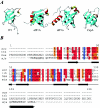
Initiation factor IF1 and structural homologues. (A) Structures of IF1 and homologues: IF1 from E. coli (PDB entry 1AH9); human eIF1A, residues 40 to 125 (PDB entry 1D7Q); aIF1A from Methanococcus jannaschii (PDB entry 1JT8); and cold shock protein A (CspA) from E. coli (PDB entry 1MJC). eIF1A and aIF1A have an additional helix located at the C terminus. (B) Sequence alignment of selected IF1 sequences. Abbreviations: eco, E. coli; tth, T. thermophilus; hsa, H. sapiens; mja, M. jannaschii. Positions with 100% identity are shown in blue. If more than 50% are identical or highly similar, the residues are highlighted in red; if more than 50% of the residues are weakly similar, they are highlighted in orange.
FIG. 8.
FIG. 8.
IF1 bound to the 30S ribosomal subunit. (A) Structure of IF1 on the 30S ribosomal subunit. IF1 is shown in blue, helix 44 is shown in magenta, the 530 loop is shown in yellow, and protein S12 is shown in green. The structure is derived from PDB entry 1HR0. (B) Close-up of the interaction of IF1 with the 30S subunit. IF1 is shown in a surface representation colored according to the electrostatic potential (positive charges, blue; negative charges, red). Helix 44 and the 530 loop of 16S rRNA are shown in magenta and yellow, respectively. Protein S12 is shown in a green ribbon representation. Bases A1492 and A1493 of the 16S rRNA are indicated in red. Note that they have flipped out of helix 44 and are buried in a pocket in IF1 and a pocket between IF1 and S12, respectively.
FIG. 9.
FIG. 9.
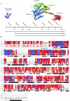
IF2 and structural homologues. (A) Schematic representation of the E. coli IF2 primary structure. The domain boundaries and the lengths of the three IF2 isoforms are indicated. Ribbon diagrams of the structures of the IF2N domain from E. coli (PDB entry 1ND9) and the IF2 homologue aIF5B from M. thermoautotrophicum (PDB entry 1G7T) are shown. The domains are indicated in different colors, and the E. coli domain nomenclature is used. (B) Sequence alignment of selected bacterial IF2 and archaeal and eukaryotic homologues. Only a small part of the N-terminal nonconserved region is shown. Abbreviations: tth; T. thermophilus, bst, B. stearothermophilus; eco, E. coli; mth, M. thermoautotrophicum; hsa, H. sapiens. Secondary-structure elements defined from the structure of aIF5B from M. thermoautotrophicum are indicated by cylinders for helical segments and arrows for segments in β-strand conformation. The domain boundaries are indicated by yellow arrows. Color codes are as in Fig. 7.
FIG. 10.
FIG. 10.
IF2 Interactions with fMet-tRNAfMet and the ribosome. (A) The 30S ribosomal subunit from T. thermophilus (PDB entry 1J5E) in complex with a P-site fMet-tRNAfMet (derived and docked based on PDB entry 1GIX). The fMet-ACC region of the tRNA that interacts with the C-terminal region of IF2 is shown in red. The corresponding region is red on the aIF5B structure shown at the bottom part (PDB entry 1G7T). Two residues in domain V of IF2 (V451 and S520 of B. stearothermophilus IF2) are shown in yellow and magenta, respectively. A nuclease at position V451 cleaves positions 38 to 40 and 498, and a nuclease at position S520 cleaves positions 538 to 540 of the E. coli 16S rRNA in a 70S ribosomal complex. The corresponding positions are indicated in yellow and magenta on the ribosomal subunit. Note that these cleavages are absent in a 30S-IF2 initiation complex. (B) The 50S ribosomal subunit from H. marismortui (PDB entry 1JJ2) is shown along with aIF5B. Red indicates the position of the fMet ACC in the decoding center of the ribosomal subunit and the corresponding region on a IF5B that interacts with the fMET ACC. Pale blue and light green on the subunit indicate two positions in helix 89 of the 23S rRNA (U2474 and A2482 in E. coli numbering) that are cleaved by nucleases attached at the residue located at the interface between domains VI-1 and VI-2 of IF2 (shown in dark green on the aIF5b structure) (E644 of the B. stearothermophilus IF2). A nuclease attached to a residue in domain VI-1 of IF2 (dark blue) (E632 of the B. stearothermophilus IF2) cleaves both the C1076 (cyan) and G1068 (cyan) positions in the L11 region. The nuclease (shown in purple) on aIF5B (position Y625 of the VI-1 domain) cleaves weakly at C1076 (cyan) and U2474 (light blue). Data were derived from reference . Another study showed that IF2 protects residues in the sarcin-ricin domain (SRD) (G2655, A2665, and G2661) against chemical modification (100). These residues are indicated in yellow on the subunit. No attempt was made to dock IF2 on the ribosomal subunit, since it is likely that conformational changes occur in the subunit as a result of IF2 binding.
FIG. 11.
FIG. 11.
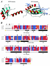
IF3 structure and alignment. (A) Structures of the IF3N domain from B. stearothermophilus (PDB entry 1TIF) and the IF3C domain from E. coli (PDB entry 2IFE). The side chains of the arginine residues in the IF3C domain are shown and labeled with the residue number. Mutations in the arginine residues that affect binding to the 30S ribosomal subunit are residue numbers 99, 112, 116, 147, and possibly 168. These roughly define the surface that binds to the 30S ribosomal subunit. Mutations of arginine residues reducing IF3 activity involved in mRNA-related functions define a surface comprising residues 129, 131, and 133. (B) Sequence alignment of selected sequences of IF3. Abbreviations, color codes, and secondary-structure nomenclature are as in Fig. 7. Secondary-structure elements are as defined in reference . Black vertical arrows indicate residues that have been identified as interacting with the 30S ribosomal subunit by mutagenesis and/or chemical modification. Grey triangles indicate residues whose intensity was most strongly affected by titration with 30S ribosomal subunits in NMR spectroscopy studies (reference and references cited therein). Yellow triangles indicate approximate domain boundaries.
FIG. 12.
FIG. 12.
Interaction of IF3 with the 30S ribosomal subunit. The interactions between IF3 and the 30S ribosomal subunit identified by hydroxyl radical footprinting and directed probing are shown (data from reference 42). (A) 30S ribosomal subunit from T. thermophilus with P-site-bound tRNA (derived from PDB entries 1J5E and 1GIX). The tRNA is shown in yellow. (B) Close-up of the ribosomal subunit. Sites in the 16S rRNA and on the tRNA that are cleaved by nucleases attached to IF3 are indicated. The IF3N and IF3C domains (PDB entries 1TIF and 2IFE) are shown in a ribbon representation, with the modified cysteines indicated by spheres. Orange spheres on the IF3 domains indicate residues where an attached nuclease does not cleave the 16S rRNA or tRNA. The magenta spheres in the IF3C domain indicate residues K97 and MI35 from where nucleases cleave in the 790 loop of the 16S rRNA (magenta) seen below the acceptor arm of the tRNA. Nucleases at these positions also cleave the side of the tRNA marked in blue (residues 3 to 5 and 12 to 24). The cyan sphere in the IF3C domain indicates E104 from which nucleases cleave in the 790 loop (magenta) and at residues 1482 to 1487, indicated in cyan on the 30S subunit. Green spheres in the linker region on the IF3N domain indicate residues (E76 and S80) on which nucleases cleave on the other side of the tRNA, marked in green (residues 26 to 29 and 35 to 37), the 790 loop region (magenta), and the 690 loop region (red) of the 16S rRNA. A nuclease attached to position R11 in the IF3N domain (red) cleaves at positions in the 690 loop (red). No attempt was made to dock IF3 on the ribosomal subunit, since conformational changes most probably take place in the subunit as a result of IF3 binding.
FIG. 13.
FIG. 13.
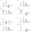
Examples of translational regulation mechanisms. (A) Repression of translation by binding of a metabolite that stabilizes an alternative mRNA secondary structure and leaves the SD sequence and initiation codon (AUG) in a base-paired region. (B) Activation of translation by binding of a metabolite that stabilizes an alternative mRNA secondary structure and leaves the SD sequence and initiation codon (AUG) in an unpaired region, thus providing ribosomal access. (C) Repression of translation by the formation of an alternative mRNA secondary structure as a result of a change in temperature. (D) Activation of translation by an increase in temperature, causing a local melting of the mRNA secondary structure covering the SD and AUG region.

Similar articles

Cited by

References

    1. Adhin, M. R., and J. van Duin. 1990. Scanning model for translational reinitiation in eubacteria. J. Mol. Biol. 213:811-818. - PubMed
    1. Antoun, A., M. Y. Pavlov, K. Andersson, T. Tenson, and M. Ehrenberg. 2003. The roles of initiation factor 2 and guanosine triphosphate in initiation of protein synthesis. EMBO J. 22:5593-5601. - PMC - PubMed
    1. Baan, R. A., J. J. Duijfjes, E. van Leerdam, P. H. van Knippenberg, and L. Bosch. 1976. Specific in situ cleavage of 16S ribosomal RNA of Escherichia coli interferes with the function of initiation factor IF-1. Proc. Natl. Acad. Sci. USA 73:702-706. - PMC - PubMed
    1. Bae, W., B. Xia, M. Inouye, and K. Severinov. 2000. Escherichia coli CspA-family RNA chaperones are transcription antiterminators. Proc. Natl. Acad. Sci. USA 97:7784-7789. - PMC - PubMed
    1. Ban, N., P. Nissen, J. Hansen, P. B. Moore, and T. A. Steitz. 2000. The complete atomic structure of the large ribosomal subunit at 2.4 A resolution. Science 289:905-920. - PubMed

Publication types

MeSH terms

LinkOut - more resources