Comparative Study
. 2005 Mar 2;25(9):2204-14.
doi: 10.1523/JNEUROSCI.3610-04.2005.

The multiple functions of cysteine-string protein analyzed at Drosophila nerve terminals

Affiliations
Comparative Study

The multiple functions of cysteine-string protein analyzed at Drosophila nerve terminals

Peter Bronk et al. J Neurosci. .

Abstract

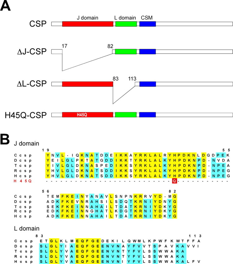
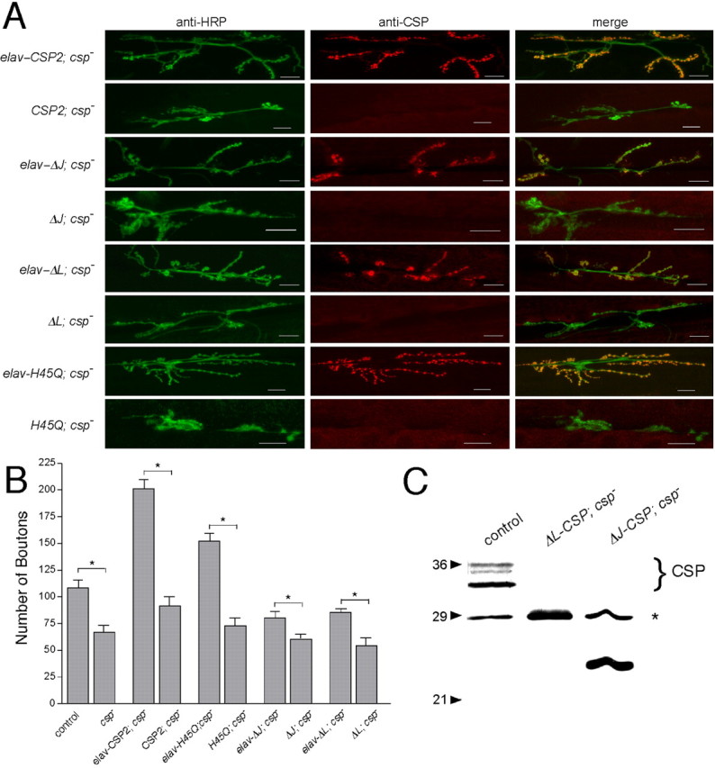
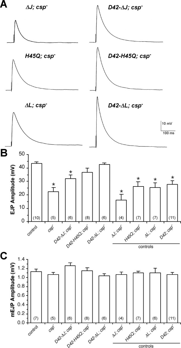
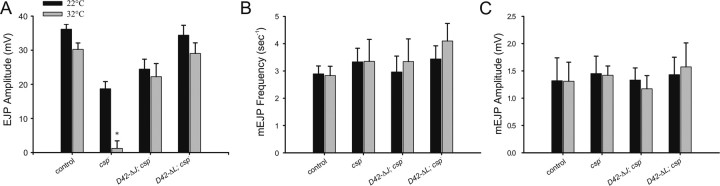
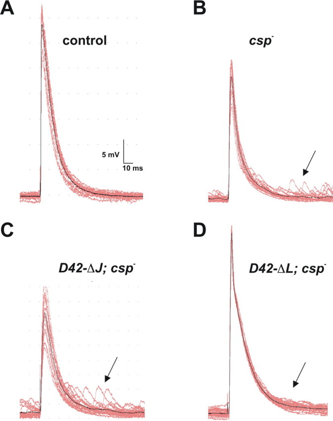
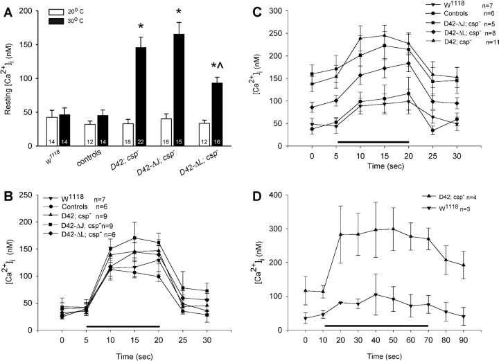
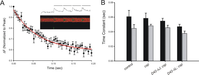
The synaptic vesicle-associated cysteine-string protein (CSP) is important for synaptic transmission. Previous studies revealed multiple defects at neuromuscular junctions (NMJs) of csp null-mutant Drosophila, but whether these defects are independent of each other or mechanistically linked through J domain mediated-interactions with heat-shock cognate protein 70 (Hsc70) has not been established. To resolve this issue, we genetically dissected the individual functions of CSP by an in vivo structure/function analysis. Expression of mutant CSP lacking the J domain at csp null-mutant NMJs fully restored normal thermo-tolerance of evoked transmitter release but did not completely restore evoked release at room temperature and failed to reverse the abnormal intraterminal Ca2+ levels. This suggests that J domain-mediated functions are essential for the regulation of intraterminal Ca2+ levels but only partially required for regulating evoked release and not required for protecting evoked release against thermal stress. Hence, CSP can also act as an Hsc70-independent chaperone protecting evoked release from thermal stress. Expression of mutant CSP lacking the L domain restored neurotransmission and partially reversed the abnormal intraterminal Ca2+ levels, suggesting that the L domain is important, although not essential, for the role of CSP in regulating intraterminal Ca2+ levels. We detected no effects of csp mutations on individual presynaptic Ca2+ signals triggered by action potentials, suggesting that presynaptic Ca2+ entry is not primarily impaired. Both the J and L domains were also required for the role of CSP in synaptic growth. Together, these results suggest that CSP has several independent synaptic functions, affecting synaptic growth, evoked release, thermal protection of evoked release, and intraterminal Ca2+ levels at rest and during stimulation.


Figures

Figure 1.
Figure 1.
Mutations in Drosophila CSP. A, Schematic representation of CSP and its three evolutionary conserved domains: the J domain (residues 19-82), the L domain (residues 83-113), and the cysteine-string motif (CSM; residues 114-138). The J domain deletion spans residues 17-82 (ΔJ-CSP), and the L domain deletion spans residues 83-113 (ΔL-CSP). The H45Q mutation substitutes histidine45 in the conserved HPD motif with a glutamine (H45Q-CSP). B, Primary sequence of the J and L domain of CSP from Caenorhabditis elegans (Ccsp), Drosophila melanogaster (Dcsp), Torpedo californica (Tcsp), rat (Rcsp), and human (Hcsp). The position of the fly H45Q mutation in the HPD motif of the J domain is indicated.
Figure 2.
Figure 2.
Transgenic expression of in vitro engineered mutant Drosophila CSPs. A, Mutant UAS-CSP transgenes are expressed in the presence but not in the absence of the neuron-specific elav-Gal4 driver. Double immunostainings of NMJs from csp null-mutant larvae containing the elav-Gal4 driver and a UAS-transgene encoding a normal CSP protein (elav-CSP2; csp-) or the mutant CSP proteins ΔJ-CSP (elav-ΔJ; csp-), ΔL-CSP (elav-ΔL; csp-), and H45Q-CSP (elav-H45Q; csp-). Negative controls contain only the UAS-transgene but not the Gal4 driver (CSP2; csp-, ΔJ; csp-, H45Q; csp-, and ΔL; csp-). The left column shows the fluorescence from anti-HRP antibodies marking neuronal membranes of the NMJ, the middle column shows the signal from anti-CSP antibodies, and the right column shows the merge of both signals. Note the variation in the number of synaptic boutons between the genotypes. Scale bar, 20 μm. B, Quantification of the total number of synaptic boutons (types 1b and 1s) at NMJs of csp mutants (genotypes as in A) and control flies (w1118; genetic background of csp mutations). Significant differences to control were found for csp nulls, csp nulls expressing normal CSP, and all silent transgenes (p < 0.01; NMJs, n > 6; larvae, n > 3; one-way ANOVA). In addition, comparison of individual pairs of silent and active transgenes showed significant differences for normal and all examined mutant CSP proteins: *p < 0.05 (Student's t test). Error bars represent SEM. C, Analysis of ΔJ-CSP and ΔL-CSP expression. Shown is an immunoblot of fly protein extracts from w1118 controls, csp null-mutant larvae expressing ΔL-CSP, and larvae expressing ΔJ-CSP. The endogenous CSP triplet in control flies is indicated. Asterisk indicates nonspecific protein staining. Note the smaller size of ΔL-CSP and ΔJ-CSP. ΔL-CSP is similar in size to the nonspecifically stained protein. Proteins were resolved by 11% SDS-PAGE, blotted, and immunostained for CSP with the monoclonal antibody ab49.
Figure 3.
Figure 3.
Effects of expressing ΔJ-CSP, ΔL-CSP, and H45Q-CSP on neurotransmitter release at csp null-mutant NMJs. A-C, Evoked EJPs and spontaneous mEJPs were recorded from muscle 6 in HL-3 solution at 22-23°C with 1 mm [Ca2+]e. Error bars represent SEM, and numbers in parentheses indicate the number of larvae recorded. A, Representative EJP traces are shown for csp null-mutant larvae containing silent transgenes (left column; ΔJ; csp-, H45Q; csp-, and ΔL; csp-) and active transgenes (right column) for ΔJ-CSP (D42-ΔJ; csp-), H45Q-CSP (D42-H45Q; csp-), and ΔL-CSP (D42-ΔL; csp-). Calibration for all voltage traces is indicated. B, Mean EJP amplitudes are shown for the same genotypes as in A, with the addition of csp null mutants (csp-), wild-type control (w1118), and the D42-Gal4 driver transgene (D42; csp-). *p < 0.05 indicates significant differences to wild-type control (one-way ANOVA). EJPs from csp null larvae expressing ΔJ-CSP, ΔL-CSP, or H45Q-CSP are also significantly different from EJPs from csp nulls and csp nulls containing the respective silent transgene (p < 0.05). All negative controls including csp nulls and csp nulls containing silent transgenes are statistically similar (p > 0.05). C, Mean mEJP amplitudes from larvae expressing any CSP mutant transgene were not significantly different from wild-type control (p > 0.05). Genotypes as in B.
Figure 4.
Figure 4.
Effects of ΔJ-CSP and ΔL-CSP expression on the thermo-intolerance of neurotransmitter release at csp null-mutant NMJs. A-C, EJPs/mEJPs were recorded from muscle 6/7 in HL-3 solution at 22 and 32°C with 1 mm [Ca2+]e. Error bars represent SEM. Mean values are from five larvae for each group. A value of p < 0.05 was considered significant (Student's t test). A, Temperature dependence (22 and 32°C) of nerve-evoked EJP amplitudes for wild-type control, csp null-mutant larvae (csp-), csp null-mutant larvae expressing ΔJ-CSP (ΔJ-CSP; csp-), and csp null-mutant larvae expressing ΔL-CSP (ΔL-CSP; csp-). Only EJPs recorded from csp nulls show a significant difference in amplitude between 22 and 32°C (p < 0.05). Amplitudes of csp nulls and csp nulls expressing ΔJ-CSP are different from wild-type controls and csp nulls expressing ΔL-CSP at both temperatures (p < 0.05). Amplitudes from wild-type controls and csp nulls expressing ΔL-CSP are statistically similar at both temperatures (p > 0.05). B, C, Temperature dependence (22 and 32°C) of mEJP frequency (B) and mEJP amplitude (C). No significant differences were observed between all groups.
Figure 5.
Figure 5.
Effects of ΔJ-CSP and ΔL-CSP expression on asynchronous neurotransmitter release at csp null-mutant NMJs. A-D, Typical EJPs and subsequent mEJPs are shown from intracellular recordings of muscle 6/7 with 1 mm [Ca2+]e at 22°C, using a 1 Hz stimulation. Calibration for voltage traces is indicated. A, Wild-type control traces show little or no mEJPs shortly after the evoked EJP. B, csp- null mutants show an increased number of mEJPs (arrow) shortly after an evoked EJP, as well as a significantly reduced EJP amplitude. C, Expression of ΔJ-CSP at csp null-mutant NMJs (ΔJ-CSP; csp-) has no obvious effect on the increased occurrence of mEJPs shortly after stimulation (arrow). D, Expression of ΔL-CSP at csp null-mutant NMJs (ΔL-CSP; csp-) reduces the increased number of mEJPs occurring shortly after stimulation (arrow) and restores normal EJP amplitudes.
Figure 6.
Figure 6.
Effects of ΔJ-CSP and ΔL-CSP expression on intraterminal Ca2+ levels at csp null-mutant NMJs. A-D, Intraterminal Ca2+ levels were measured with the ratiometric Ca2+ indicator fura dextran excited at two separate wavelengths (340 and 380 nm) at rest and during stimulation in type 1b boutons. Error bars represent SEM, and n represents number of larvae. A, Ca2+ resting levels at 20 and 30°C are similar in larval motor nerve terminals of wild-type control (genetic background, w1118) and csp heterozygous siblings (controls) of larvae containing mutant transgenes but not in csp null mutants carrying only the D42-Gal4 driver (D42; csp-). At 30°C, expression of ΔL-CSP but not of ΔJ-CSP partially reversed the elevated resting [Ca2+]i of csp null mutants. Significant differences from wild-type control (*) and from csp null mutants (^) are indicated. Error bars represent SEM. B, Effects of ΔJ-CSP and ΔL-CSP expression at csp null-mutant NMJs on stimulus-induced Ca2+ levels at 20°C. Measurements were obtained before, during, and after stimulation at 30 Hz for 15 s (bar). No significant differences were observed. Genotypes as in A. Error bars represent SEM. C, Effects of ΔJ-CSP and ΔL-CSP expression at csp null-mutant NMJs on stimulus-induced Ca2+ levels at 30°C. Stimulation was as in B. Whereas expression of ΔJ-CSP had no significant effects on [Ca2+]i in csp null-mutant motor nerve terminals, expression of ΔL-CSP significantly reduced the abnormally increased Ca2+ levels. Genotypes as in A. Error bars represent SEM. D, Effects of prolonged stimulation on posttetanic Ca2+ resting levels in csp null-mutant (D42; csp-) and control (w1118) motor nerve terminals. Measurements were obtained before, during, and after stimulation at 30 Hz for 60 s (bar) at 30°C. After prolonged stimulation, posttetanic Ca2+ resting levels of csp null-mutant terminals did not return to pretetanic levels, which contrasts the similar pretetanic and posttetanic Ca2+ resting levels during shorter periods of stimulation (see C). Error bars represent SEM.
Figure 7.
Figure 7.
Effects of csp mutations on intraterminal Ca2+ signals derived from single action potentials. A, Dextran-conjugated Oregon Green 488 BAPTA-1 fluorescence changes in response to single pulse nerve stimulation in control. After normalizing amplitudes for each animal (n = 1), a line plot for the decay phase was derived from 200-ms-long Ca2+ traces, which was fitted with a single-exponential decay equation [y = y0 + ae(-bx)] ±SEM. The time constant of decay (1/b) was compared with other groups. The inset shows the raw data trace of a confocal line scan from a synaptic bouton on muscle 6/7 and its translation into a trace. B, Average time constants of decay derived from single Ca2+ signals at 22°C (dark) and 32°C (light), as described in A. There are no significant differences in the time constants of the decay found at either 22 or 32°C between control larvae (n = 7, n = 17), csp null-mutant larvae (csp-, n = 7, n = 13), and csp null-mutant larvae expressing ΔJ-CSP (D42-ΔJ; csp-, n = 6, n = 18) or ΔL-CSP (D42-ΔL; csp-, n = 5, n = 14) in motor neurons. Error bars represent SEM.

Similar articles

Cited by

References

    1. Arnold C, Reisch N, Leibold C, Becker S, Prufert K, Sautter K, Palm D, Jatzke S, Buchner S, Buchner E (2004) Structure-function analysis of the cysteine string protein in Drosophila: cysteine string, linker and C terminus. J Exp Biol 207: 1323-1334. - PubMed
    1. Barclay JW, Atwood HL, Robertson RM (2002) Impairment of central pattern generation in Drosophila cysteine string protein mutants. J Comp Physiol 188: 71-78. - PubMed
    1. Brand AH, Perrimon N (1993) Targeted gene expression as a means of altering cell fates and generating dominant phenotypes. Development 118: 401-415. - PubMed
    1. Braun J, Wilbanks SM, Scheller RH (1996) The cysteine string secretory vesicle protein activates Hsc70 ATPase. J Biol Chem 271: 25989-25993. - PubMed
    1. Bronk P, Wenniger JJ, Dawson-Scully K, Guo X, Hong S, Atwood HL, Zinsmaier KE (2001) Drosophila Hsc70-4 is critical for neurotransmitter exocytosis in vivo. Neuron 30: 475-488. - PubMed

Publication types

MeSH terms

LinkOut - more resources