. 2005 Mar;25(6):2273-87.
doi: 10.1128/MCB.25.6.2273-2287.2005.

Association with class IIa histone deacetylases upregulates the sumoylation of MEF2 transcription factors

Affiliations

Association with class IIa histone deacetylases upregulates the sumoylation of MEF2 transcription factors

Serge Grégoire et al. Mol Cell Biol. 2005 Mar.

Erratum in

  • Mol Cell Biol. 2006 Apr;26(8):3335

Abstract

The myocyte enhancer factor-2 (MEF2) family of transcription factors plays an important role in regulating cellular programs like muscle differentiation, neuronal survival, and T-cell apoptosis. Multisite phosphorylation is known to control the transcriptional activity of MEF2 proteins, but it is unclear whether other modifications are involved. Here, we report that human MEF2D, as well as MEF2C, is modified by SUMO2 and SUMO3 at a motif highly conserved among MEF2 proteins from diverse organisms. This motif is located within the C-terminal transcriptional activation domain, and its sumoylation inhibits transcription. As a transcriptional corepressor of MEF2, histone deacetylase 4 (HDAC4) potentiates sumoylation. This potentiation is dependent on the N-terminal region but not the C-terminal deacetylase domain of HDAC4 and is inhibited by the sumoylation of HDAC4 itself. Moreover, HDAC5, HDAC7, and an HDAC9 isoform also stimulate sumoylation of MEF2. Opposing the action of class IIa deacetylases, the SUMO protease SENP3 reverses the sumoylation to augment the transcriptional and myogenic activities of MEF2. Similarly, the calcium/calmodulin-dependent kinases [corrected] and extracellular signal-regulated kinase 5 signaling pathways negatively regulate the sumoylation. These results thus identify sumoylation as a novel regulatory mechanism for MEF2 and suggest that this modification interplays with phosphorylation to promote intramolecular signaling for coordinated regulation in vivo.


Figures
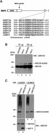
FIG.1.
FIG.1.
Sumoylation of endogenous MEF2D. (A) Domain organization of MEF2. The MADS box and MEF2-specific domain (rectangles) are highly conserved, whereas the C-terminal region (solid line) is divergent. The small box in the C-terminal region represents a conserved sequence motif. Shown in detail is the sequence alignment of this motif found on MEF2 proteins from human (h), mouse (m), Xenopus (x), Drosophila (d), and C. elegans (c), with invariant residues boxed. At the bottom of the alignment is the consensus sumoylation motif ψKxE, where ψ is any aliphatic residue and x is any amino acid (55). (B) HEK293 and HeLa extracts were prepared in buffer S and analyzed by Western blotting with anti-MEF2D antibody. (C) HEK293 cells were washed and lysed in buffer S for extract preparation. Extracts were subject to immunoprecipitation with anti-MEF2D antibody and immunoblotting with anti-SUMO and anti-MEF2D antibodies. The band detected by the anti-SUMO2 antibody (lane 4) is specific (see also Fig. 9E).
FIG. 2.
FIG. 2.
Specific sumoylation of MEF2C and MEF2D. (A to C) In vivo sumoylation assays. Expression plasmids for the indicated proteins were transfected into HEK293 cells. Extracts were prepared in buffer S for immunoprecipitation on M2 agarose, and bound proteins were eluted with Flag peptide and analyzed by Western blotting (WB) with anti-HA (top) or anti-Flag (bottom) antibody. Like HA-SUMO2, HA-SUMO3 was also conjugated to MEF2D (data not shown). (D) Extracts from HEK293 cells expressing Flag-MEF2D or Flag-K439R were used for immunoprecipitation on M2 agarose. Bound proteins were eluted with Flag peptide and subjected to Western blotting analysis with anti-Flag (left) or anti-MEF2D (right) antibody.
FIG. 3.
FIG. 3.
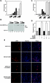
Effect of sumoylation on MEF2D activities. (A) The luciferase reporter Gal4-tk-Luc (200 ng) was transfected into HEK293 cells along with a β-galactosidase expression plasmid (50 ng) and increasing amounts (25, 50, and 100 ng) of expression plasmids for the Gal4 DNA-binding domain (residues 1 to 147), Gal4-MEF2D, and Gal4-K439R, as indicated. The normalized luciferase activity from the transfection without any effector plasmids was arbitrarily set to 1.0. Average values of at least three independent experiments are shown with standard deviation. (B) Extracts from HEK293 cells transfected as in panel A were prepared for Western blotting with anti-Gal4 antibody (RK5C1; Santa Cruz Biotech). (C) Reporter gene assays were performed as in panel A except that the amounts of expression plasmids for Gal4, Gal4-MEF2D, and Gal4-K439R were kept constant (50 ng) and the amount of the expression plasmid for Ubc9 varied (50 and 200 ng). (D and E) A MyoD expression plasmid (400 ng) was transfected into C3H10T1/2 cells along with the expression plasmid for Flag-MEF2D, Flag-K439R, or HA-SUMO2 (each, 500 ng). Myotubes were detected by indirect immunofluorescence with anti-MHC antibody. Average values of at least three independent experiments are illustrated with standard deviation in panel D, and representative images are shown in panel E.
FIG. 4.
FIG. 4.
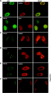
Colocalization of MEF2D and HDAC4 with SUMO2 and Ubc9. HeLa cells were transfected with expression plasmids for GFP-MEF2D, GFP-HDAC4, HA-SUMO2, and HA-Ubc9 as indicated. Transfected cells were subjected to green fluorescence microscopy to detect GFP. HA-tagged proteins were visualized by immunostaining with anti-HA antibody and Cy3-labeled secondary antibody. In the experiments shown in panels E and F, an expression plasmid for Flag-HDAC4 was cotransfected. Note that expression of SUMO2, Ubc9, or HDAC4 led to uneven distribution of MEF2D, which itself is known to be uniform in the nucleoplasm.
FIG. 5.
FIG. 5.
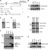
Stimulation of MEF2 sumoylation by class IIa HDACs. (A) Schematic representation of HDAC4 and mutants, with the ability to stimulate MEF2 sumoylation shown at right. (B) In vivo sumoylation assays. HEK293 cells were transfected with expression plasmids for HA-SUMO2, Flag-MEF2D, and HDAC4 proteins as indicated. HDAC4 and 118-488 were HA tagged, whereas 1-666 and 1-326 were Flag tagged. Extracts were subjected to immunoprecipitation on M2 agarose, and bound proteins were eluted with Flag peptide for Western blotting analysis with anti-HA (top) or anti-Flag (bottom) antibody. (C) HEK293 cells were transfected with expression plasmids for Flag-MEF2D and HA-tagged HDAC4 and 118-488. Extracts were prepared for Western blotting analysis with anti-HA antibody. (D) As in panel C, except that HDAC4 mutants were Flag tagged and extracts were analyzed by immunoblotting with anti-Flag antibody. (E) As in panel B, except that Flag-tagged HDAC4, mutant L175A, MEF2C, and MEF2D were expressed as indicated. (F) As in panel B, except that different class IIa HDAC members were expressed, with HDAC4 and HDAC5 HA tagged, HDAC7 tagged with both HA and Flag epitopes, and MITR Flag tagged. (G) Same as in panel B, except that Flag-MEF2C was used. Note that the mobility shift of MEF2 proteins (panels B, F, and G) may be due to HDAC-induced phosphorylation.
FIG. 6.
FIG. 6.
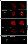
Subcellular distribution of MEF2D, HDAC4, and mutants. (A) GFP-K559R was expressed along with HA-SUMO2 or HA-Ubc9 in HeLa cells, followed by fluorescence microscopy to detect GFP. HA-tagged proteins were detected by immunostaining with anti-HA antibody and Cy3-labeled secondary antibody. (B) GFP-MEF2D and Flag-K559R were expressed along with HA-SUMO2 or HA-Ubc9 in HeLa cells. Expressed proteins were detected by immunofluorescence microscopy as in panel A. (C) GFP-K559R and HA-K439R were expressed in HeLa cells and detected as in panel A.
FIG. 7.
FIG. 7.
Effect of the HDAC4 mutant K559R on MEF2 sumoylation. (A and B) Expression plasmids for Flag-MEF2D and HA-SUMO2 were transfected into HEK293 cells along with the expression construct for Myc-HDAC4 or Myc-K559R. Extracts were subjected to Western blotting analysis with anti-Myc antibody (A) and immunoprecipitation on M2 agarose (B). For the immunoprecipitation, bound proteins were eluted with Flag peptide and subjected to Western blotting analysis with anti-HA or anti-Flag antibody as indicated. (C) Expression plasmids for Flag-MEF2C and HA-SUMO2 were transfected into HEK293 cells along with the expression construct for Myc-HDAC4 or Myc-K559R. Extracts were prepared for immunoprecipitation and immunoblotting as above.
FIG. 8.
FIG. 8.
SENP3 reverses the sumoylation of MEF2D and augments its transcriptional activity. (A) HEK293 cells were transfected with plasmids expressing Flag-MEF2D (2 μg) and HA-SUMO-2 (2 μg), along with HA-SENP3 (6 μg) or its mutant C524S (6 μg). Extracts were used for immunoprecipitation on M2 agarose, and bound proteins were eluted with Flag peptide and subjected to Western blotting analysis with anti-HA (top) or anti-Flag (bottom) antibody. (B) HeLa cells were transfected with expression plasmids for GFP-MEF2D and HA-SENP3, followed by fluorescence microscopy to detect GFP. HA-SENP3 was detected by immunostaining with anti-HA antibody and Cy3-labeled secondary antibody. (C) The luciferase reporter Gal4-tk-Luc (200 ng) and a β-galactosidase expression plasmid (50 ng) were transfected into HEK293 cells along with the expression plasmid for indicated Gal4 proteins (25 ng) and increased amounts of the HA-SENP3 expression plasmid (100 and 200 ng). The normalized luciferase activity from the transfection without any effector plasmids was arbitrarily set to 1.0. Average values of at least three independent experiments are shown with standard deviation. (D and E) The MyoD expression plasmid (400 ng) was transfected into C3H10T1/2 cells along with expression plasmids for Flag-MEF2D (500 ng) and HA-SENP3 (500 ng) as indicated. Myotubes were detected by indirect immunofluorescence with anti-MHC antibody. Average values of at least three independent experiments are illustrated with standard deviation (D) and representative images are shown (E).
FIG. 9.
FIG. 9.
Signal-dependent sumoylation of MEF2D. (A) Expression plasmids for Flag-MEF2D and HA-SUMO2 were transfected into C2C12 cells. After transfection, cells were washed once with PBS and fed with medium containing 20% FBS (lane 1), 2% horse serum (lane 2), or no serum (lane 3). After 48 h, extracts were prepared in buffer S for immunoprecipitation on M2 agarose. Bound proteins were eluted with Flag peptide and subjected to Western blotting analysis with anti-HA or anti-Flag antibody. (B) Expression plasmids for Flag-MEF2D and HA-SUMO2 were transfected into HEK293 cells along with constructs for MEK5(D) and ERK5 as indicated. Extracts were prepared for immunoprecipitation and immunoblotting as above. Note that phosphorylation by MEK5(D) and ERK5 often retards the migration of MEF2D, but it is not clear on this particular gel. (C) Reporter gene assays were performed as described in the legend to Fig. 3A, except that expression plasmids for Gal4-MEF2D, Gal4-K439R, MEK5(D), and ERK5 (each, 100 ng) were used as indicated. (D) As in panel B, except that an expression plasmid for a constitutively active form of CaMKIV was cotransfected as indicated. Nuclear extracts were prepared for immunoprecipitation and immunoblotting. (E) After treatment with or without PMA (10 ng/ml) and ionomycin (0.5 μM) for 4 h, HEK293 cells were subject to nucleus isolation. Nuclear extracts were then used for immunoprecipitation with anti-MEF2D antibody and immunoblotting with anti-SUMO and anti-MEF2D antibodies.
FIG. 10.
FIG. 10.
Cartoon showing how sumoylation of MEF2 is regulated. Class IIa deacetylases, such as HDAC4, associate with MEF2 to potentiate the sumoylation, whereas the phosphorylation-dependent nuclear export of these HDACs precludes them from upregulating MEF2 sumoylation. This modification is also regulated by phosphorylation at distant and adjacent sites. For example, sumoylation of human MEF2D at Lys439 is inhibited by ERK5-mediated phosphorylation of Ser179. The underlying mechanisms remain elusive. Whether phosphorylation at Ser444 by Cdk5 positively regulates the sumoylation of MEF2 is an interesting question awaiting further exploration. P, phosphorylation; S, sumoylation.

Similar articles

Cited by

References

    1. Appella, E., and C. W. Anderson. 2001. Post-translational modifications and activation of p53 by genotoxic stresses. Eur. J. Biochem. 268:2764-2772. - PubMed
    1. Ayaydin, F., and M. Dasso. 2004. Distinct in vivo dynamics of vertebrate SUMO paralogues. Mol. Biol. Cell 15:5208-5218. - PMC - PubMed
    1. Bakin, R. E., and M. O. Jung. 2004. Cytoplasmic sequestration of HDAC7 from mitochondrial and nuclear compartments upon initiation of apoptosis. J. Biol. Chem. 279:51218-51225. - PubMed
    1. Bernier-Villamor, V., D. A. Sampson, M. J. Matunis, and C. D. Lima. 2002. Structural basis for E2-mediated SUMO conjugation revealed by a complex between ubiquitin-conjugating enzyme Ubc9 and RanGAP1. Cell 108:345-356. - PubMed
    1. Best, J. L., S. Ganiatsas, S. Agarwal, A. Changou, P. Salomoni, O. Shirihai, P. B. Meluh, P. P. Pandolfi, and L. I. Zon. 2002. SUMO-1 protease-1 regulates gene transcription through PML. Mol. Cell 10:843-855. - PubMed

Publication types

MeSH terms