. 2005 Mar;17(3):836-48.
doi: 10.1105/tpc.104.029710. Epub 2005 Feb 18.

Identification and dynamics of two classes of aurora-like kinases in Arabidopsis and other plants

Affiliations

Identification and dynamics of two classes of aurora-like kinases in Arabidopsis and other plants

Dmitri Demidov et al. Plant Cell. 2005 Mar.

Abstract

Aurora-like kinases play key roles in chromosome segregation and cytokinesis in yeast, plant, and animal systems. Here, we characterize three Arabidopsis thaliana protein kinases, designated AtAurora1, AtAurora2, and AtAurora3, which share high amino acid identities with the Ser/Thr kinase domain of yeast Ipl1 and animal Auroras. Structure and expression of AtAurora1 and AtAurora2 suggest that these genes arose by a recent gene duplication, whereas the diversification of plant alpha and beta Aurora kinases predates the origin of land plants. The transcripts and proteins of all three kinases are most abundant in tissues containing dividing cells. Intracellular localization of green fluorescent protein-tagged AtAuroras revealed an AtAurora-type specific association mainly with dynamic mitotic structures, such as microtubule spindles and centromeres, and with the emerging cell plate of dividing tobacco (Nicotiana tabacum) BY-2 cells. Immunolabeling using AtAurora antibodies yielded specific signals at the centromeres that are coincident with histone H3 that is phosphorylated at Ser position10 during mitosis. An in vitro kinase assay demonstrated that AtAurora1 preferentially phosphorylates histone H3 at Ser 10 but not at Ser 28 or Thr 3, 11, and 32. The phylogenetic analysis of available Aurora sequences from different eukaryotic origins suggests that, although a plant Aurora gene has been duplicated early in the evolution of plants, the paralogs nevertheless maintained a role in cell cycle-related signal transduction pathways.


Figures

Figure 1.
Figure 1.
Sequence, Structure, and Similarity of AtAurora1, 2, and 3. (A) Comparison of AtAuroras' kinase catalytic domains, deduced from AtAurora cDNAs with the domains of the Aurora/Ipl1p protein related kinases, IPL1p of Saccaromyces cerevisiae (accession number U07163), Aurora of Drosophila melanogaster (accession number X83465), and AIE1 of Mus musculus (accession number AF054620). Residues that are similar (conservative substitutions mode) in all six proteins are shaded black; those 80% similar are shaded gray. Dashes indicate gaps introduced to maximize alignment. The asterisks indicate the consensus sequence of the proposed activation motif that is a conserved signature for Aurora/Ipl1p family members (Giet and Prigent, 1999; Giet et al., 1999b). The plus signs indicate the conserved degradation box in the C-terminal domain RXXLXX(V)XXHPW (Arlot-Bonnemains et al., 2001). (B) Diagrammatic representation of AtAuroras and other Aurora/IPL1p protein kinases. aa, amino acids. (C) Intron–exon structure of AtAuroras. Introns are shown as black boxes. Nucleotide positions of exons were deduced by comparing genomic and cDNA sequences. Positions of primers employed in subsequent experiments are arrowed.
Figure 2.
Figure 2.
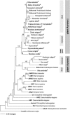
The Relationships between the Kinase Domains of the Aurora Kinases Revealed by Neighbor-Joining Analysis. Numbers depict bootstrap values (above 50%) derived from 500 bootstrap resamples. Nearly the same tree topology resulted from parsimony analysis. Auroras derived from EST sequences are indicated by asterisks. The accession numbers of the proteins in the tree are as follows: C. reinhardtii (AAF97501), S. cerevisiae (U07163), D. melanogaster (A56220 and AAF53026), L. major (AAD00707), C. elegans (AF071206 and AF071207), X. laevis (Z17207), H. sapiens (AF008551, AF054621, and AF008552), M. musculus (U69106, AF054620, and U69107), R. norvegicus (D89731), A. thaliana (At4g32830, At2g25880, and At2g45490), C. sinensis (CF263820), M. domestica (CN879930), G. raimondii (CO130728), G. arboreum (BQ411481), P. coccineus (CA908888), L. albus (CA411358), P. tremula × P. tremuloides (BU8303), A. majus (AJ795958), N. benthamiana (CK289306), Z. elegans (AU308986), H. vulgare (BF263829 and BF260084), T. aestivum (BJ305786), Z. mays (BI319148 and AY106302), S. officinarum (CA114185), O. sativa (BAA99439 and BE40040), P. patens (AJ566700), C. rumphii (CB091521), M. vestita (CD651861), A. cepa (CF444489), and V. aestivalis (CF355370).
Figure 3.
Figure 3.
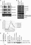
Transcriptional Analysis of AtAurora1, 2, and 3 and Expression Pattern of AtAurora Proteins. (A) RNA gel blot analysis of AtAurora1, 2, and 3 transcripts isolated from Arabidopsis seedlings, flower buds, roots, leaves, flowers, and stems. Ethidium bromide staining of the gel is shown in the bottom panel. (B) AtAurora mRNA abundance assessed by semiquantitative RT-PCR in undifferentiated tissue culture cells undergoing rapid (young callus) or stationary growth phase (old callus) in differentiated tissues of fast-growing flower buds and mature leaves. In parallel, to correlate AtAurora expression with active cell division, the activity of the cell cycle–specific genes CDC6 and AtE2F was monitored. Each sample contained approximately the same amount of RNA as revealed by the nearly homogeneous amplification of constitutive actin and elongation factor 1B α-subunit transcripts. (C) mRNA profiles of the AtAuroras during the mitotic cell cycle deduced from publicly available microarray data of synchronized Arabidopsis tissue culture cells (Menges et al., 2003). AtAuroras are coactivated with cycB1;3 at the onset of mitosis, 8 h after release from the aphidicolin block. (D) SDS-PAGE separation of purified recombinant AtAurora1 protein used for immunization after Coomassie blue (CBB) staining (first lane) or after immunoblotting using the antisera produced against recombinant AtAurora1 (second lane). The position of the recombinant AtAurora1 (40 kD) protein is indicated. The smaller extra band (asterisk) is background specific to bacteria. (E) Forty micrograms of total protein isolated from seedlings, young and old roots, stems, leaves of stems and rosettes, flowers, flower buds, and siliques of Arabidopsis were separated by SDS-PAGE and probed by immunoblotting with antirecombinant AtAurora1. To demonstrate the uniformity of loading, the various protein samples were probed by immunoblotting with antihistone H3.
Figure 4.
Figure 4.
Subcellular Localization of GFP-Tagged AtAurora1, AtAurora2, and AtAurora3 in Transgenic Mitotic Tobacco BY-2 Cells. AtAurora1, 2, and 3 are present in the cytoplasm and in the nucleus during interphase ([A], [K], and [U]). Upon entry into the mitotic cell cycle phase, AtAurora1 and 2 concentrated at the perinuclear region ([B] and [L]), whereas AtAurora3 concentrated at dots around the nucleolus (U). AtAurora1 and 2 associated with the spindle microtubules and moved toward the polar sides during later stages of the metaphase ([C] to [F] and [O] to [Q]). AtAurora3 moved to the midzone of the spindle, where it appeared as discrete spots, presumably corresponding to the centromeric region of chromosomes ([V] and [W]). At the start of anaphase, AtAurora3 was dispersed or destroyed ([X] and [Y]). At the end of mitosis, AtAurora1 and 2 localized at the cell plate (arrowheads; [G] to [J] and [R] to [T]). This was most pronounced for AtAurora1. In addition, some accumulation occurred at the nuclear rim ([H] to [J], [S], and [T]; arrows). Bars = 20 μm.
Figure 5.
Figure 5.
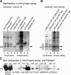
Phosphorylation of Histone H3 at Ser 10 by Recombinant AtAurora1. (A) and (B) Radioactive in vitro kinase assay performed with immunoprecipitated His-tagged AtAurora1 using recombinant Arabidopsis-type histone H3 (A) or a total histone mix isolated from calf thymus as substrate (B). The amounts of histones in each reaction were monitored by Coomassie staining (lanes 1 to 4 of [A] and lanes 1 and 2 of [B]). (A) With anti-AtAurora antibodies, immunoprecipitated recombinant AtAurora1 shows kinase activity toward recombinant histone H3 (lane 6). Negative control experiments were performed with proteins of noninfected baculovirus insect cells and substrate (lane 5), with AtAurora antibody, and substrate (lane 7) or substrate only (lane 8). (B) The kinase assay with recombinant AtAurora1 precipitated with anti-AtAurora (lane 3) or anti-6xHisTag antibodies (lane 4) indicates a preferential phosphorylation of H3. The arrows indicate the bands corresponding to the position of phosphorylated H3. A weak kinase activity was observed toward histone H4 (arrowhead). (C) Nonradioactive in vitro kinase assay performed with protein extracts from recombinant AtAurora1-infected baculovirus insect cells (inf) and uninfected baculovirus insect cells (con; negative control) using recombinant histone H3 as substrate. Specific phosphorylation at Ser 10 of histone H3 is indicated with an asterisk. Samples were transferred onto polyvinylidene difluoride membranes, and then the membranes were incubated with antibodies against histone H3 (anti-H3, as control), phosphorylated histone H3 at Thr 3 [p(T3)H3], Ser 10 [p(S10)H3], Thr 11 [p(T11)H3], Ser 28 [p(S28)H3], or at Thr 32 [p(T32)H3]. The arrow indicates the band corresponding to the position of histone H3.
Figure 6.
Figure 6.
Detailed Analysis of AtAurora in Prophase Nuclei and Mitotic Metaphase Chromosomes of Field Bean. (A) to (F) At prophase and metaphase, immunosignals of the anti-AtAurora antibody are restricted to the pericentromeric regions where sister chromatids cohere chromosomes. DAPI, 4′,6-diamidino-2-phenylindole. (G) to (J) Metaphase chromosome after simultaneous immunostaining with anti-AtAurora and with anti-α tubulin antibodies. (K) to (M) Immunodetection of phosphorylated histone H3 at Ser 10 [p(S10)H3] yields centromeric signals, coinciding with the chromosomal position of AtAurora. Further enlarged pericentromeric domains are shown in insets in (B), (D), and (J).

Similar articles

Cited by

References

    1. Adams, R.R., Carmena, M., and Earnshaw, W.C. (2001. a). Chromosomal passengers and the (aurora) ABCs of mitosis. Trends Cell Biol. 11, 49–54. - PubMed
    1. Adams, R.R., Carmena, M., and Earnshaw, W.C. (2001. b). Chromosomal passengers and the (aurora) ABCs of mitosis. Trends Cell Biol. 11, 49–54. - PubMed
    1. Adams, R.R., Maiato, H., Earnshaw, W.C., and Carmena, M. (2001. c). Essential roles of Drosophila inner centromere protein (INCENP) and aurora B in histone H3 phosphorylation, metaphase chromosome alignment, kinetochore disjunction, and chromosome segregation. J. Cell Biol. 153, 865–880. - PMC - PubMed
    1. Andrews, P.D., Knatko, E., Moore, W.J., and Swedlow, J.R. (2003). Mitotic mechanics: The auroras come into view. Curr. Opin. Cell Biol. 15, 672–683. - PubMed
    1. Arlot-Bonnemains, Y., Klotzbucher, A., Giet, R., Uzbekov, R., Bihan, R., and Prigent, C. (2001). Identification of a functional destruction box in the Xenopus laevis aurora-A kinase pEg2. FEBS Lett. 508, 149–152. - PubMed

MeSH terms

Associated data