. 2005 Feb 14;168(4):643-53.
doi: 10.1083/jcb.200407060.

Inhibition of endothelial cell migration by thrombospondin-1 type-1 repeats is mediated by beta1 integrins

Affiliations

Inhibition of endothelial cell migration by thrombospondin-1 type-1 repeats is mediated by beta1 integrins

Sarah M Short et al. J Cell Biol. .

Erratum in

  • J Cell Biol. 2005 May 9;169(3):541

Abstract

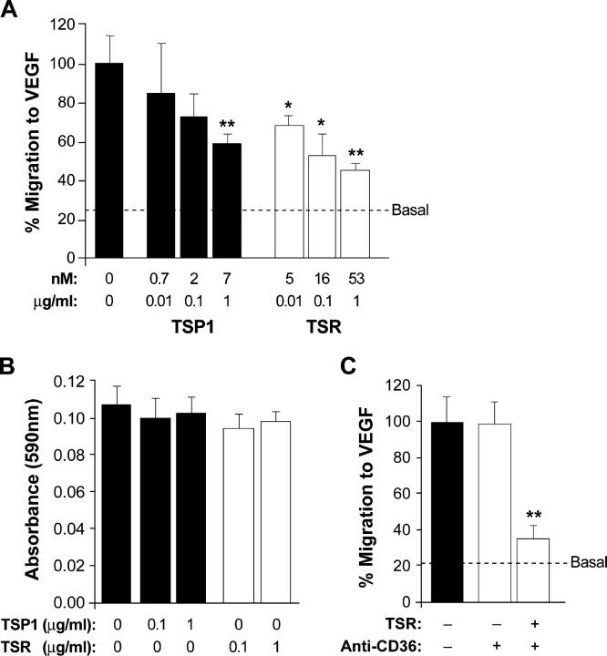
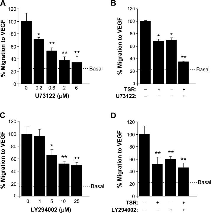
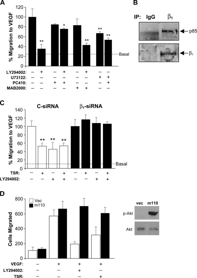
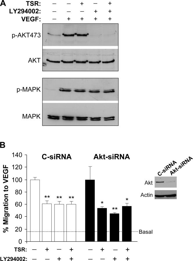
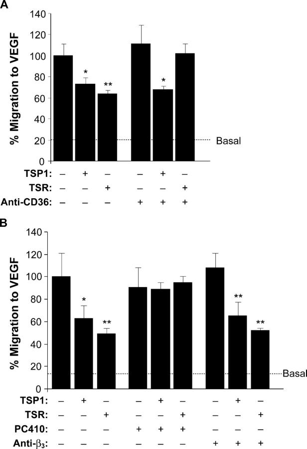
The anti-angiogenic effect of thrombospondin-1 has been shown to be mediated through binding of the type-1 repeat (TSR) domain to the CD36 transmembrane receptor. We now report that the TSR domain can inhibit VEGF-induced migration in human umbilical vein endothelial cells (HUVEC), cells that lack CD36. Moreover, we identified beta1 integrins as a critical receptor in TSR-mediated inhibition of migration in HUVEC. Using pharmacological inhibitors of downstream VEGF receptor effectors, we found that phosphoinositide 3-kinase (PI3k) was essential for TSR-mediated inhibition of HUVEC migration, but that neither PLCgamma nor Akt was necessary for this response. Furthermore, beta1 integrins were critical for TSR-mediated inhibition of microvascular endothelial cells, cells that express CD36. Together, our results indicate that beta1 integrins mediate the anti-migratory effects of TSR through a PI3k-dependent mechanism.


Figures

Figure 1.
Figure 1.
TSP1 and TSR inhibit VEGF-induced HUVEC migration. (A) A modified Boyden chamber assay was used to characterize VEGF-induced (3–5 ng/ml) migration of HUVEC. Cell migration is expressed as percentage of the maximal migration induced by VEGF. Dashed lines indicate basal migration levels, in the absence of VEGF. TSP1 and TSR concentrations are presented as both nM and μg/ml. Where indicated, suspended cells were treated with TSP1 (solid bars) or TSR (open bars) for 30 min, placed in migration chambers, and allowed to migrate to VEGF for 4 h. (B) The effect of cell adhesion in TSP1/TSR-treated cells was measured. HUVEC were pretreated with either TSP1 (solid bars) or TSR (open bars) for 30 min before plating on fibronectin-coated wells for 1.5 h. Adhesion was determined by measuring the absorbance at 590 nm of crystal violet–stained cells. (C) HUVEC were treated with an antibody against CD36 (5 μg/ml) for 10 min, followed by the addition of TSR (16 nM) for 30 min and were allowed to migrate to VEGF for 4 h. Error bars indicate SDs. *, P < 0.05, **, P < 0.01 compared with VEGF alone as determined using t test for unpaired data.
Figure 2.
Figure 2.
TSR inhibits migration through β1 integrins. (A) Cells were treated with increasing concentrations of an antibody (clone PC410) against β1 and were allowed to migrate to VEGF for 4 h. (B) Cells were treated with antibodies against β1 (2 μg/ml) followed by addition of TSR (16 nM) before migration to VEGF for 4 h. Treatment with the PC410 β1 antibody prevented TSR from inhibiting migration, whereas MAB2000 β1 antibody had no effect. (C) Treatment with the β3 antibody had no effect on TSR-mediated inhibition of migration. Cells were treated with an antibody against β3 (2 μg/ml) followed by addition of TSR (16 nM) before migration to VEGF for 4 h. (D) HUVEC were transfected with either control siRNA (C-siRNA) or β1-or β3-siRNA. 72 h after transfection, cells were lysed and integrin β1 and β3 levels were analyzed by Western blotting. Membranes were stripped and probed for actin to show equal protein loading. (E) siRNA-transfected cells were used in the migration assay 72 h after transfection. Where indicated, control siRNA cells were treated with the PC410 β1-activating antibody followed by treatment with TSR (16 nM) for 30 min and were then allowed to migrate to VEGF for 4 h. (F) Ligand-coated Spherotech beads were incubated with suspended HUVEC, beads were isolated, and protein resolved by SDS-PAGE. Beads coated with TSR and RGD (but not HSA) show specific binding to β1 integrins. *, P < 0.05, **, P < 0.01 compared with relevant control (VEGF alone or antibody alone, respectively) as determined using a t test for unpaired data.
Figure 3.
Figure 3.
TSR complexes with and functions through α5β1 integrins. (A) Cells were treated with α subunit antibodies (2 μg/ml) for 15 min, treated with TSR (16 nM) for 30 min, and allowed to migrate toward VEGF for 4 h. (B) Spherotech magnetic substrate-coated beads were used to prepare supramolecular complexes from suspended HUVEC. Western blotting (WB) analysis using an α5β1 integrin antibody shows major bands in fractions prepared from beads coated with TSR, RGD, and β1 antibodies BD15 and MAB2000 (β1), but not for HSA (top). The blot was stripped and probed for β1 (bottom). (C) Spherotech magnetic substrate-coated beads were used to prepare supramolecular complexes from HUVEC lysate using 1% NP-40 lysis buffer. Membranes were probed with an α5β1 antibody. (D) Fibronectin-adherent HUVEC treated with Spherotech magnetic substrate-coated beads were immunostained for α5β1 (bottom). Arrows show a representative bead by phase contrast (top). The arrowhead denotes a cell margin (top). (E) Quantification of the percent positive beads presented in D. *, P < 0.05, **, P < 0.01 compared with VEGF alone as determined using t test for unpaired data.
Figure 4.
Figure 4.
TSR inhibition of migration is dependent on PI3k, but independent of PLCγ. (A) HUVEC were treated with the indicated concentrations of the PLCγ inhibitor U73122 and were allowed to migrate to VEGF (5 ng/ml) for 4 h. (B) HUVEC were treated with U73122 (2 μM) for 10 min followed by treatment with TSR (16 nM) for 30 min and allowed to migrate to VEGF for 4 h. (C) Cells were pretreated with increasing amounts of a PI3k inhibitor, LY294002, and allowed to migrate to VEGF (5 ng/ml) for 4 h. (D) Cells were treated with LY294002 (5 μM) for 10 min followed by treatment with TSR (16 nM) for 30 min, and allowed to migrate to VEGF for 4 h. *, P < 0.05, **, P < 0.01 compared with VEGF alone as determined using t test for unpaired data.
Figure 5.
Figure 5.
TSR requires PI3k and β1 integrin for inhibiting migration. (A) Where indicated, cells were treated with antibodies against β1 (PC410 or MAB2000; 2 μg/ml) for 10 min, followed by treatment with either LY294002 (5 μM) or U73122 (2 nM) for 15 min and were allowed to migrate to VEGF for 4 h. (B) The p85 subunit of PI3k coimmunoprecipitates with β1 integrins. Near-confluent HUVEC were lysed and 200 μg of protein was immunoprecipitated with either anti-β1 antibody (MAB2000) or a mouse IgG control antibody. Immunocomplexes were separated by SDS-PAGE and transferred to nitrocellulose. The membrane was probed with an mAb against the p85 subunit of PI3k, stripped, and reprobed with an mAb against β1. (C) TSR requires β1 integrins and PI3k to inhibit migration. Cells were transfected with either control- or β1-siRNA. Cells were treated with LY294002 (5 μM) for 10 min, then treated with TSR (16 nM) for 30 min and allowed to migrate to VEGF for 4 h. (D) Cells were infected with pLNCX retrovirus, either empty (vec) or containing myristoylated p110 (m110). Cells were split 24 h after infection, and 48 h later subjected to a migration assay or lysed for Western analysis. For the migration assay, cells were treated with either LY294002 (10 μM) or TSR (16 nM) and allowed to migrate to VEGF for 4 h. *, P < 0.05, **, P < 0.01 compared with relevant control (VEGF alone or antibody alone, respectively) as determined using a t test for unpaired data.
Figure 6.
Figure 6.
TSR does not require Akt to inhibit migration. (A) Where indicated, near-confluent and serum-starved HUVEC were treated with LY294002 (5 μM) for 10 min, followed by treatment with TSR (16 nM) for 2 h and then with VEGF (5 ng/ml) for 5 min before cell lysis. Lysates were separated by SDS-PAGE and transferred to nitrocellulose. Membranes were probed for either phospho-ser473Akt or phospho-MAPK, stripped, and probed for total Akt or MAPK. (B) Cells were transfected with Akt siRNA as described in Materials and methods. Where indicated, cells were treated with LY294002 (5 μM) for 10 min, then treated with TSR (16 nM) for 30 min and allowed to migrate to VEGF (5 ng/ml) for 4 h. The inset is a Western blot showing Akt-siRNA treatment greatly reduces total Akt protein levels. *, P < 0.05; **, P < 0.01 compared with VEGF alone as determined using a t test for unpaired data.
Figure 7.
Figure 7.
TSP1 and TSR inhibit migration through β1 integrins in cells expressing CD36. (A) HMVEC-d were treated with a blocking antibody against CD36 (5 μg/ml) for 10 min, followed by the addition of either TSP1 (7 nM) or TSR (5 nM) for 30 min. Cells were then placed in Transwell chambers and allowed to migrate to VEGF for 4 h. (B) HMVEC-d were treated with antibodies (2 μg/ml) against either β1 (PC410) or β3 integrins for 10 min followed by the addition of either TSP1 (7 nM) or TSR (5 nM) for 30 min. Cells were allowed to migrate to VEGF (5 ng/ml) for 4 h. Error bars indicate SDs. *, P < 0.05, **, P < 0.01 compared with relevant control (VEGF alone or antibody alone, respectively) as determined using a t test for unpaired data.

Similar articles

Cited by

References

    1. Adams, J.C., and J. Lawler. 2004. The thrombospondins. Int. J. Biochem. Cell Biol. 36:961–968. - PMC - PubMed
    1. Armulik, A., T. Velling, and S. Johansson. 2004. The integrin β1 subunit transmembrane domain regulates phosphatidylinositol 3-kinase-dependent tyrosine phosphorylation of Crk-associated substrate. Mol. Biol. Cell. 15:2558–2567. - PMC - PubMed
    1. Asch, A.S., S. Silbiger, E. Heimer, and R.L. Nachman. 1992. Thrombospondin sequence motif (CSVTCG) is responsible for CD36 binding. Biochem. Biophys. Res. Commun. 182:1208–1217. - PubMed
    1. Berrier, A.L., A.M. Mastrangelo, J. Downward, M. Ginsberg, and S.E. LaFlamme. 2000. Activated R-ras, Rac1, PI 3-kinase and PKCε can each restore cell spreading inhibited by isolated integrin β1 cytoplasmic domains. J. Cell Biol. 151:1549–1560. - PMC - PubMed
    1. Bjorndahl, M., R. Cao, A. Eriksson, and Y. Cao. 2004. Blockage of VEGF-induced angiogenesis by preventing VEGF secretion. Circ. Res. 94:1443–1450. - PubMed

Publication types

MeSH terms