. 2005 Feb 7;201(3):473-84.
doi: 10.1084/jem.20040934.

RAGE limits regeneration after massive liver injury by coordinated suppression of TNF-alpha and NF-kappaB

Affiliations

RAGE limits regeneration after massive liver injury by coordinated suppression of TNF-alpha and NF-kappaB

Guellue Cataldegirmen et al. J Exp Med. .

Abstract

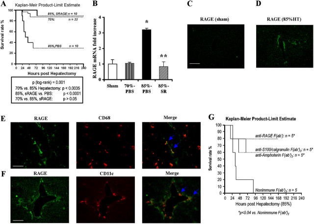
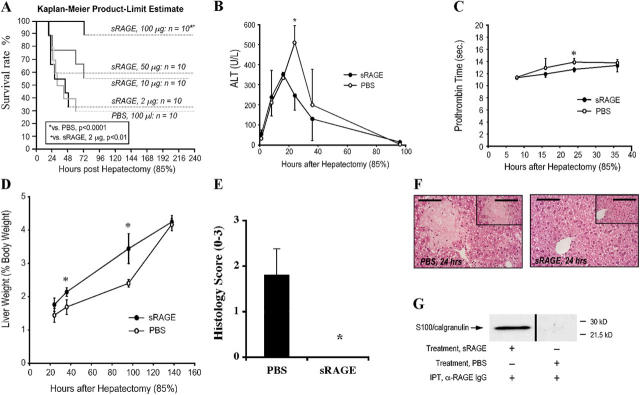
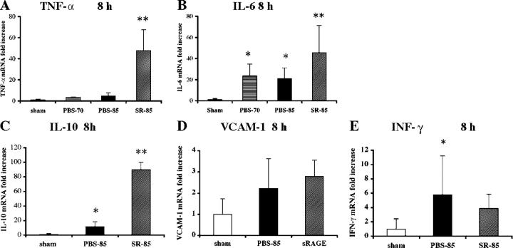
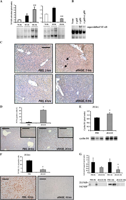
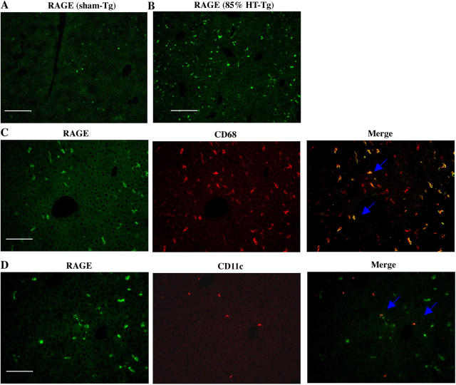
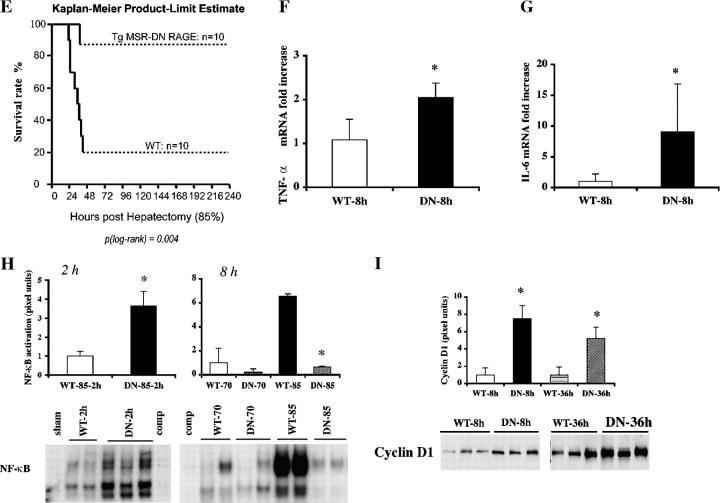
The exquisite ability of the liver to regenerate is finite. Identification of mechanisms that limit regeneration after massive injury holds the key to expanding the limits of liver transplantation and salvaging livers and hosts overwhelmed by carcinoma and toxic insults. Receptor for advanced glycation endproducts (RAGE) is up-regulated in liver remnants selectively after massive (85%) versus partial (70%) hepatectomy, principally in mononuclear phagocyte-derived dendritic cells (MPDDCs). Blockade of RAGE, using pharmacological antagonists or transgenic mice in which a signaling-deficient RAGE mutant is expressed in cells of mononuclear phagocyte lineage, significantly increases survival after massive liver resection. In the first hours after massive resection, remnants retrieved from RAGE-blocked mice displayed increased activated NF-kappaB, principally in hepatocytes, and enhanced expression of regeneration-promoting cytokines, TNF-alpha and IL-6, and the antiinflammatory cytokine, IL-10. Hepatocyte proliferation was increased by RAGE blockade, in parallel with significantly reduced apoptosis. These data highlight central roles for RAGE and MPDDCs in modulation of cell death-promoting mechanisms in massive hepatectomy and suggest that RAGE blockade is a novel strategy to promote regeneration in the massively injured liver.


Figures

Figure 1.
Figure 1.
Up-regulation of RAGE in hepatic remnants after massive (85%) hepatectomy: cellular localization and impact of ligand/RAGE blockade. Male C57BL/6 mice were subjected to partial (70%) or massive (85%) hepatectomy or sham surgery. Mice subjected to 85% resection were treated with murine sRAGE, 100 μg per day, or vehicle, PBS daily until death. (A) Kaplan-Meier Product Limit Estimate. The times of death were recorded for mice undergoing hepatectomy, and survival was plotted. (B) Quantitative PCR. Quantitative PCR was performed on liver remnants retrieved from mice undergoing sham, 70% or 85% liver resection, or 85% resection in the presence of sRAGE. After normalization to internal controls, levels of murine RAGE transcripts in sham-treated mice livers were arbitrarily defined as “1.” In this experiment, n = 5 mice per group. *, P < 0.05 versus 70% hepatectomy. **, P < 0.05 versus PBS treatment/85% hepatectomy. (C–F) Immunofluorescence microscopy. Immunofluorescence for detection of RAGE antigen was performed in sections prepared 8 h after sham surgery (C) or massive resection (D, 85%). In tissues prepared from remnants after massive resection, double immunofluorescence staining was performed with anti-RAGE IgG (green) and an anti-CD68 IgG (E, red), or with anti-RAGE IgG (green) and anti-CD11c IgG (F, red) as described before. Single and merged images are shown. (C, E, and F) Bars, 50 μm. (G) Kaplan-Meier Product Limit Estimate. Male C57BL/6 mice were subjected to massive hepatic resection and treated with the indicated F(ab′)2 fragments of rabbit anti-RAGE, anti-S100/calgranulin, antiamphoterin, or nonimmune IgG. The times of death were recorded for mice undergoing hepatectomy, and survival was plotted.
Figure 2.
Figure 2.
Administration of sRAGE improves survival and enhances regeneration after massive hepatectomy. Male C57BL/6 mice were subjected to 85% hepatectomy in the presence of the indicated once daily dose of sRAGE or vehicle, PBS. (A) Kaplan-Meier Product Limit Estimate. The times of death were recorded for mice undergoing hepatectomy in the presence of sRAGE versus vehicle, and survival was plotted. Survival curves for sRAGE, 100 μg/d- and PBS-treated mice, reflect the same animals as in Fig. 1 A. (B–D) Indices of injury and regeneration. At the indicated times, plasma or hepatic remnant was retrieved and analyzed for ALT (B) and Prothrombin Time (C). (D) Liver weight/body weight ratios are shown. (B and C) n = 3–8 mice per condition. (D) n = 10 (PBS) or n = 27 (sRAGE) mice per condition. (B–D) *, P < 0.05. (E and F) Histology. Hepatic remnants were retrieved and grading was performed based on the percentage of necrosis as described before; mean ± standard error is shown. (E) n = 5 mice per condition. *, P < 0.05. (F) Representative sections from PBS- and sRAGE-treated mice at 24 h are illustrated. Bar, 80 μm (inset) 160 μm. (G) Immunoprecipitation. 16 h after 85% hepatectomy, plasma from sRAGE- or PBS-treated mice was retrieved and subjected to immunoprecipitation using rabbit anti-RAGE IgG. Immunoprecipitated material was subjected to blotting using anti-S100/calgranulin IgG. Plasma was pooled from n = 3 sRAGE- or n = 3 PBS-treated plasma for immunoprecipitation studies.
Figure 3.
Figure 3.
Blockade of RAGE modulates expression of inflammatory and regenerative mediators after massive hepatectomy. 8 h after sham surgery, 70 or 85% resection in the presence of vehicle or sRAGE treatment, hepatic remnants were retrieved and subjected to quantitative PCR for detection of transcripts for the following: A, TNF-α; B, IL-6; C, IL-10; D, VCAM-1; and E, IFN-γ. After normalization to internal controls, fold changes relative to sham surgery remnant transcript levels (“1.0”) were reported. n = 5 mice per condition. *, P < 0.05 versus sham control. **, P < 0.05 versus PBS treatment.
Figure 4.
Figure 4.
Blockade of RAGE modulates activation of NF-κB after massive hepatectomy. (A) EMSA. Nuclear extracts were prepared from the remnants of the indicated mice and the EMSA was performed. The illustrated bands are representative of n = 4–6 mice per condition. **, P < 0.05 versus PBS treatment/85% resection. (B) Supershift assay. Remnants retrieved from mice undergoing 85% hepatectomy in the presence of sRAGE at 2 h were retrieved and subjected to incubation with anti-p50, anti-p65, or both anti-p50 and anti-p65 IgG before EMSA. (C) Immunohistochemistry. Hepatic remnants at 2 and 8 h were retrieved and subjected to immunohistochemistry with anti-p65 NF-κB subunit antibodies. Bar, 50 μm. (D) Proliferation. Hepatic remnants were retrieved and subjected to immunohistochemistry with anti-PCNA IgG and mean numbers of PCNA+ cells were determined from n = 10 fields per section/mouse. The indicated results are representative of n = 5 mice per condition. Bars, 80 μm. (inset) 160 μm. (E) Western blotting of hepatic remnant lysates was performed at the indicated times using anti-cyclin D1 IgG. The illustrated bands are representative of n = 3–4 mice per condition. (F) Apoptosis. Hepatic remnants were retrieved and subjected to TUNEL assay and mean numbers of TUNEL+ cells determined from n = 10 fields per section/mouse. The indicated results are representative of n = 5 mice per condition. Bar, 40 μm. (G) Western blotting on remnants was performed using anti-activated caspase-3 IgG. The illustrated bands are representative of n = 5 mice per condition. In all cases, bands were scanned into a densitometer and relative pixel units of band density reported. *, P < 0.05 versus PBS/85% resection.
Figure 5.
Figure 5.
Transgenic mice expressing DN RAGE in cells of MP lineage display enhanced survival and liver regeneration after massive hepatectomy. Transgenic mice expressing human DN RAGE selectively in cells of MP lineage using the scavenger receptor type A promoter were used for these studies. (A–D) Immunofluorescence microscopy. Immunofluorescence for detection of RAGE antigen was performed in sections prepared from the remnants of transgenic mice 8 h after sham surgery (A) or massive resection (B, 85%). Double immunofluorescence staining was performed on sections prepared from transgenic mice 8 h after massive resection using the following antibodies: anti-RAGE IgG (C and D, green) and anti-CD68 IgG (C, red) or anti-CD11c IgG (D, red). Single and merged images are shown. (A and B) Bars, 100 μm; (C and D) Bars, 50 μm. (E) Kaplan-Meier Product Limit Estimate. The times of death were recorded for transgenic versus littermate control mice undergoing massive hepatectomy and survival was plotted. (F and G) Quantitative PCR. Massive hepatectomy was performed in transgenic (DN) and wild-type littermate mice (WT). Hepatic remnants were retrieved at 8 h after resection and subjected to quantitative PCR for detection of transcripts for TNF-α (F) and IL-6 (G). After normalization to internal controls, fold changes relative to WT mice remnant transcript levels (“1.0”) are reported. n = 5 mice per condition. *, P < 0.05 versus WT mice. (H) EMSA. Massive hepatectomy was performed in transgenic (DN) and wild-type littermate mice (WT). Remnants were retrieved at 2 and 8 h, and nuclear extracts were prepared for EMSA. Bands were scanned into a densitometer and relative pixel units of band density reported. *, P < 0.05 versus WT mice/85% resection. (I) Western blotting. At the indicated times after massive resection, hepatic remnants were retrieved and subjected to Western blotting using anti-cyclin D1 IgG. (H and I) The illustrated bands are representative of n = 3–4 mice per condition.
Figure 5.
Figure 5.
Transgenic mice expressing DN RAGE in cells of MP lineage display enhanced survival and liver regeneration after massive hepatectomy. Transgenic mice expressing human DN RAGE selectively in cells of MP lineage using the scavenger receptor type A promoter were used for these studies. (A–D) Immunofluorescence microscopy. Immunofluorescence for detection of RAGE antigen was performed in sections prepared from the remnants of transgenic mice 8 h after sham surgery (A) or massive resection (B, 85%). Double immunofluorescence staining was performed on sections prepared from transgenic mice 8 h after massive resection using the following antibodies: anti-RAGE IgG (C and D, green) and anti-CD68 IgG (C, red) or anti-CD11c IgG (D, red). Single and merged images are shown. (A and B) Bars, 100 μm; (C and D) Bars, 50 μm. (E) Kaplan-Meier Product Limit Estimate. The times of death were recorded for transgenic versus littermate control mice undergoing massive hepatectomy and survival was plotted. (F and G) Quantitative PCR. Massive hepatectomy was performed in transgenic (DN) and wild-type littermate mice (WT). Hepatic remnants were retrieved at 8 h after resection and subjected to quantitative PCR for detection of transcripts for TNF-α (F) and IL-6 (G). After normalization to internal controls, fold changes relative to WT mice remnant transcript levels (“1.0”) are reported. n = 5 mice per condition. *, P < 0.05 versus WT mice. (H) EMSA. Massive hepatectomy was performed in transgenic (DN) and wild-type littermate mice (WT). Remnants were retrieved at 2 and 8 h, and nuclear extracts were prepared for EMSA. Bands were scanned into a densitometer and relative pixel units of band density reported. *, P < 0.05 versus WT mice/85% resection. (I) Western blotting. At the indicated times after massive resection, hepatic remnants were retrieved and subjected to Western blotting using anti-cyclin D1 IgG. (H and I) The illustrated bands are representative of n = 3–4 mice per condition.

Similar articles

Cited by

References

    1. Fausto, N. 2000. Liver regeneration. J. Hepatol. 32:19–31. - PubMed
    1. Schulte-Hermann, R., B. Grasl-Kraupp, and W. Bursch. 1995. Apoptosis and hepatocarcinogenesis. In Liver Regeneration and Carcinogenesis. Molecular and Cellular Mechanisms. R.L. Jirtle, editor. Academic Press, San Diego, CA. 141–178.
    1. Panis, Y., D.M. McMullan, and J.C. Emond. 1997. Progressive necrosis after hepatectomy and the pathophysiology of liver failure after massive resection. Surgery. 121:142–149. - PubMed
    1. Francavilla, A., Q. Zeng, and L. Polimeno. 1994. Small-for-size liver transplanted into larger recipient: a model of hepatic regeneration. Hepatology. 19:210–216. - PMC - PubMed
    1. Emond, J.C., J.F. Renz, R.C. Lim, J.P. Roberts, and N.L. Ascher. 1996. Functional characterization of liver grafts from living donors: implications for the treatment of older children and adults. Ann. Surg. 224:544–552. - PMC - PubMed

Publication types

MeSH terms