. 2005 Jan 31;168(3):453-63.
doi: 10.1083/jcb.200409078.

Receptor tyrosine phosphatase-dependent cytoskeletal remodeling by the hedgehog-responsive gene MIM/BEG4

Affiliations

Receptor tyrosine phosphatase-dependent cytoskeletal remodeling by the hedgehog-responsive gene MIM/BEG4

Rosa Gonzalez-Quevedo et al. J Cell Biol. .

Abstract

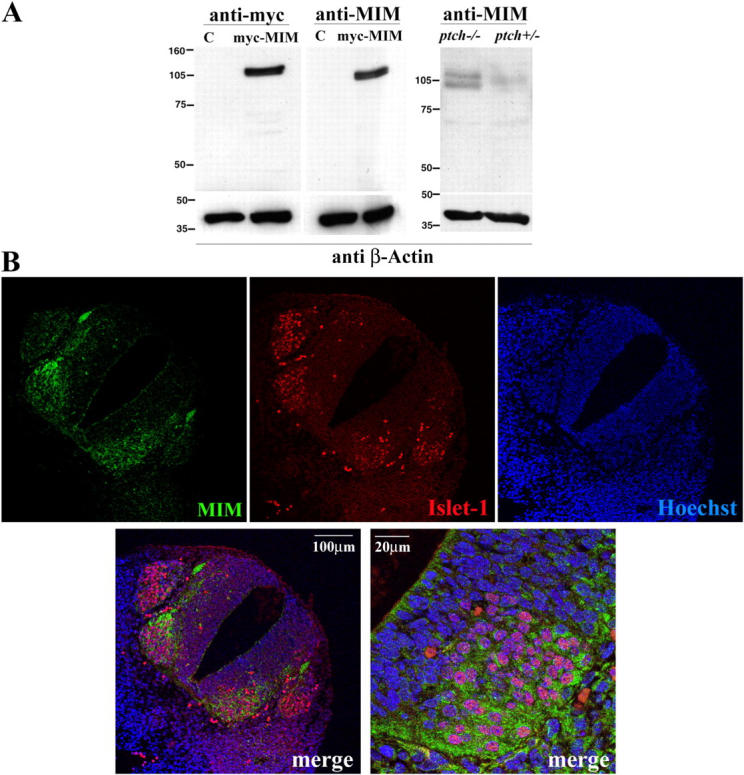
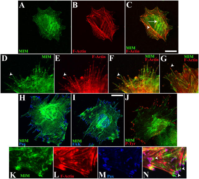
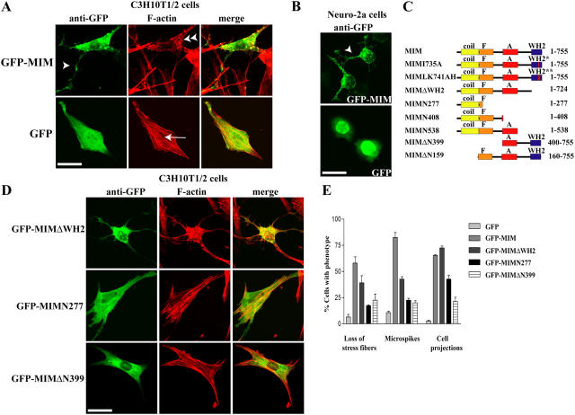
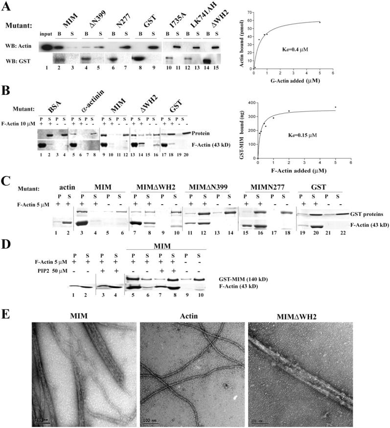
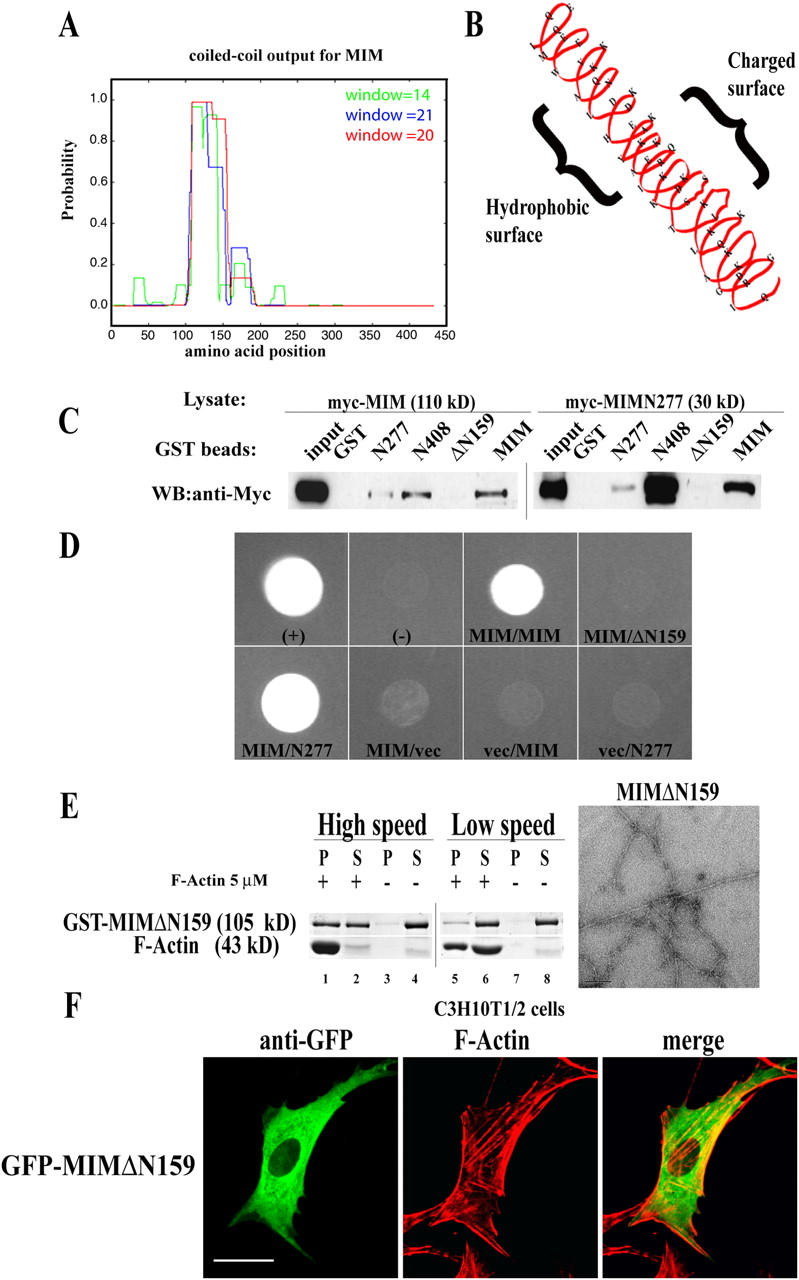
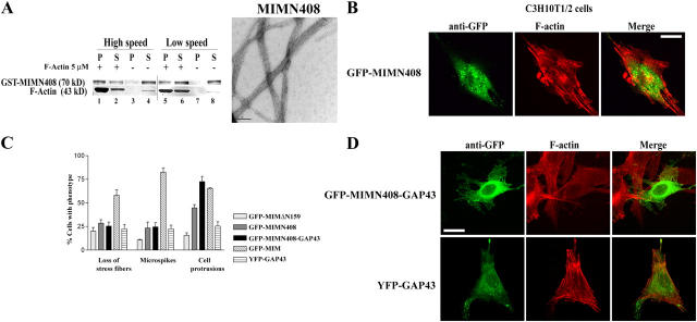
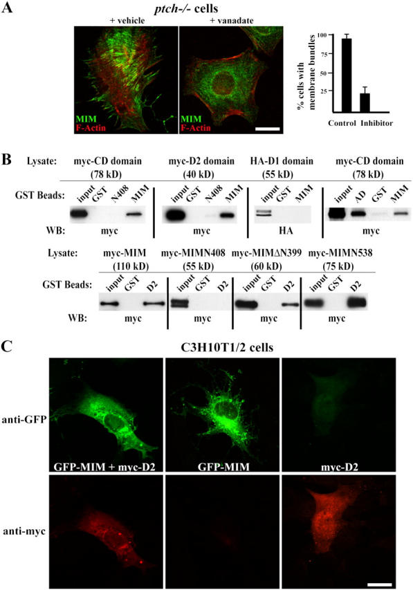
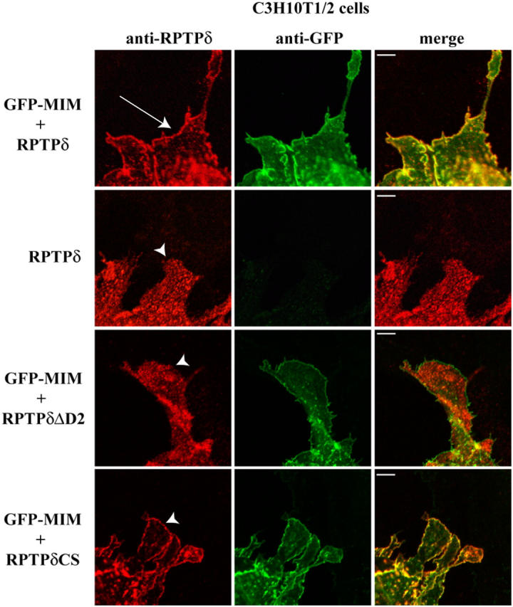
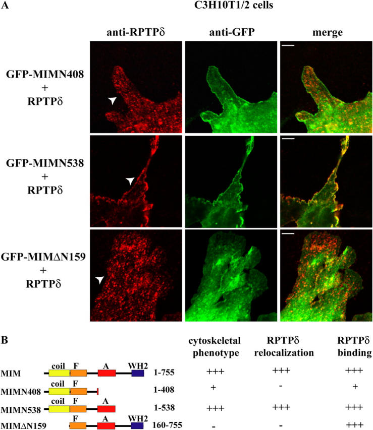
During development, dynamic remodeling of the actin cytoskeleton allows the precise placement and morphology of tissues. Morphogens such as Sonic hedgehog (Shh) and local cues such as receptor protein tyrosine phosphatases (RPTPs) mediate this process, but how they regulate the cytoskeleton is poorly understood. We previously identified Basal cell carcinoma-enriched gene 4 (BEG4)/Missing in Metastasis (MIM), a Shh-inducible, Wiskott-Aldrich homology 2 domain-containing protein that potentiates Gli transcription (Callahan, C.A., T. Ofstad, L. Horng, J.K. Wang, H.H. Zhen, P.A. Coulombe, and A.E. Oro. 2004. Genes Dev. 18:2724-2729). Here, we show that endogenous MIM is induced in a patched1-dependent manner and regulates the actin cytoskeleton. MIM functions by bundling F-actin, a process that requires self-association but is independent of G-actin binding. Cytoskeletal remodeling requires an activation domain distinct from sequences required for bundling in vitro. This domain associates with RPTPdelta and, in turn, enhances RPTPdelta membrane localization. MIM-dependent cytoskeletal changes can be inhibited using a soluble RPTPdelta-D2 domain. Our data suggest that the hedgehog-responsive gene MIM cooperates with RPTP to induce cytoskeletal changes.


Figures

Figure 1.
Figure 1.
MIM is a hedgehog-responsive gene. (A) Characterization of the anti-MIM antibody. (left) Lysates of 293T cells untransfected (C, control) or transfected with myc-MIM and immunoblotted with an anti-myc monoclonal antibody or anti-MIM polyclonal antibody. (right) Lysates from ptch−/− and ptch+/− MEFs immunoblotted with the anti-MIM antibody. (B) MIM is expressed in a hedgehog-responsive tissue in vivo during spinal cord development. Paraffin sections of mouse neural tube at day 11.5 stained using the anti-MIM antibody. MIM (green) localizes to the Shh-responsive ventral part of the neural tube, including motor neurons (Islet-1, red). (bottom) Magnification of an area showing localization of MIM and Islet-1 to motor neurons. Nuclei (blue) are labeled with Hoechst staining.
Figure 2.
Figure 2.
Endogenous MIM localizes to actin bundles that support focal adhesion complexes. Characterization of endogenous MIM localization in ptch−/− MEFs. (A–C) In ptch−/− cells, MIM (A, green) accumulates on a subset of stress fibers (B, red) near the center of the cell (C, arrow), but not with more peripheral cortical actin fibers (arrowhead). (D–G) MIM (D, green) decorates the length of long actin cables (E, red) at sites of membrane projections. MIM staining continues along actin bundles but is excluded from the tip of the bundle (note difference in staining near arrowheads). (H–J) MIM (green) accumulates on actin bundles that support focal adhesions complexes. Focal adhesion complexes are marked by staining with monoclonal antibodies to paxillin (H, blue), FAK (I, blue), and phosphotyrosine (J, red). (K–N) Magnified view of 2H showing that MIM (K, green) decorates the actin bundle (L, red) and is enriched in the area adjacent to paxillin-labeled focal adhesions (M, blue, and arrowheads in the merged image N). Bars, 30 μm.
Figure 3.
Figure 3.
MIM induces cytoskeletal changes independently of the WH2 domain. Cytoskeletal remodeling activity of wild-type and mutant MIM proteins expressed in C3H10T1/2 cells. Cells were labeled with an anti-GFP Alexa Fluor 488 antibody (green) or phalloidin-TRITC (red, F-actin) and imaged by confocal microscopy. (A) Full-length GFP-MIM (top) or GFP (bottom). MIM induces loss of stress fibers, microspikes (double arrowhead), and actin-based cell projections (arrowhead). GFP control does not alter the cytoskeleton, and control-treated cells have well-defined stress fibers (arrow). Bar, 30 μm. (B) A similar phenotype is observed when MIM is expressed in the mouse neuroblastoma cell line Neuro-2a. The cytoskeleton is dramatically reorganized, which results in the induction of numerous cell projections (top, arrowhead), whereas the GFP control (bottom) has no effect. Bar, 35 μm. (C) Diagram of predicted domains (coil, coiled-coil; F, F-actin binding; A, activation; and WH2, WH2 domain) of MIM and the mutants used in this study. Asterisks represent the point mutations in the WH2 domain (*, substitution of I735A; **, substitution LK741,742AH). (D) The WH2 domain, but not the NH2 terminus of MIM, is dispensable for the induction of cell projections. Confocal images of C3H10T1/2 cells transfected with GFP-MIMΔWH2 (1–724), GFP-MIMN277 (1–277), or GFP-MIMΔN399 (400–755). Bar, 30 μm. (E) Quantification of the phenotypes observed. Means ± SEM (n = 3) are shown.
Figure 4.
Figure 4.
MIM is a novel actin-bundling protein. (A) The WH2 domain of MIM specifically binds G-actin. Pull-down assay from a solution of G-actin using different GST-tagged MIM proteins. Beads (B) and supernatants (S) were separated by SDS-PAGE and visualized by antiactin or anti-GST antibodies. MIM binds to monomeric actin (lanes 2 and 3), whereas substitution of critical residues (lanes 10–13) or deletion of the WH2 domain (lanes 14 and 15) completely abrogates binding. (right) Binding data determining the dissociation constant (K d = 0.4 μM) of the interaction between MIM and monomeric actin. (B) MIM binds F-actin in vitro. Purified GST-MIM proteins were incubated in the presence (+) or absence (−) of F-actin and cosedimented at 155,000 g. Comparative aliquots of pellet (P) and supernatant (S) were separated and gels were stained with Coomassie blue. Both MIM and MIMΔWH2 coprecipitate with F-actin (lanes 9, 10, 13, and 14). (right) Curve to establish the dissociation constant for the MIM and F-actin interaction (K d = 0.15 μM). (C) MIM cross-links actin filaments in vitro. Coprecipitation assay using purified GST-MIM proteins and F-actin at 10,000 g. Same aliquots of pellet (P) and supernatant (S) were separated by SDS-PAGE and stained with Coomassie blue. Apparent molecular masses of used proteins are as follows: MIM, 140 kD; MIMΔWH2, 120 kD; MIMΔN399, 75 kD; MIMN277, 50 kD; and GST, 27 kD. (D) PIP2 inhibits actin bundling mediated by MIM. GST-MIM was incubated with (+) or without (−) F-actin, in the presence (+) or absence (−) of PIP2. Comparable aliquots of pellet (P) and supernatant (S) were separated by SDS-PAGE and stained with Coomassie blue. In the absence of PIP2, F-actin appears mostly in the pellet fraction (lanes 5 and 6), whereas in the presence of PIP2, F-actin is shifted to the supernatant fraction (lanes 7 and 8). (E) Electron micrographs of actin structures in the absence or presence of equimolar amounts of 6XHis-MIM proteins, showing that MIM and MIMΔWH2 bundle actin filaments. Bars, 100 nm.
Figure 5.
Figure 5.
MIM NH2-terminal self-association domain is required for the induction of cytoskeletal changes. (A) Diagram of the predicted coiled-coil region of MIM (http://pbil.ibcp.fr/html/pbil_index.html). (B) The coiled-coil domain is predicted to have distinct hydrophobic and hydrophilic surfaces. (C) MIM self-associates through the NH2-terminal coiled-coil region. Pull-down assay from myc-MIM– or myc-MIMN277–transfected 293T cell lysates using purified GST-MIM proteins or GST bound to beads. Proteins were visualized by Western blot using an anti-myc antibody. MIM binds to GST-MIMN277 but not the control GST or MIMΔN159. (D) Yeast two-hybrid interaction assay showing single colonies from cells transformed with indicated bait/prey plasmids grown on selective media. Full-length MIM interacts with itself (MIM/MIM) or the coil domain (MIM/N277), but not with MIM lacking the coiled-coil domain (MIM/ΔN159). +, P53/Large T; −, P53/vector. MIM/vector, vector/MIM, and vector/N277 demonstrate specificity of the interaction. (E, left) Coprecipitation assay of purified GST-MIMΔN159 in the presence (+) or absence (−) of F-actin to study binding (high speed, 155,000 g) and bundling (low speed, 10,000 g). MIMΔN159 binds F-actin (lanes 1 and 2) but does not bundle (5 and 6) in vitro. (right) Electron micrographs of purified F-actin incubated with MIMΔN159. This mutant, which lacks the self-association domain, does not induce ordered bundles of filamentous actin. Bar, 100 nm. (F) Overexpression of MIMΔN159 (green) in C3H10T1/2 cells. The self-association domain is required for the induction of cytoskeletal changes. Note the presence of stress fibers (F-actin, red) and minimal cellular projections. Bar, 25 μm.
Figure 6.
Figure 6.
Characterization of MIM membrane activation. MIMN408 bundles actin in vitro but cannot reconstitute the phenotype of wild-type MIM in C3H10T1/2 cells. (A, left) Coprecipitation assay of purified GST-MIMN408 in the presence (+) or absence (−) of F-actin at high speed (155,000 g) or low speed (10,000 g) demonstrates both binding (lanes 1 and 2) and bundling (5 and 6). (right) Electron micrograph of MIMN408 incubated with F-actin by TEM. Bar, 100 nm. (B) Confocal microscopy of C3H10T1/2 cells expressing MIMN408. Cells show increased punctate staining in the cytosol and fewer cell projections compared with the wild type. Bar, 20 μm. (C) Quantification of phenotypes. Means ± SEM (n = 3) are shown. (D) Addition of a GAP-43 tag to MIMN408 rescues the cytoskeletal phenotype. Confocal analysis of C3H10T1/2 cells expressing GFP-MIMN408-GAP43 (top, green) or control YFP-GAP43 (bottom, green). Cells were labeled with an anti-GFP antibody and F-actin (red) was visualized using phalloidin-TRITC. Bar, 25 μm.
Figure 7.
Figure 7.
MIM cytoskeletal remodeling requires interaction with the RPTPδ-D2 domain. (A, left) Phosphatase treatment with orthovanadate (+ vanadate) inhibits endogenous MIM membrane bundles in ptch−/− cells but not in cells treated with control (+ vehicle). Confocal images of cells stained for MIM (green) and F-actin (red). Bar, 30 μm. (right) Quantification. Means ± SEM (n = 3) are shown. (B) MIM amino acids 400–538 bind the substrate-binding D2 domain of RPTPδ. (top) GST pull-down assay using the indicated GST-MIM beads and cell lysates containing myc-CD (cytoplasmic domain; apparent molecular mass is 78 kD), myc-D2 (substrate binding domain 2; runs at 40 kD), or HA-D1 (catalytic domain; runs as a doublet at 55 kD). AD, MIM activation domain amino acids 401–541. (bottom) GST pull-down assay using GST-D2 or control GST beads and the indicated MIM-containing lysates. (C) Coexpression of soluble myc-D2 domain (red) with GFP-MIM (green) blocks cell projections in C3H10T1/2. Bar, 25 μm. Representative cells are shown with anti-GFP (green; top) and anti-myc (red) staining (bottom).
Figure 8.
Figure 8.
MIM alters RPTPδ localization and both colocalize on the membrane. Confocal images of C3H10T1/2 cells transfected with the indicated constructs. Cells are labeled with anti-RPTPδ antibody and anti-GFP Alexa Fluor 488. Cells coexpressing RPTPδ (red, arrow) and GFP-MIM (green) show that MIM dramatically enhances RPTPδ membrane insertion compared with cells expressing RPTP alone (arrowhead) and colocalizes with it (top two rows). (third row) MIM (green) does not colocalize or alter RPTPδ-ΔD2 distribution (red, arrowhead). (bottom) The phosphatase activity of RPTPδ is not required for the membrane relocalization as a phosphatase-dead mutant (RPTPδCS; red, arrowhead) is still inserted at the plasma membrane when coexpressed with MIM (green). Bars, 5 μm.
Figure 9.
Figure 9.
MIM activation domain is required for RPTPδ membrane relocalization. (A, top) Coexpression of GFP-MIMN408 (green) with RPTPδ (red). MIMN408 does not colocalize or affect RPTPδ distribution (top, arrowhead). However, GFP-MIMN538, a mutant that contains the activation domain, does (middle, arrowhead). (bottom) Coexpression of GFP-MIMΔN159 (green) with RPTPδ (red, arrowhead) shows that the activation domain is necessary but not sufficient for relocalizing RPTPδ, and that the bundling activity is also required. Bars, 5 μm. (B) Summary of activities of MIM and MIM mutants on the cytoskeleton and RPTPδ binding and localization.

Similar articles

Cited by

References

    1. Bear, J.E., M. Krause, and F.B. Gertler. 2001. Regulating cellular actin assembly. Curr. Opin. Cell Biol. 13:158–166. - PubMed
    1. Bretscher, A., and K. Weber. 1980. Villin is a major protein of the microvillus cytoskeleton which binds both G and F actin in a calcium-dependent manner. Cell. 20:839–847. - PubMed
    1. Callahan, C.A., T. Ofstad, L. Horng, J.K. Wang, H.H. Zhen, P.A. Coulombe, and A.E. Oro. 2004. MIM/BEG4, a Sonic hedgehog-responsive gene that potentiates Gli-dependent transcription. Genes Dev. 18:2724–2729. - PMC - PubMed
    1. Charron, F., E. Stein, J. Jeong, A.P. McMahon, and M. Tessier-Lavigne. 2003. The morphogen sonic hedgehog is an axonal chemoattractant that collaborates with netrin-1 in midline axon guidance. Cell. 113:11–23. - PubMed
    1. Dent, E.W., F. Tang, and K. Kalil. 2003. Axon guidance by growth cones and branches: common cytoskeletal and signaling mechanisms. Neuroscientist. 9:343–353. - PubMed

Publication types

MeSH terms

Substances