. 2005 Feb;79(4):2151-9.
doi: 10.1128/JVI.79.4.2151-2159.2005.

Inhibition of coxsackievirus B3 replication by small interfering RNAs requires perfect sequence match in the central region of the viral positive strand

Affiliations

Inhibition of coxsackievirus B3 replication by small interfering RNAs requires perfect sequence match in the central region of the viral positive strand

Ji Yuan et al. J Virol. 2005 Feb.

Abstract

Coxsackievirus B3 (CVB3) is the most common causal agent of viral myocarditis, but existing drug therapies are of limited value. Application of small interfering RNA (siRNA) in knockdown of gene expression is an emerging technology in antiviral gene therapy. To investigate whether RNA interference (RNAi) can protect against CVB3 infection, we evaluated the effects of RNAi on viral replication in HeLa cells and murine cardiomyocytes by using five CVB3-specific siRNAs targeting distinct regions of the viral genome. The most effective one is siRNA-4, targeting the viral protease 2A, achieving a 92% inhibition of CVB3 replication. The specific RNAi effects could last at least 48 h, and cell viability assay revealed that 90% of siRNA-4-pretreated cells were still alive and lacked detectable viral protein expression 48 h postinfection. Moreover, administration of siRNAs after viral infection could also effectively inhibit viral replication, indicating its therapeutic potential. Further evaluation by combination found that no enhanced inhibitory effects were observed when siRNA-4 was cotransfected with each of the other four candidates. In mutational analysis of the mechanisms of siRNA action, we found that siRNA functions by targeting the positive strand of virus and requires a perfect sequence match in the central region of the target, but mismatches were more tolerated near the 3' end than the 5' end of the antisense strand. These findings reveal an effective target for CVB3 silencing and provide a new possibility for antiviral intervention.


Figures

FIG. 1.
FIG. 1.
CVB3-specific siRNAs inhibit CVB3 replication in HeLa cells. (a) Western blot analysis of CVB3 capsid protein VP1. HeLa cells were transfected with siRNAs at a final concentration of 300 nM using Oligofectamine and then were infected with CVB3 at an MOI of 10. At 8 hpi, cell lysates were collected for VP1 detection by Western blot and supernatants were used for detecting infectious viral particles by plaque assay. The VP1 expression levels were quantified by densitometry and were normalized to the level of β-actin, which served as a loading control. The ratios of VP1 to β-actin were calculated and are expressed in the graph. (−), mock transfection. (b) Plaque assays of infectious viral particles. The assay was conducted on HeLa cell monolayers as described in Materials and Methods. Data are presented as log10 values of virus titer. (c) In situ hybridization of CVB3 RNA. After treatments and infections in chamber slides, CVB3 positive-strand RNAs were detected by in situ hybridization using antisense riboprobes (red). Cell nuclei were counterstained with hematoxylin (blue). Images 1 to 5 and C (control) represent different siRNAs treatments. Two negative controls were used: cells transfected with siRNA-C and infected with CVB3 were detected by sense probes (C-S), and cells transfected with siRNA-4 but sham infected with DMEM were detected with antisense probes (4-sham). Magnification, ×200. Data shown are from one of two independent experiments.
FIG. 1.
FIG. 1.
CVB3-specific siRNAs inhibit CVB3 replication in HeLa cells. (a) Western blot analysis of CVB3 capsid protein VP1. HeLa cells were transfected with siRNAs at a final concentration of 300 nM using Oligofectamine and then were infected with CVB3 at an MOI of 10. At 8 hpi, cell lysates were collected for VP1 detection by Western blot and supernatants were used for detecting infectious viral particles by plaque assay. The VP1 expression levels were quantified by densitometry and were normalized to the level of β-actin, which served as a loading control. The ratios of VP1 to β-actin were calculated and are expressed in the graph. (−), mock transfection. (b) Plaque assays of infectious viral particles. The assay was conducted on HeLa cell monolayers as described in Materials and Methods. Data are presented as log10 values of virus titer. (c) In situ hybridization of CVB3 RNA. After treatments and infections in chamber slides, CVB3 positive-strand RNAs were detected by in situ hybridization using antisense riboprobes (red). Cell nuclei were counterstained with hematoxylin (blue). Images 1 to 5 and C (control) represent different siRNAs treatments. Two negative controls were used: cells transfected with siRNA-C and infected with CVB3 were detected by sense probes (C-S), and cells transfected with siRNA-4 but sham infected with DMEM were detected with antisense probes (4-sham). Magnification, ×200. Data shown are from one of two independent experiments.
FIG. 2.
FIG. 2.
siRNA-4 interferes with CVB3 replication in HeLa cells. (a and b) Dose-dependent inhibition of CVB3 production by siRNA-4. HeLa cells were transfected with siRNA-4 at a final concentration as indicated and followed by infection with a CVB3 MOI of 10 for 8 h. Supernatants were used for viral plaque assay (a), and cell lysates were collected for viral VP1 detection by Western blotting (b). β-Actin served as the loading control. (c and d) Time course of inhibition of CVB3 replication by siRNA-4. Cells were transfected with siRNA-4 at a final concentration of 300 nM and followed by CVB3 infection at an MOI of 10. Supernatants and cell lysates were collected at the indicated time points for plaque assay (c) and Western blotting (d), respectively. Data shown are representatives of two independent experiments.
FIG. 3.
FIG. 3.
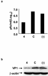
siRNA-4 inhibits ongoing CVB3 replication in HeLa cells. Cells were infected with CVB3 at an MOI of 0.01 for 1 h and then were transfected with siRNA-4 at a final concentration of 300 nM. Forty hours after infection, supernatants and cell lysates were collected for detection of virus titer by plaque assay (a) and VP1 by Western blotting (b), respectively. Data shown are representative of two independent experiments.
FIG. 4.
FIG. 4.
siRNAs protect cells against CVB3-induced cytopathic effects. (a) Morphological changes of HeLa cells following infection. Cells were transfected with each siRNA at a final concentration of 300 nM and then infected with CVB3 at an MOI of 0.01. Cell morphology was observed under a phase-contrast microscope at 48 hpi (magnification, ×100). S, sham infected; (−), mock transfected. (b) MTS cell viability assay. The assay was performed as described in Materials and Methods. Cell viability of each sample was expressed relative to that of the sham-infected control, which was defined as 100% survival. Values shown here are means ± standard deviations of three independent experiments. P < 0.005. (c and d) Western blot analysis of CVB3 VP1 in the cell lysates (c) and supernatants (d). Note that cells transfected with siRNA-2 or control, as well as mock-transfected cells, were dead after CVB3 infection for 48 h. Thus, no intact cells were remaining 48 hpi for preparing cell lysates used for Western blot analysis (c). Data shown are representatives of three independent experiments.
FIG. 5.
FIG. 5.
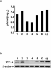
CVB3-specific siRNAs inhibit CVB3 replication in HL-1 cells. Cells were transfected with each siRNA at a final concentration of 300 nM by the Oligofectamine method overnight and then were infected with CVB3 at an MOI of 10. At 48 hpi, supernatants were used for detecting virus titer by plaque assay (a), and cell lysates were collected for VP1 detection by Western blot (b). β-Actin was used as the loading control. Data shown are representatives of two independent experiments.
FIG. 6.
FIG. 6.
Effects of mismatches on antiviral activity of siRNA. (a) Sequences of the mutated siRNA-4. The targeting sequence for siRNA-4 is listed at the top, with the center base boxed. The sequences of wild-type and mutated siRNAs are shown, with mismatched nucleotides underlined. (b and c) siRNA-4mAS with one point mutation in the center of the antisense strand failed to inhibit virus replication, which was detected by viral plaque assay (b) and Western blotting (c). (d) siRNA-4 with one nucleotide mismatch near the 5′ end of the antisense strand but not with one near the 3′ end partially reduced its antiviral activity. HeLa cells were transfected with wild-type and mutated siRNA-4 at a final concentration of 300 nM, followed by CVB3 infection at an MOI of 10 for 8 h. Virus titer in the supernatants and VP1 protein expression in the cell lysates were analyzed by plaque assay (b) and Western blot (c and d). β-Actin served as the loading control. Data shown are representatives of two independent experiments.
FIG. 7.
FIG. 7.
Effect of combined siRNAs on antiviral activity. HeLa cells were cotransfected with siRNA-4 and each of the other four siRNAs at a common final concentration as indicated, followed by CVB3 infection at an MOI of 10 for 8 h. Cell lysates and supernatants in cultures were used for detecting viral protein expression by Western blot. β-Actin served as the loading control. Note that no enhanced inhibitory effect on CVB3 infection was observed by cotransfection of each siRNA with siRNA-4. Data shown are representatives of two independent experiments.

Similar articles

Cited by

References

    1. Baulcombe, D. 2002. RNA silencing. Curr. Biol. 12:R82—R84. - PubMed
    1. Bertrand, J. R., M. Pottier, A. Vekris, P. Opolon, A. Maksimenko, and C. Malvy. 2002. Comparison of antisense oligonucleotides and siRNAs in cell culture and in vivo. Biochem. Biophys. Res. Commun. 296:1000-1004. - PubMed
    1. Bitko, V., and S. Barik. 2001. Phenotypic silencing of cytoplasmic genes using sequence-specific double-stranded short interfering RNA and its application in the reverse genetics of wild type negative-strand RNA viruses. BMC Microbiol. 1:34. - PMC - PubMed
    1. Boden, D., O. Pusch, F. Lee, L. Tucker, and B. Ramratnam. 2003. Human immunodeficiency virus type 1 escape from RNA interference. J. Virol. 77:11531-11535. - PMC - PubMed
    1. Cetta, F., and V. V. Michels. 1995. The autoimmune basis of dilated cardiomyopathy. Ann. Med. 27:169-173. - PubMed

Publication types

MeSH terms

LinkOut - more resources