. 2005 Feb;115(2):247-57.
doi: 10.1172/JCI22037.

Pathogenesis of persistent lymphatic vessel hyperplasia in chronic airway inflammation

Affiliations

Pathogenesis of persistent lymphatic vessel hyperplasia in chronic airway inflammation

Peter Baluk et al. J Clin Invest. 2005 Feb.

Abstract

Edema occurs in asthma and other inflammatory diseases when the rate of plasma leakage from blood vessels exceeds the drainage through lymphatic vessels and other routes. It is unclear to what extent lymphatic vessels grow to compensate for increased leakage during inflammation and what drives the lymphangiogenesis that does occur. We addressed these issues in mouse models of (a) chronic respiratory tract infection with Mycoplasma pulmonis and (b) adenoviral transduction of airway epithelium with VEGF family growth factors. Blood vessel remodeling and lymphangiogenesis were both robust in infected airways. Inhibition of VEGFR-3 signaling completely prevented the growth of lymphatic vessels but not blood vessels. Lack of lymphatic growth exaggerated mucosal edema and reduced the hypertrophy of draining lymph nodes. Airway dendritic cells, macrophages, neutrophils, and epithelial cells expressed the VEGFR-3 ligands VEGF-C or VEGF-D. Adenoviral delivery of either VEGF-C or VEGF-D evoked lymphangiogenesis without angiogenesis, whereas adenoviral VEGF had the opposite effect. After antibiotic treatment of the infection, inflammation and remodeling of blood vessels quickly subsided, but lymphatic vessels persisted. Together, these findings suggest that when lymphangiogenesis is impaired, airway inflammation may lead to bronchial lymphedema and exaggerated airflow obstruction. Correction of defective lymphangiogenesis may benefit the treatment of asthma and other inflammatory airway diseases.


Figures
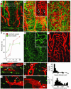
Figure 1
Figure 1
Lymphangiogenesis and angiogenesis after M. pulmonis infection. (AC, E, and F) Confocal micrographs of tracheal whole mounts stained for lymphatic vessels (red) and blood vessels (green). (A) Pathogen-free C3H mouse. (B) C3H mouse infected for 14 days; inset shows lymphatic sprouts (arrowheads) and filopodia (arrows). (C) C3H mouse infected for 28 days. (D) Proliferation of lymphatic vessels (red) and blood vessels (green) in tracheas of C3H mice over 28 days of infection. (E) In a C57BL/6 mouse infected for 14 days, blood vessels (CD31) exhibit sprouting (arrow) and enlargement. (F) Same region as shown in E. Lymphatic vessels (LYVE-1) have a growth pattern similar to that of C3H mice. (G, H, J, and K) Dividing endothelial cells stained for phosphohistone H3 (PH3, green) in tracheal lymphatic vessels (red) of C3H mice infected for 14 days. (G) Section of trachea showing dividing cells, which are sparse in lymphatic vessels (arrow) and numerous in epithelial cells and leukocytes (asterisks). (H, J, and K) Tracheal whole mounts. (H) Dividing lymphatic endothelial cells (arrows) in stalks of medium-sized sprouts. (I) Size distribution of 100 dividing lymphatic endothelial cells. Most dividing cells are near sprout tips (J) or in larger lymphatic vessels (K). (L) Distribution of distances of dividing lymphatic endothelial cells from sprout tips. Scale bar in K applies to all figures: 100 μm in AC, E and F, and 20 μm in G, H, J, and K.
Figure 2
Figure 2
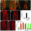
VEGF receptor distribution and inhibition in tracheal vessels. (A) Strong VEGFR-2 immunoreactivity on blood vessels (arrowheads) and weaker staining of lymphatic vessels (arrows) in pathogen-free mouse. (B) Immunoreactivities for VEGFR-3 and LYVE-1 colocalize on lymphatic vessels (arrows) in pathogen-free mouse. (C and D) Lymphatic vessels in mice infected with M. pulmonis for 7 days, either untreated (C) or pretreated with soluble VEGFR-3–Ig (D). Sprouts (arrows) emerge from enlarged lymphatic vessels after infection (C) but not if pretreated with soluble VEGFR-3–Ig (D). (E) Number of lymphatic sprouts in tracheas from pathogen-free mice, or from mice after 7 days of infection, with or without soluble VEGFR-3–Ig pretreatment. (F and G) After 14 days of infection, lymphangiogenesis is not affected by concurrent treatment with antibody against VEGFR-1 (F) but is blocked by antibody against VEGFR-3 (G). (H) Area density of lymphatic vessels (red) and blood vessels (green) showing inhibition of infection-induced lymphangiogenesis by anti–VEGFR-3 (R3) antibody but not by anti–VEGFR-1 (R1) or anti–VEGFR-2 (R2). Anti–VEGFR-2 did not augment the effect of anti–VEGFR-3 (R2 + R3). None of the antibodies reduced angiogenesis. *P < 0.05 vs. pathogen-free group; P < 0.05 vs. infected control group. Scale bar in G applies to all figures: 100 μm in A and B and 50 μm in C, D, F, and G.
Figure 3
Figure 3
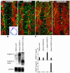
Overexpression of VEGF family growth factors by adenoviral vectors. Tracheal lymphatic vessels (red) and blood vessels (green) 10 days after intranasal inoculation of nude mice with adenoviral vectors. (A) Transduction of airway epithelial cells by adenovirus encoding LacZ (Adeno-LacZ), shown by X-gal staining (inset, blue), had no apparent effect on lymphatic vessels (arrows) or blood vessels (arrowheads). (B) Adenoviral VEGF induced angiogenesis (arrowheads) but no detectable lymphangiogenesis (arrows). (C) Adenoviral VEGF-C induced extensive lymphangiogenesis (arrows) but no angiogenesis (arrowheads). (D) Adenoviral VEGF-D induced widespread lymphangiogenesis but no apparent change in blood vessels (arrowheads); lymphatic vessels appear to coalesce (arrows). Scale bar in D (100 μm) applies to all figures. (E) Northern blot showing human VEGF-C (hVEGF-C) and VEGF-D mRNA in extracts of lungs 10 days after inoculation with adenoviral LacZ, human VEGF-C, or human VEGF-D ΔNΔC. (F) Results of ELISA showing VEGF-C and VEGF-D protein in lungs 10 days after inoculation with adenoviral LacZ, human VEGF165, VEGF-C, or VEGF-D ΔNΔC.
Figure 4
Figure 4
VEGF-C and VEGF-D in M. pulmonis–infected airways. Immunohistochemical staining of VEGF-C (AC) and VEGF-D (DF) in mouse airways and lung 14 days after infection. (A) VEGF-C (green) in epithelium, peribronchial inflammatory cells, and type II alveolar epithelial cells (asterisks) of lung. Blood vessels stained for CD31 (red). (B) VEGF-C (green) in inflammatory cells (arrows) near sprouting lymphatic vessels (red) in tracheal whole mount. (C) Colocalization of VEGF-C immunoreactivity (green) and F4/80 immunoreactivity (red) in macrophages (yellow, arrows). (D) Strong VEGF-D staining (green) of neutrophils in airway lumen. (E) VEGF-D–positive neutrophils (boxed area in D) shown at higher magnification in airway lumen (arrows, green) and airway smooth muscle cells (arrowheads). Blood vessels stained for CD31 (red). (F) VEGF-D immunoreactivity in neutrophils (arrows, green) in tracheal mucosa near sprouting lymphatic vessels (arrowheads) stained for VEGFR-3 (red). Scale bar in F applies to all figures: 200 μm in A and D and 50 μm in B, C, E, and F. (G) Results of RT-PCR analysis showing higher expression of mouse mRNA for VEGF-C (mVEGF-C) and VEGF-D in tracheas after 14 days of infection.
Figure 5
Figure 5
Consequences of blocking lymphangiogenesis in inflamed airways. Comparison of 3 groups of C3H mice: pathogen-free, infected with M. pulmonis for 14 days, and infected with lymphangiogenesis blocked by concurrent treatment with soluble VEGFR-3–Ig delivered by adenovirus. (A) Evans blue accumulation in trachea over 30 minutes showing significantly increased dye accumulation after infection (*P < 0.05) but even greater accumulation when lymphangiogenesis was blocked by soluble VEGFR-3–Ig. (B) Lymph nodes were enlarged after infection (*P < 0.05), but significantly less so when lymphangiogenesis was inhibited by soluble VEGFR-3–Ig ( P < 0.05).
Figure 6
Figure 6
Persistence of new lymphatic vessels after treatment. (A and B) Weight of lungs and bronchial lymph nodes (A) and abundance of tracheal lymphatic vessels and blood vessels (B) in pathogen-free mice (0), mice infected with M. pulmonis for 2 or 4 weeks (2 and 4), and mice infected for 2 weeks and then treated with oxytetracycline for 2 to 12 weeks (2 + 2 through 2 + 12). The weight of both organs increased after infection and decreased toward normal after treatment. Airway blood vessels (green) showed a similar pattern, whereas lymphatic vessels (red) proliferated after infection but regressed little, even after 12 weeks of treatment. *P < 0.05 vs. pathogen-free group; P < 0.05 vs. 2-week-infected group without treatment. (C and D) Confocal micrographs showing tracheal lymphatic vessels (red) and blood vessels (green) after infection for 2 weeks and then oxytetracycline treatment for 8 weeks. Blood vessels regressed almost to the pathogen-free state. Lymphatic vessels showed some changes but regressed only slightly; lymphatic vessels with constrictions and no LYVE-1 immunoreactivity in some cells are indicated by arrows. (D) Enlargement of the boxed area in C. Scale bar in D applies to both figures: 100 μm in C, 25 μm in D.
Figure 7
Figure 7
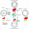
Role of lymphangiogenesis in airway inflammation. The diagram shows relationships of changes in lymphatic vessels (red), blood vessels (green), inflammatory cells (blue), and plasma leakage (short arrows) in inflamed airway mucosa. Cross-sections of bronchi and vessel schematics compare 4 conditions. (I) In normal airways, blood vessels have little or no leukocyte traffic; baseline leakage drains via lymphatic vessels. (II) After infection, activated antigen-presenting cells traffic from airways to local lymph nodes, evoking an immune response. Mucosal capillaries remodel into activated venules that mediate leukocyte influx. These cells release VEGF-C and VEGF-D, which drive lymphangiogenesis via VEGFR-3 signaling in lymphatic endothelial cells. The lymphatic network expands by sprouting from existing lymphatic vessels to accommodate the increased leakage from venules and increased trafficking of immune cells to lymph nodes. (III) If lymphangiogenesis does not occur, leakage exceeds drainage, bronchial lymphedema develops, trafficking of immune cells to lymph nodes decreases, and the immune response is reduced. (IV) Treatment decreases the inflammatory stimulus, allowing blood vessels to return to the baseline state. Leukocyte influx decreases, and the stimulus for lymphangiogenesis diminishes, but lymphatic vessels that formed during the infection persist – ready for the next infection.

Similar articles

Cited by

References

    1. Wiggs BR, Bosken C, Pare PD, James A, Hogg JC. A model of airway narrowing in asthma and in chronic obstructive pulmonary disease. Am. Rev. Respir. Dis. 1992;145:1251–1258. - PubMed
    1. Wilson J. The bronchial microcirculation in asthma. Clin. Exp. Allergy. 2000;30:51–53. - PubMed
    1. Pullinger BD, Florey HW. Proliferation of lymphatics in inflammation. J. Pathol. Bact. 1937;45:157–170.
    1. Jussila L, Alitalo K. Vascular growth factors and lymphangiogenesis. Physiol. Rev. 2002;82:673–700. - PubMed
    1. Saaristo A, et al. Adenoviral VEGF-C overexpression induces blood vessel enlargement, tortuosity, and leakiness but no sprouting angiogenesis in the skin or mucous membranes. FASEB J. 2002;16:1041–1049. - PubMed

Publication types

MeSH terms

Substances