. 2005 Jan 19;25(3):594-603.
doi: 10.1523/JNEUROSCI.4011-04.2005.

Distinct gamma2 subunit domains mediate clustering and synaptic function of postsynaptic GABAA receptors and gephyrin

Affiliations

Distinct gamma2 subunit domains mediate clustering and synaptic function of postsynaptic GABAA receptors and gephyrin

Melissa J Alldred et al. J Neurosci. .

Abstract

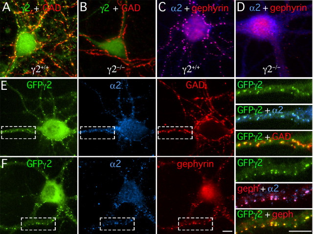
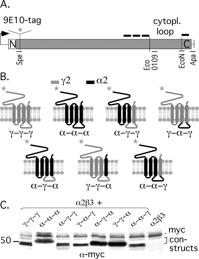
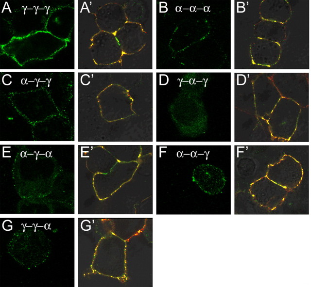
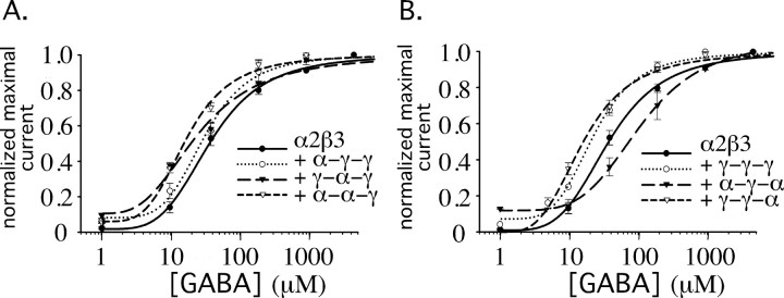
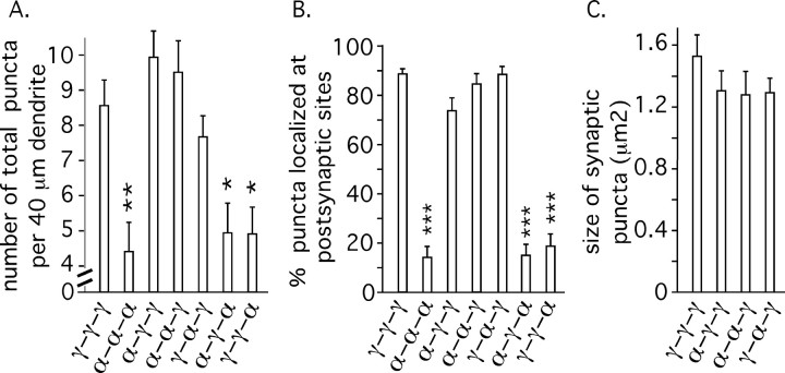
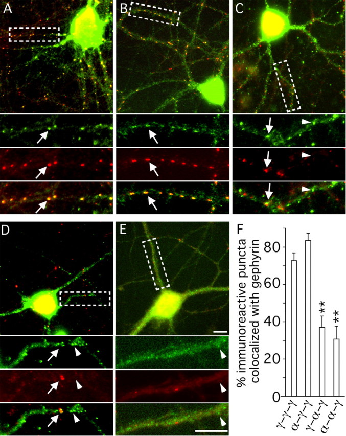
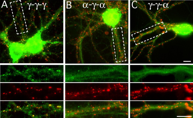
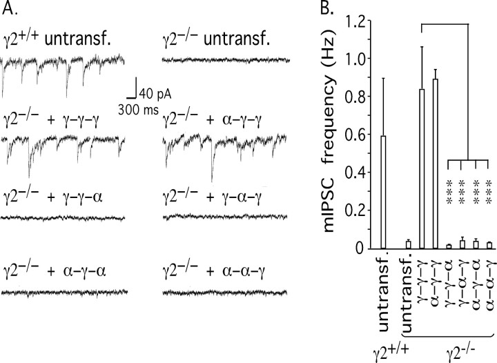
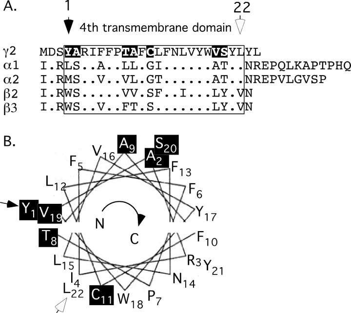
Modulation of the concentration of postsynaptic GABA(A) receptors contributes to functional plasticity of inhibitory synapses. The gamma2 subunit of GABA(A) receptor is specifically required for clustering of these receptors, for recruitment of the submembrane scaffold protein gephyrin to postsynaptic sites, and for postsynaptic function of GABAergic inhibitory synapses. To elucidate this mechanism, we here have mapped the gamma2 subunit domains required for restoration of postsynaptic clustering and function of GABA(A) receptors in gamma2 subunit mutant neurons. Transfection of gamma2-/- neurons with the gamma2 subunit but not the alpha2 subunit rescues postsynaptic clustering of GABA(A) receptors, results in recruitment of gephyrin to postsynaptic sites, and restores the amplitude and frequency of miniature inhibitory postsynaptic currents to wild-type levels. Analogous analyses of chimeric gamma2/alpha2 subunit constructs indicate, unexpectedly, that the fourth transmembrane domain of the gamma2 subunit is required and sufficient for postsynaptic clustering of GABA(A) receptors, whereas cytoplasmic gamma2 subunit domains are dispensable. In contrast, both the major cytoplasmic loop and the fourth transmembrane domain of the gamma2 subunit contribute to efficient recruitment of gephyrin to postsynaptic receptor clusters and are essential for restoration of miniature IPSCs. Our study points to a novel mechanism involved in targeting of GABA(A) receptors and gephyrin to inhibitory synapses.


Figures

Figure 1.
Figure 1.
Restoration of postsynaptic GABAA receptors and gephyrin clusters in γ2-/- neurons by transfection of GFP-tagged γ2 subunit. A-D, Cortical neurons cultured from embryonic day 14.5 γ2+/+ (A, C) or γ2-/- embryos (B, D) (20 DIV) were double stained with antibodies specific for the γ2 subunit (shown in green) and GAD (red) (A, B) or the α2 subunit (blue) and gephyrin (red) (C, D); colocalization is shown in yellow (A, B) and pink (C, D), respectively. Note the dramatic loss of punctate staining for the γ2 and α2 subunits as well as gephyrin in γ2-/- neurons, whereas presynaptic GAD staining is unchanged. E, F, Cortical neurons cultured from γ2-/- embryos were transfected at 18 DIV with GFPγ2. They were fixed, permeabilized, and processed for immunofluorescent staining at 21 DIV with an antiserum specific for the GABAA receptor α2 subunit (blue) and antibodies specific for either GAD (red) (E), indicating GABAergic terminals, or gephyrin (red) (F), as indicated. Boxed dendritic segments are shown enlarged in the panels on the right for either GFPγ2 alone (green) or as merged images. Colocalization in the merged enlargement of the cell in A representing GFPγ2 (green) and the α2 subunit (blue) is shown in cyan blue and colocalization of GFPγ2 and GAD (red) is shown in yellow. Colocalization in the merged enlargement of the cell in B between gephyrin and the α2 subunit is shown in magenta, and colocalization of GFPγ2 and gephyrin is shown in yellow. Scale bars, 10 μm.
Figure 2.
Figure 2.
Schematic representation of chimeric subunit constructs and analysis of their expression after transfection into HEK 293T cells. A, Schematic representation of the myc 9E10 epitope-tagged γ2 subunit with the position of silent restriction sites inserted flanking the large cytoplasmic loop domain. The γ2 subunit translational open reading frame is shown as a gray box, with N and C termini indicated. The positions of the four transmembrane domains are indicated by short black lines above this box. The 9E10 epitope tag and an adjacent SpeI site are inserted between the fourth and fifth amino acid of the open reading frame. B, Schematic representation of chimeric constructs, with the γ2 subunit-derived portion shown in gray and the α2 subunit-derived portion shown in black. The nomenclature for chimeric construct indicated underneath each drawing is explained in Results. C, Western blot analysis of chimeric constructs cotransfected with α2 and β3 subunits into HEK 293T cells. Equal amounts of protein (40 μg) were loaded on the gel, and the blot was developed using an antiserum raised against the 9E10 myc epitope. The ubiquitous protein band running as a band of ∼60 kDa represents endogenous myc and serves as a gel loading control. The constructs and subunits transfected in each lane are indicated.
Figure 3.
Figure 3.
Analysis of surface expression of chimeric subunit constructs transfected into HEK 293T cells. The 9E10-tagged constructs indicated were transfected either alone (A-G) or together with α2 and β3 subunits (A′-G′). Nonpermeabilized cells were stained with antibody specific for the 9E10-tagged construct indicated (A-G) or double labeled for this construct and the α2 subunit (A′-G′). Both antibodies are directed against N-terminal epitopes and selectively recognize subunits inserted into plasma membrane. The staining was developed with fluorescent secondary antibody for imaging. Note the efficient expression of the γ2 subunit (9E10γ-γ-γ) in the plasma membrane independent of whether it is expressed alone (A) or together with α2 and β3 subunits (A′). In contrast, efficient incorporation of the α2 subunit (B, B′) or chimeric constructs (C-G, C′-G′) depends on coexpression of the α2 or β3 subunit, or both.
Figure 4.
Figure 4.
GABA dose-response curves of GABAA receptors containing chimeric subunits expressed in HEK 293T cells. A, B, The α2 and β3 subunits were transfected either alone or in combination with the 9E10γ-γ-γ, 9E10α-γ-α, or 9E10γ-γ-α constructs (A) or in combination with the 9E10α-γ-γ, 9E10γ-α-γ, or 9E10α-α-γ constructs (B). GABA-evoked whole-cell currents were normalized to the maximal responses obtained at 1-5 mm GABA application. For each concentration tested, the data were averaged from 3-11 cells.
Figure 5.
Figure 5.
Restoration of postsynaptic GABAA receptor clusters in γ2-/- neurons transfected with chimeric γ2/α2 subunit constructs. A-G, Cortical neurons isolated from γ2-/- embryos were transfected at 18 DIV with (9E10) epitope-tagged constructs indicated and processed for immunofluorescence analysis at 21 DIV for the 9E10 epitope (shown in green) and presynaptic GAD (red). Shown are merged images with colocalization represented in yellow. Boxed dendritic segments of the images are shown enlarged in separate panels below each image. Note the faithful formation of clusters revealed in the form of punctate staining for the 9E10γ-γ-γ (A), 9E10α-γ-γ (C), 9E10α-α-γ (D), and γ-α-γ (E) constructs that were closely apposed to presynaptic GAD, whereas the 9E10α-α-α (B), 9E10α-γ-α (F), and 9E10γ-γ-α (G) constructs failed to form clusters and showed no juxtaposition to GAD immunoreactivity. Arrows point to clusters apposed to GAD; arrowheads indicate 9E10 immunoreactivity that is diffuse or punctate but not apposed to GAD immunoreactivity. Scale bars, 10 μm.
Figure 6.
Figure 6.
Quantitative analyses of postsynaptic clusters formed by chimeric constructs transfected into γ2-/- neurons. Cortical cultures isolated from γ2-/- embryos were transfected and double labeled for the 9E10 epitope tag of the transfected construct and for the GABAergic terminal marker GAD as shown in Figure 5. Immunoreactive puncta on dendritic segments that were innervated by a GABAergic axon as revealed by GAD immunoreactivity were quantified from digitized video images as described in Materials and Methods. A, The average number of 9E10 immunoreactive puncta per 40 μm dendritic segment determined for each of the chimeric constructs including 9E10α-α-α was compared with the value determined for the γ2 subunit (9E10γ-γ-γ). Note the similar number of puncta observed for the 9E10γ-γ-γ, 9E10α-γ-γ, 9E10α-α-γ, and 9E10γ-α-γ constructs (n = 13, 15, 19, 16 cells, respectively). In contrast, the number of puncta observed for the 9E10α-γ-α (n = 14) and 9E10γ-γ-α (n = 16) constructs was similar to that observed for the α2 subunit (9E10α-α-α; n = 17) and greatly reduced compared with the γ2 subunit (9E10γ-γ-γ). B, For comparison of the number of clusters localized to postsynaptic sites, the fraction of puncta apposed to punctate GAD immunoreactivity was determined for each chimeric construct and compared with the value determined for 9E10γ-γ-γ. Note that the same constructs that showed a high propensity to cluster in A similar to the γ2 subunit are also indistinguishable from the γ2 subunit in that they result in a high percentage of clusters that are postsynaptic. In contrast, the percentage of immunoreactive puncta for the 9E10α-γ-α and 9E10γ-γ-α constructs and the α2 subunit (9E10α-α-α) are significantly reduced compared with the value for the γ2 subunit. C, The average size of postsynaptic clusters induced by the 9E10α-γ-γ, 9E10α-α-γ, and 9E10γ-α-γ constructs is indistinguishable from the value observed for the γ2 subunit (9E10γ-γ-γ). Error bars represent SE. *p < 0.05; **p < 0.01; ***p < 0.001.
Figure 7.
Figure 7.
Recruitment of gephyrin to GABAA receptor clusters. The γ2 subunit (A) and the three other γ2 subunit TM4-containing chimeric constructs shown in Figure 5 to form clusters (B, 9E10α-γ-γ; C, 9E10γ-α-γ; D, 9E10α-α-γ) were transfected into γ2-/- neurons and analyzed for colocalization of punctate 9E10 immunoreactivity (green) with endogenous gephyrin (red). In addition, analysis of the diffusely expressed construct 9E10α-γ-α (E), which is not concentrated at synapses (Fig. 5F), is shown for comparison. Boxed dendritic segments are shown enlarged in color-separated and merged panels underneath each image with arrows pointing to colocalized clusters (yellow) and arrowheads indicating punctate 9E10 immunoreactivity that was not colocalized (green). Note the similar high degree of colocalization seen for the γ2 subunit (A) and for the 9E10α-γ-γ construct (B) with only few 9E10-immunoreactive puncta that were not colocalized with gephyrin (green puncta in the merged enlargement). In contrast, only a fraction of the 9E10-immunoreactive puncta that were formed by the 9E10γ-α-γ and 9E10α-α-γ constructs were colocalized with gephyrin (C, D). This visual impression was confirmed by quantitative analyses (F). The percentage of 9E10α-γ-γ puncta colocalized with gephyrin was similar to values seen with the γ2 subunit. In contrast, the percentage of 9E10γ-α-γ or 9E10α-α-γ puncta colocalized was significantly reduced compared with the γ2 subunit, consistent with the notion that GABAA receptors can form clusters in the absence of gephyrin. Colocalization of 9E10α-γ-α and gephyrin (E) could not be quantified because expression of this construct was mostly diffuse. Scale bars, 10 μm.
Figure 8.
Figure 8.
Assessment of dysfunctional constructs in wild-type neurons. The 9E10γ-γ-γ, 9E10α-γ-α, and 9E10γ-γ-α constructs were transfected into wild-type neurons and double labeled for the 9E10 epitope and endogenous gephyrin to test for negative effects on postsynaptic differentiation. A, The 9E10γ-γ-γ construct formed clusters colocalized with gephyrin as expected. The 9E10α-γ-α (B) and 9E10γ-γ-α (C) constructs failed to cluster, but endogenous gephyrin clusters remained unaffected, indicating that failure of these constructs to accumulate at synapses was not associated with dominant-negative effects on synapse formation. Scale bars, 10 μm.
Figure 9.
Figure 9.
Functional rescue of inhibitory synapses in γ2-/- neurons requires both the major intracellular loop and the fourth transmembrane domain of the γ2 subunit. A, Representative traces are shown of mIPSCs recorded from γ2+/+ and γ2-/- neurons and of γ2-/- neurons transfected with the γ2 subunit (9E10γ-γ-γ) or chimeric subunits, as indicated. B, Summary of data showing mIPSC frequencies confirming that transfection of either 9E10γ-γ-γ or 9E10α-γ-γ restored the function of inhibitory synaptic transmission in γ2-/- neurons to values similar to those found in wild-type neurons. Note that none of the 9E10γ-γ-α, 9E10γ-α-γ, 9E10α-γ-α, and 9E10α-α-γ constructs were able to restore mIPSCs of γ2-/- neurons, indicating that both the γ2 subunit cytoplasmic loop and TM4 domain are required for restoration of synaptic function in γ2-/- neurons. Error bars represent SE. ***p < 0.001.
Figure 10.
Figure 10.
Structural features of the γ2 TM4 domain. A, Alignment of the C-terminal domains of major GABAA receptor subunits known to constitute postsynaptic GABAA receptors reveals seven amino acids that are uniquely present in the γ2 subunit TM4 domain (shown as white text on black background). B, A helical wheel representation of the putative α-helix of the γ2 subunit TM4 domain predicts that amino acids uniquely present in the γ2 subunit TM4 domain map to two narrow faces of the putative TM4 α-helix, suggesting potential contact sites that are uniquely present in this subunit. Amino acids in B are denoted in single-letter code with numeric subscripts indicating the position of the amino acid in the transmembrane domain, as shown in A. Amino acids that are unique for the γ2 subunit TM4 domain are shown as white text on black background.

Similar articles

Cited by

References

    1. Angelotti TP, Macdonald RL (1993) Assembly of GABAA receptor subunits: α1β1 and α1β1γ2S subunits produce unique ion channels with dissimilar single-channel properties. J Neurosci 13: 1429-1440. - PMC - PubMed
    1. Baer K, Essrich C, Benson JA, Benke D, Bluethmann H, Fritschy J-M, Luscher B (1999) Postsynaptic clustering of GABAA receptors by the γ3 subunit in vivo Proc Natl Acad Sci USA 96: 12860-12865. - PMC - PubMed
    1. Banker G, Goslin K (1998) Culturing nerve cells. Cambridge, MA: MIT.
    1. Benson JA, Low K, Keist R, Mohler H, Rudolph U (1998) Pharmacology of recombinant γ-aminobutyric acidA receptors rendered diazepam-insensitive by point-mutated α-subunits. FEBS Lett 431: 400-404. - PubMed
    1. Blanton MP, Cohen JB (1994) Identifying the lipid-protein interface of the Torpedo nicotinic acetylcholine receptor: secondary structure implications. Biochemistry 33: 2859-2872. - PubMed

Publication types

MeSH terms