Morphogenesis of the rat forebrain
- PMID: 15628985
- PMCID: PMC7245524
- DOI: 10.17305/bjbms.2004.3467
Morphogenesis of the rat forebrain
Abstract
Background and purpose: Developmental process that leads to final forebrain shaping is a result of complex histogenetic and morphogenetic events. Comprehensions about brain development are based on observations carried out on onthogenetic successive stages. Microscopic analysis of brain together with analysis of serial sections gives information about shape the of some forebrain parts and basic relations between them. The aim of this study was to analyse morphogenesis in the earliest stages of rat's forebrain development.
Material and methods: Rat brains used in this study were obtained from Fisher inbred rats with accurately timed pregnancies. The investigation was carried out on serial frontal sections of rat embryonic heads from the 12th (E12) to the 16th (E16) day of gestation. Gestation was considered to have begun early in the morning when sperm was found in the vaginal smear. Histological paraffin and plastic sections were systematically inspected with regard to morphogenetic changes of the forebrain parts telencephalon and diencephalon.
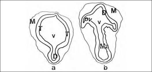
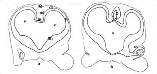
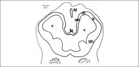
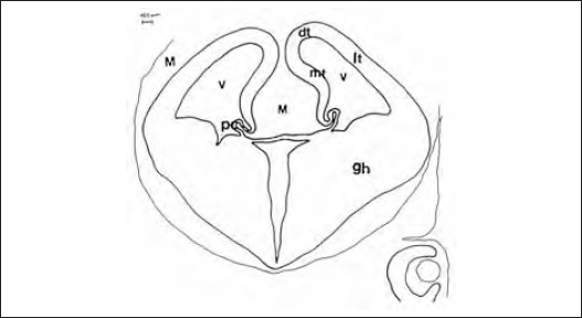
Results: E12: neural tube is completely closed in its cranial part. Rostral part of forebrain shows telencephalons vesicles origins as slightly paired enlargements of neuroepithelial wall. Between telencephalic vesicles origin and in direction to caudal there is an origin of diencephalon. E13: rostral part of forebrain shows well expressed and divided areas of telencephalons vesicles as basal, basolateral, dorsal and medial telencephalon. Central area between paired vesicles is a telencephalon impar. In diencephalon optic vesicles appeared. Epithalamus, thalamus and hypothalamus origins are slight enlargements of its neuroepithelial wall. E14: telencephalic vesicles spread above telencephalon impar into rostral direction and above diencephalon in rostrodorsal direction. Their basolateral parts of are very thickened and become folded. Sulcus telodiencephalicus appears. E15: the main event is the appearance of the origins of plexus choroideus in the area of telencephalon impar as fingerlike processes. E16: all forebrain parts, especially telencephalic vesicles-origin of brain hemispheres and processes of plexus choroideus, are progressively growing and shaping.
Conclusions: Our morphologic analysis describes significant morphogenetic changes in the forebrain shape. The forebrain changes from a relatively simple tubular structure with thin walls surrounding a large ventricular system to a thick-walled brain with a highly convoluted but reduced ventricular system.
Figures

Similar articles
-
Three-dimensional reconstructions of the developing forebrain in rat embryos.Neuroimage. 1994 Nov;1(4):296-307. doi: 10.1006/nimg.1994.1014. Neuroimage. 1994. PMID: 9343579
-
Coordinate expression of Fgf8, Otx2, Bmp4, and Shh in the rostral prosencephalon during development of the telencephalic and optic vesicles.Neuroscience. 2001;108(2):183-206. doi: 10.1016/s0306-4522(01)00411-0. Neuroscience. 2001. PMID: 11734354
-
Development of the rat telencephalon--volumetric analysis.Bosn J Basic Med Sci. 2004 Jul;4(3):11-4. doi: 10.17305/bjbms.2004.3375. Bosn J Basic Med Sci. 2004. PMID: 15629006 Free PMC article.
-
The forebrain of gnathostomes: in search of a morphotype.Brain Behav Evol. 1995;46(4-5):275-318. doi: 10.1159/000113279. Brain Behav Evol. 1995. PMID: 8564468 Review.
-
Genes and signaling events that establish regional patterning of the mammalian forebrain.Semin Cell Dev Biol. 2009 Jun;20(4):378-86. doi: 10.1016/j.semcdb.2009.02.005. Epub 2009 Mar 3. Semin Cell Dev Biol. 2009. PMID: 19560042 Review.
Cited by
-
Embryogenesis of the rat telencephalon--a morphologic and stereologic analysis.Bosn J Basic Med Sci. 2005 May;5(2):59-64. doi: 10.17305/bjbms.2005.3286. Bosn J Basic Med Sci. 2005. PMID: 16053457 Free PMC article.
References
-
- Karfunkel P. The mechanisms of neural tube formation. Int Rev Cytol. 1974;38:245–271. - PubMed
-
- Waterman R.E. Topographical changes along the neural fold associated with neurulation in the hamster and mouse. Am J Anat. 1976;146:151–172. - PubMed
-
- Morriss-Kay G.M. Growth and development of pattern in the cranial neural epithelium of rat embryos during neurulation. J Embryol Exp Morphol. 1981;65:225–241. - PubMed
-
- Schoenwolf G.C. On the morphogenesis of the early rudiments of the developing central nervous system. Scanning Elec Microsc. 1982;1:289–308. - PubMed
-
- Geelen J.A.G, Langman J. Closure of the neural tube in the cephalic region of the mouse embryo. Anat Rec. 1977;189:625–640. - PubMed