. 2005 Jan;25(1):389-402.
doi: 10.1128/MCB.25.1.389-402.2005.

Lysosomal targeting of E-cadherin: a unique mechanism for the down-regulation of cell-cell adhesion during epithelial to mesenchymal transitions

Affiliations

Lysosomal targeting of E-cadherin: a unique mechanism for the down-regulation of cell-cell adhesion during epithelial to mesenchymal transitions

Felipe Palacios et al. Mol Cell Biol. 2005 Jan.

Abstract

A hallmark characteristic of epithelial tumor progression as well as some processes of normal development is the loss of the epithelial phenotype and acquisition of a motile or mesenchymal phenotype. Such epithelial to mesenchymal transitions are accompanied by the loss of E-cadherin function by either transcriptional or posttranscriptional mechanisms. Here we demonstrate that, upon v-Src expression, a potent trigger of epithelial to mesenchymal transitions, E-cadherin is internalized and then shuttled to the lysosome instead of being recycled back to the lateral membrane. Thus, while E-cadherin internalization facilitates the dissolution of adherens junctions, its subsequent traffic to the lysosome serves as a means to ensure that cells do not reform their cell-cell contacts and remain motile. We also show that ubiquitin tagging of E-cadherin is essential for its sorting to the lysosome. The lysosomal targeting of E-cadherin is mediated by hepatocyte growth factor-regulated tyrosine kinase substrate (Hrs) and v-Src-induced activation of the Rab5 and Rab7 GTPases. Our studies reveal that the lysosomal targeting of E-cadherin is an important posttranscriptional mechanism to deplete cellular E-cadherin during Src-induced epithelial to mesenchymal transitions.


Figures

FIG. 1.
FIG. 1.
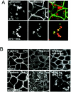
v-Src expression promotes the formation of enlarged vesicles that contain E-cadherin. (A) MDCKpp60v-Src cells at nonpermissive or permissive temperatures for 4 h and parental MDCK I cells stably expressing ARF6(Q67L) were fixed and labeled for E-cadherin. Expression of v-Src in MDCK cells induces the formation of enlarged vesicles that contain E-cadherin that are morphologically distinct from those observed in cells expressing the ARF6-GTP mutant. The overall levels of E-cadherin in v-Src-expressing cells appear to be reduced compared to those in untransfected MDCK cells. (B) MDCKpp60v-Src cells stably expressing HA-tagged ARF6(T27N) were maintained at the permissive temperature of 35°C for 4 h and then fixed and labeled for E-cadherin (red) and HA (green). Expression of ARF6(T27N) blocks E-cadherin internalization and the formation of Src-induced enlarged vesicles.
FIG. 2.
FIG. 2.
Src activation induces the shuttling of E-cadherin to the lysosome and prevents its recycling to the basolateral plasma membrane. (A) MDCKpp60v-Src cells transiently expressing GFP-tagged E-cadherin at the nonpermissive temperature of 41°C (top panels) or at the permissive temperature of 35°C (bottom panels) were incubated with LysoTracker to label lysosomes. v-Src activation induces the shuttling of E-cadherin to the lysosome. (B) Parental MDCK I cells or MDCKpp60v-Src cells were incubated at 37 and 41°C, respectively (left panels), followed by incubation at 18°C for 2 h (middle panels) and then for 30 min at 37 or 35°C as indicated (right panels). Cells were fixed at each temperature and labeled for E-cadherin. In normal cells, E-cadherin gets internalized into the cytoplasm and then recycles back to the plasma membrane. Src activation blocks the recycling of E-cadherin to the plasma membrane and instead shuttles E-cadherin to lysosomes.
FIG. 3.
FIG. 3.
Src activation promotes the lysosome-mediated degradation of E-cadherin. (A) MDCKpp60v-Src cells grown at nonpermissive temperatures were switched to 35°C for times indicated to allow expression of v-Src. Cells were then lysed in RIPA buffer and examined for E-cadherin degradation by resolving proteins by SDS-PAGE and then labeling with anti-E-cadherin antibodies by Western blotting procedures followed by autoradiography. At low exposure a major degradation product of 35 kDa was observed. At an increased time (5 min) of autoradiography another degradation product of approximately 20 kDa became apparent. Cell lysates were probed for γ-tubulin expression as a control for equal loading of protein on SDS gels. Numbers at left are molecular masses in kilodaltons. (B) From left to right in each panel, respectively, MDCKpp60v-Src cells were grown at the nonpermissive temperature of 41°C and then switched to 35°C for 4 h to allow expression of v-Src in medium alone or medium containing herbimycin A, chloroquine, or lactacystin. E-cadherin degradation was monitored as described above. The major degradation product of 35 kDa was quantitated by densitometry as a measure of E-cadherin degradation (right). Herbimycin A and chloroquine blocked the degradation of E-cadherin, but lactacystin had no effect. Numbers at left are molecular masses in kilodaltons. (C) MDCKpp60v-Src cells were grown at the nonpermissive temperature of 41°C (lane 1) and then switched to 35°C for 6 h to allow expression of v-Src. In experiments done in parallel, cells were treated with type IV trypsin prior to collection of growth medium. The 90-kDa fragment released by trypsin treatment was quantitated by densitometry. E-cadherin fragments were not detected in the growth medium upon Src activation; however, 90-kDa fragments of the E-cadherin extracellular domain were observed in the growth medium upon trypsin treatment. (D) MDCKpp60v-Src cells were grown at the nonpermissive temperature of 41°C (lane 1) and then switched to 35°C as indicated to allow expression of v-Src. The cell surface was biotinylated, and surface levels of biotinylated E-cadherin were determined as previously described (32). Surface E-cadherin is significantly decreased upon v-Src expression. (E) Epithelial cell colonies at nonpermissive temperatures, with and without treatment with chloroquine for 1 h, were switched to permissive temperatures for times indicated. Cells were fixed, labeled with phalloidin and examined by confocal immunofluorescence microscopy. Chloroquine-treated cells were significantly hampered in their ability to scatter relative to untreated cells.
FIG. 3.
FIG. 3.
Src activation promotes the lysosome-mediated degradation of E-cadherin. (A) MDCKpp60v-Src cells grown at nonpermissive temperatures were switched to 35°C for times indicated to allow expression of v-Src. Cells were then lysed in RIPA buffer and examined for E-cadherin degradation by resolving proteins by SDS-PAGE and then labeling with anti-E-cadherin antibodies by Western blotting procedures followed by autoradiography. At low exposure a major degradation product of 35 kDa was observed. At an increased time (5 min) of autoradiography another degradation product of approximately 20 kDa became apparent. Cell lysates were probed for γ-tubulin expression as a control for equal loading of protein on SDS gels. Numbers at left are molecular masses in kilodaltons. (B) From left to right in each panel, respectively, MDCKpp60v-Src cells were grown at the nonpermissive temperature of 41°C and then switched to 35°C for 4 h to allow expression of v-Src in medium alone or medium containing herbimycin A, chloroquine, or lactacystin. E-cadherin degradation was monitored as described above. The major degradation product of 35 kDa was quantitated by densitometry as a measure of E-cadherin degradation (right). Herbimycin A and chloroquine blocked the degradation of E-cadherin, but lactacystin had no effect. Numbers at left are molecular masses in kilodaltons. (C) MDCKpp60v-Src cells were grown at the nonpermissive temperature of 41°C (lane 1) and then switched to 35°C for 6 h to allow expression of v-Src. In experiments done in parallel, cells were treated with type IV trypsin prior to collection of growth medium. The 90-kDa fragment released by trypsin treatment was quantitated by densitometry. E-cadherin fragments were not detected in the growth medium upon Src activation; however, 90-kDa fragments of the E-cadherin extracellular domain were observed in the growth medium upon trypsin treatment. (D) MDCKpp60v-Src cells were grown at the nonpermissive temperature of 41°C (lane 1) and then switched to 35°C as indicated to allow expression of v-Src. The cell surface was biotinylated, and surface levels of biotinylated E-cadherin were determined as previously described (32). Surface E-cadherin is significantly decreased upon v-Src expression. (E) Epithelial cell colonies at nonpermissive temperatures, with and without treatment with chloroquine for 1 h, were switched to permissive temperatures for times indicated. Cells were fixed, labeled with phalloidin and examined by confocal immunofluorescence microscopy. Chloroquine-treated cells were significantly hampered in their ability to scatter relative to untreated cells.
FIG. 4.
FIG. 4.
Ubiquitination-deficient E-cadherin is internalized but not trafficked to the lysosomes for degradation. (A) MDCKpp60v-Src cells transiently expressing a Myc-tagged ubiquitination-deficient mutant of E-cadherin, E-cadherin(Y755F-Y756F), at nonpermissive temperatures or at 35°C for 4 h were labeled for actin (red) and Myc (green). The ubiquitination-deficient mutant of E-cadherin localizes to cell-cell contacts and to cytoplasmic vesicles. (B) Cells were transiently transfected with retrovirus encoding ARF6(T27N) and GFP for 24 h followed by transient transfection with the Myc-tagged ubiquitination-deficient mutant of E-cadherin. Cells were fixed and labeled for Myc and with Draq5, a nuclear stain. Expression of ARF6(T27N) prevents the cytoplasmic distribution of mutant E-cadherin, suggesting that internalized mutant E-cadherin was derived by endocytosis from the cell surface. (C) MDCKpp60v-Src cells transiently expressing Myc-tagged wild-type E-cadherin or E-cadherin(Y755F-Y756F) at the permissive temperature of 35°C for 4 h were lysed in RIPA buffer, and equal amounts of the cell lysates were probed with antibodies to Myc, by Western blotting procedures. Myc-tagged E-cadherin(Y755F-Y756F) is not degraded upon expression of v-Src.
FIG. 5.
FIG. 5.
Hrs regulates the traffic of ubiquitinated E-cadherin to the lysosome. (A to D) MDCKpp60v-Src cells transiently transfected with Myc-tagged wild-type Hrs or Hrs(S270E) as indicated were grown at the nonpermissive temperature of 41°C and then switched to 35°C for 8 h. Cells were fixed and labeled for Myc (green) and E-cadherin (A and C, red) or LAMP-1 (B and D, red). Coincident staining appears yellow in the merged images. At nonpermissive temperatures, wild-type Hrs colocalizes with E-cadherin on vesicles that do not label for LAMP-1. Upon expression of v-Src, wild-type Hrs and E-cadherin are shuttled toward the late endosomal-lysosomal compartments as seen by the colocalization with LAMP-1 antibodies. However, Hrs(S270E) and E-cadherin are retained at vesicular compartments that do not colocalize with LAMP-1. Thus, expression of Hrs(S270E) blocks the shuttling of E-cadherin to the lysosome upon Src activation. (E) Lysates of MDCKpp60v-Src cells expressing Myc-tagged wild-type Hrs or Hrs(S270E) at permissive temperatures were immunoprecipitated with anti-Myc monoclonal antibody. Immunoprecipitates were resolved on SDS gels and probed with anti-E-cadherin antibody. Only wild-type Hrs, not the Hrs mutant lacking a functional UIM domain, coprecipitates with E-cadherin upon Src activation.
FIG. 6.
FIG. 6.
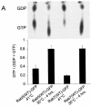
Expression of v-Src induces the activation of Rab5 and Rab7 to facilitate the shuttling of E-cadherin to the lysosome. (A) MDCKpp60v-Src cells transiently transfected with GFP-tagged wild-type Rab5 (lanes 1 and 2) and GFP-tagged wild-type Rab7 (lanes 3 and 4) were incubated at either 41 or 35°C for 4 h as indicated, and the nucleotide bound to Rab5 and Rab7 was determined. Upon Src activation the cellular levels of GTP-bound Rab5 and Rab7 are markedly enhanced. (B and C) MDCKpp60v-Src cells transiently transfected with GFP-tagged wild-type Rab5 at the nonpermissive temperature of 41°C (B) or at the permissive temperature of 35°C (C) were fixed and labeled for E-cadherin (red). Coincident staining appears yellow in the merged images. Upon Src activation, wild-type Rab5 localizes to enlarged endosomes that also contain E-cadherin. Some Rab5-positive endosomes did not contain E-cadherin; these vesicles could represent endosomes that are derived from the apical membrane, which also contain Rab5 (7). (D) MDCKpp60v-Src cells expressing ARF6(T27N) were transiently transfected with GFP-tagged wild-type Rab5, incubated at 35°C for 4 h, and labeled for E-cadherin (red). Coexpression of dominant-negative ARF6, ARF6(T27N), with wild-type Rab5 blocked the internalization of E-cadherin but did not prevent the fusion of Rab5-positive endosomes in response to Src activation. (E) Parental MDCK cells transiently transfected with HA-tagged Rab5(Q79L) were labeled for E-cadherin. Activated Rab5 expression had no effect on the adherens junctions. (F) MDCKpp60v-Src cells transiently transfected with GFP-tagged Rab5(S34N) were grown at 35°C for 4 h and labeled for E-cadherin (red). Expression of dominant-negative Rab5, Rab5(S34N), blocks the formation of enlarged endosomes upon Src activation. The arrow points to an E-cadherin-positive enlarged endosome formed by expression of v-Src in an untransfected cell adjacent to a Rab5(S34N)-transfected cell where enlarged endosomes are not observed. (G and H) MDCKpp60v-Src cells transiently transfected with GFP-tagged wild-type Rab7 at 41 or 35°C were fixed and labeled for E-cadherin (red). In the absence of Src activation, wild-type Rab7 localizes to late endosomes. Upon Src activation, wild-type Rab7 localizes to enlarged vesicles that also contain E-cadherin and are likely formed by the fusion of early and late endosomes. (I) MDCKpp60v-Src cells transiently transfected with GFP-tagged Rab7(T22N) at 41°C were incubated at the permissive temperature of 35°C for 4 h and labeled for E-cadherin (blue) and LysoTracker (red) to visualize lysosomes. Expression of dominant-negative Rab7, Rab7(T22N), prevents the trafficking of E-cadherin to the lysosome. (J) Untransfected MDCKpp60v-Src cells (lanes 1 and 2) and those transiently transfected with GFP-tagged Rab5(S34N) (lane 3) and GFP-tagged Rab7(T22N) (lane 4) were maintained at 41 or 35°C for 4 h as indicated. Cells were then lysed in RIPA buffer, and the cell lysates were analyzed by SDS-PAGE and blotted with antibodies against E-cadherin. The 35-kDa E-cadherin degradation product was quantified using a densitometer and plotted in the graph shown below. Transient expression of Rab5(S34N) and Rab7(T22N) blocked the degradation of E-cadherin in response to Src activation.
FIG. 6.
FIG. 6.
Expression of v-Src induces the activation of Rab5 and Rab7 to facilitate the shuttling of E-cadherin to the lysosome. (A) MDCKpp60v-Src cells transiently transfected with GFP-tagged wild-type Rab5 (lanes 1 and 2) and GFP-tagged wild-type Rab7 (lanes 3 and 4) were incubated at either 41 or 35°C for 4 h as indicated, and the nucleotide bound to Rab5 and Rab7 was determined. Upon Src activation the cellular levels of GTP-bound Rab5 and Rab7 are markedly enhanced. (B and C) MDCKpp60v-Src cells transiently transfected with GFP-tagged wild-type Rab5 at the nonpermissive temperature of 41°C (B) or at the permissive temperature of 35°C (C) were fixed and labeled for E-cadherin (red). Coincident staining appears yellow in the merged images. Upon Src activation, wild-type Rab5 localizes to enlarged endosomes that also contain E-cadherin. Some Rab5-positive endosomes did not contain E-cadherin; these vesicles could represent endosomes that are derived from the apical membrane, which also contain Rab5 (7). (D) MDCKpp60v-Src cells expressing ARF6(T27N) were transiently transfected with GFP-tagged wild-type Rab5, incubated at 35°C for 4 h, and labeled for E-cadherin (red). Coexpression of dominant-negative ARF6, ARF6(T27N), with wild-type Rab5 blocked the internalization of E-cadherin but did not prevent the fusion of Rab5-positive endosomes in response to Src activation. (E) Parental MDCK cells transiently transfected with HA-tagged Rab5(Q79L) were labeled for E-cadherin. Activated Rab5 expression had no effect on the adherens junctions. (F) MDCKpp60v-Src cells transiently transfected with GFP-tagged Rab5(S34N) were grown at 35°C for 4 h and labeled for E-cadherin (red). Expression of dominant-negative Rab5, Rab5(S34N), blocks the formation of enlarged endosomes upon Src activation. The arrow points to an E-cadherin-positive enlarged endosome formed by expression of v-Src in an untransfected cell adjacent to a Rab5(S34N)-transfected cell where enlarged endosomes are not observed. (G and H) MDCKpp60v-Src cells transiently transfected with GFP-tagged wild-type Rab7 at 41 or 35°C were fixed and labeled for E-cadherin (red). In the absence of Src activation, wild-type Rab7 localizes to late endosomes. Upon Src activation, wild-type Rab7 localizes to enlarged vesicles that also contain E-cadherin and are likely formed by the fusion of early and late endosomes. (I) MDCKpp60v-Src cells transiently transfected with GFP-tagged Rab7(T22N) at 41°C were incubated at the permissive temperature of 35°C for 4 h and labeled for E-cadherin (blue) and LysoTracker (red) to visualize lysosomes. Expression of dominant-negative Rab7, Rab7(T22N), prevents the trafficking of E-cadherin to the lysosome. (J) Untransfected MDCKpp60v-Src cells (lanes 1 and 2) and those transiently transfected with GFP-tagged Rab5(S34N) (lane 3) and GFP-tagged Rab7(T22N) (lane 4) were maintained at 41 or 35°C for 4 h as indicated. Cells were then lysed in RIPA buffer, and the cell lysates were analyzed by SDS-PAGE and blotted with antibodies against E-cadherin. The 35-kDa E-cadherin degradation product was quantified using a densitometer and plotted in the graph shown below. Transient expression of Rab5(S34N) and Rab7(T22N) blocked the degradation of E-cadherin in response to Src activation.
FIG. 7.
FIG. 7.
Working model for the regulation of the E-cadherin turnover rate in response to Src activation. As previously described, the activation of ARF6 in response to extracellular stimuli and/or Src activation promotes the internalization of E-cadherin to endosomes vis a process mediated by Nm23-H1 and dynamin (32). Src also promotes the ubiquitination of E-cadherin (11), although it is unclear whether this occurs at the plasma membrane or on internal vesicular compartments. Src-mediated activation of Rab5 promotes early-endosome fusion to induce the formation of E-cadherin-positive enlarged endosomes and perhaps accelerates the rate of E-cadherin internalization. Ubiquitinated E-cadherin is sorted in early endosomes via its recognition by Hrs. Thus, while nonubiquitinated E-cadherin is recycled back to the plasma membrane, ubiquitinated E-cadherin is trafficked to the lysosome for degradation. The latter step is facilitated by Src-induced activation of Rab7. It is unclear at this time if Src induces the phosphorylation of Hrs to promote the lysosomal targeting of E-cadherin. HGF, hepatocyte growth factor.

Similar articles

Cited by

References

    1. Behrens, J., L. Vakaet, R. Friis, E. Winterhager, F. Van Roy, M. M. Mareel, and W. Birchmeier. 1993. Loss of epithelial differentiation and gain of invasiveness correlates with tyrosine phosphorylation of the E-cadherin/beta-catenin complex in cells transformed with a temperature-sensitive v-SRC gene. J. Cell Biol. 120:757-766. - PMC - PubMed
    1. Berx, G., A. M. Cleton-Jansen, F. Nollet, W. J. de Leeuw, M. van de Vijver, C. Cornelisse, and F. van Roy. 1995. E-cadherin is a tumour/invasion suppressor gene mutated in human lobular breast cancers. EMBO J. 14:6107-6115. - PMC - PubMed
    1. Birchmeier, W., J. Behrens, K. M. Weidner, J. Hulsken, and C. Birchmeier. 1996. Epithelial differentiation and the control of metastasis in carcinomas. Curr. Top. Microbiol. Immunol. 213:117-135. - PubMed
    1. Bolos, V., H. Peinado, M. A. Perez-Moreno, M. F. Fraga, M. Esteller, and A. Cano. 2003. The transcription factor Slug represses E-cadherin expression and induces epithelial to mesenchymal transitions: a comparison with Snail and E47 repressors. J. Cell Sci. 116:499-511. - PubMed
    1. Boyer, B., Y. Bourgeois, and M. F. Poupon. 2002. Src kinase contributes to the metastatic spread of carcinoma cells. Oncogene 21:2347-2356. - PubMed

Publication types

MeSH terms

Substances