Golgi inheritance in small buds of Saccharomyces cerevisiae is linked to endoplasmic reticulum inheritance
- PMID: 15596717
- PMCID: PMC539800
- DOI: 10.1073/pnas.0408256102
Golgi inheritance in small buds of Saccharomyces cerevisiae is linked to endoplasmic reticulum inheritance
Abstract
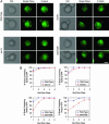
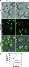
According to the cisternal maturation hypothesis, endoplasmic reticulum (ER)-derived membranes nucleate new Golgi cisternae. The yeast Saccharomyces cerevisiae offers a unique opportunity to test this idea because small buds contain both ER and Golgi structures early in the cell cycle. We previously predicted that mutants defective in ER inheritance also would show defects in Golgi inheritance. Surprisingly, studies of S. cerevisiae have not revealed the expected link between ER and Golgi inheritance. Here, we revisit this issue by generating mutant strains in which many of the small buds are devoid of detectable ER. These strains also show defects in the inheritance of both early and late Golgi cisternae. Strikingly, virtually all of the buds that lack ER also lack early Golgi cisternae. Our results fit with the idea that membranes exported from the ER coalesce with vesicles derived from existing Golgi compartments to generate new Golgi cisternae. This basic mechanism of Golgi inheritance may be conserved from yeast to vertebrate cells.
Figures


Similar articles
-
Live imaging of yeast Golgi cisternal maturation.Nature. 2006 Jun 22;441(7096):1007-10. doi: 10.1038/nature04737. Epub 2006 May 14. Nature. 2006. PMID: 16699523
-
Maturation-driven transport and AP-1-dependent recycling of a secretory cargo in the Golgi.J Cell Biol. 2019 May 6;218(5):1582-1601. doi: 10.1083/jcb.201807195. Epub 2019 Mar 11. J Cell Biol. 2019. PMID: 30858194 Free PMC article.
-
A role for actin, Cdc1p, and Myo2p in the inheritance of late Golgi elements in Saccharomyces cerevisiae.J Cell Biol. 2001 Apr 2;153(1):47-62. doi: 10.1083/jcb.153.1.47. J Cell Biol. 2001. PMID: 11285273 Free PMC article.
-
Organization of transport from endoplasmic reticulum to Golgi in higher plants.Biochem Soc Trans. 2000;28(4):505-12. Biochem Soc Trans. 2000. PMID: 10961949 Review.
-
Differential ER exit in yeast and mammalian cells.Curr Opin Cell Biol. 2004 Aug;16(4):350-5. doi: 10.1016/j.ceb.2004.06.010. Curr Opin Cell Biol. 2004. PMID: 15261666 Review.
Cited by
-
Scp160-dependent mRNA trafficking mediates pheromone gradient sensing and chemotropism in yeast.Cell Rep. 2012 May 31;1(5):483-94. doi: 10.1016/j.celrep.2012.03.004. Epub 2012 Apr 20. Cell Rep. 2012. PMID: 22832273 Free PMC article.
-
Different polarisome components play distinct roles in Slt2p-regulated cortical ER inheritance in Saccharomyces cerevisiae.Mol Biol Cell. 2013 Oct;24(19):3145-54. doi: 10.1091/mbc.E13-05-0268. Epub 2013 Aug 7. Mol Biol Cell. 2013. PMID: 23924898 Free PMC article.
-
An Ssd1 Homolog Impacts Trehalose and Chitin Biosynthesis and Contributes to Virulence in Aspergillus fumigatus.mSphere. 2019 May 8;4(3):e00244-19. doi: 10.1128/mSphere.00244-19. mSphere. 2019. PMID: 31068436 Free PMC article.
-
Alternative modes of organellar segregation during sporulation in Saccharomyces cerevisiae.Eukaryot Cell. 2007 Nov;6(11):2009-17. doi: 10.1128/EC.00238-07. Epub 2007 Sep 28. Eukaryot Cell. 2007. PMID: 17905927 Free PMC article.
-
Capacity of the Golgi apparatus for cargo transport prior to complete assembly.Mol Biol Cell. 2006 Sep;17(9):4105-17. doi: 10.1091/mbc.e05-12-1112. Epub 2006 Jul 12. Mol Biol Cell. 2006. PMID: 16837554 Free PMC article.
References
-
- Warren, G. & Wickner, W. (1996) Cell 84, 395–400. - PubMed
-
- Shorter, J. & Warren, G. (2002) Annu. Rev. Cell Dev. Biol. 18, 379–420. - PubMed
-
- Colanzi, A., Suetterlin, C. & Malhotra, V. (2003) Curr. Opin. Cell Biol. 15, 462–467. - PubMed
-
- Rossanese, O. W. & Glick, B. S. (2001) Traffic 2, 589–596. - PubMed
-
- Zaal, K. J., Smith, C. L., Polishchuk, R. S., Altan, N., Cole, N. B., Ellenberg, J., Hirschberg, K., Presley, J. F., Roberts, T. H., Siggia, E., et al. (1999) Cell 99, 589–601. - PubMed
Publication types
MeSH terms
Substances
Grants and funding
LinkOut - more resources
Full Text Sources
Molecular Biology Databases

