. 2004 Nov;168(3):1293-306.
doi: 10.1534/genetics.104.029827.

The identities of sym-2, sym-3 and sym-4, three genes that are synthetically lethal with mec-8 in Caenorhabditis elegans

Affiliations

The identities of sym-2, sym-3 and sym-4, three genes that are synthetically lethal with mec-8 in Caenorhabditis elegans

John Yochem et al. Genetics. 2004 Nov.

Abstract

On the basis of synthetic lethality, five genes in Caenorhabditis elegans are known to be redundant with the mec-8 gene, which encodes a protein that contains two copies of an RNA recognition motif (RRM) and affects alternative RNA splicing. The molecular identities of two of the redundant genes, sym-1 and sym-5, were previously reported. The remaining three genes have now been cloned, and their synthetically lethal phenotypes with mec-8 are described in more detail. Animals homozygous for mec-8 and sym-2 loss-of-function mutations die during late embryogenesis. The SYM-2 predicted protein contains three RRMs; we propose that SYM-2 and MEC-8 can substitute for each other in promoting the maturation of the transcripts of a vital gene. Animals homozygous for mutations in mec-8 and in either sym-3 or sym-4 have the same striking defect: they arrest development just prior to or just after hatching with a pharynx that appears fully formed but is not properly attached to the body cuticle. sym-3 encodes a protein of unknown function with orthologs in Drosophila and mammals. sym-4 encodes a WD-repeat protein and may also have orthologs in Drosophila and mammals. We propose that SYM-3 and SYM-4 contribute to a common developmental pathway that is redundant with a MEC-8-dependent pathway.


Figures

F<sc>igure</sc> 1.—
Figure 1.—
The synthetic lethality of mec-8 and sym-2. (A) Nomarski image of an embryo arrested at the twofold stage that segregated from a strain having the genotype mec-8(u74); rol-6 unc-4/sym-2. (B) Image of a developmentally arrested embryo that segregated from a mec-8(u314) homozygote following injection of dsRNA derived from part of the first and third exons and all of the second exon of the sym-2 gene. Injection of mec-8(u74) homozygotes with the same dsRNA produced the same effect. Bar, 25 μm.
F<sc>igure</sc> 2.—
Figure 2.—
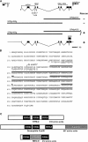
The sym-2 gene and its predicted product. (A) Two subclones of ZK1067 rescued when co-injected but not when individually injected, implicating ZK1067.6 as sym-2. The genomic positions of the eight exons of the gene, determined from cDNA sequences from 5′-RLM-RACE and from 3′-RACE, are indicated relative to the 5′ and 3′ flanking genes, the terminal exons of which are shown at the extreme left and right. The position of the sym-2(mn617) mutation, the regions encoding copies of the RRM motif, and the region used for deriving dsRNA are also indicated. The ability of subclones to rescue the mec-8; sym-2 synthetic lethality is summarized below the schematic of the gene. A schematic of a GFP fusion (SP#jy424b) that rescued sym-2 is also shown. (B) The amino acid sequence predicted for the translation of sym-2. Thick lines over the sequence delimit the three copies of the RNA recognition motif, and the amino acid change associated with mn617 is indicated. Shown beneath parts of the third and fourth lines of the C. elegans sequence is the predicted amino acid sequence, based on data in WormBase, for the region of SYM-2 from C. briggsae that flanks the tyrosine affected by mn617, indicating that the tyrosine is conserved in C. briggsae. Identical amino acids are underlined. (C) A schematic comparison of SYM-2 with the Drosophila hnRNP F-like protein Fusilli (Wakabayashi-Ito et al. 2001) and with MEC-8 (Lundquist et al. 1996), which has a region rich in alanines and glutamines that is not present in SYM-2. The carboxyl-terminal third of Fusilli is also rich in these amino acids.
F<sc>igure</sc> 3.—
Figure 3.—
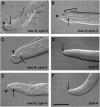
The phenotype of sym-3 or sym-4 in combination with mec-8(u74) or mec-8(+). (A–D) Nomarski images of sym-3 homozygotes. (E and F) Nomarski images of sym-4 homozygotes. In A–F, an arrow indicates the anterior end of the buccal cavity of the pharynx. The walls of the cavity appear as parallel lines in these optical sections, which are longitudinal, because of strong refraction of light caused by cuticle that forms the wall of the cavity. (A–C) mec-8; sym-3 double mutants. (A) The anterior end of the pharynx appears to be fully formed but is not properly connected to the anterior tip (arrowhead) of the animal. Overall, the morphology of the pharynx, which is shown in its entirety, appears to be normal. (B) The “bulbous nose” (indicated with a bracket) associated with mec-8; sym-3 (and mec-8; sym-4) double mutants is particularly apparent in this animal. As in A, the pharynx is not properly attached to the body. (C) An example of a malformed, asymmetric anterior end of the body, which is sometimes seen in the double mutant. The arrowhead indicates refraction caused by an accumulation of material. The anterior end of the pharynx (arrow), which is in a different focal plane, is not properly attached to the body. (D) The wild-type phenotype of a sym-3 homozygote in a mec-8(+) background. Note the orifice formed by the proper junction of the anterior end of the pharynx with the body cuticle. (E) The failure of proper attachment of the pharynx to the outer body in a mec-8; sym-4 double mutant. As in C, the anterior end of the body is asymmetric. (F) The wild-type phenotype of a sym-4 homozygote in a mec-8(+) background. Bar (pertains to A–F), 25 μm.
F<sc>igure</sc> 4.—
Figure 4.—
Abnormal morphology at the junction of the hypodermis and pharyngeal primordium in embryos. (A) A pronounced invagination (arrow) at the anterior end of a mec-8; sym-3 mutant embryo. The basement membrane of the pharyngeal primordium is evident (arrowheads). (B) An apparently normal invagination (arrow) at the anterior end of a mec-8(); sym-3(+) sibling of the embryo in A. The focal plane with the maximum invagination is shown. The dorsal part of the pharyngeal primordium can be seen (arrowhead). (C) The invagination of a wild-type N2 embryo. (D) A mec-8; sym-3 embryo later in development exhibiting extreme abnormality at the anterior end. The buccal cavity (arrowhead) is grossly recessed from the anterior tip of the embryo (arrow). A canal may exist between the anterior tip and the buccal cavity. In contrast, the anterior ends of normal embryos at this stage resemble those of the larvae shown in Figure 3, D and F. Bar, 25 μm.
F<sc>igure</sc> 5.—
Figure 5.—
Identification of the sym-3 gene (C54H2.1). (A) The ability of regions of the cosmid C54H2 (C. elegans Sequencing Consortium 1998) to rescue sym-3 on the basis of micro-injection of long-range PCR products. (B) The relative positions of the exons for the two forms of the gene and the location of sym-3(mn618) in sym-3a are indicated. The exons used for derivation of dsRNA are also indicated. (C) On the basis of DNA sequencing of RT-PCR products, the mn618 mutation of the splice acceptor of exon 7 results in the use of a cryptic acceptor and subsequent truncation of the open reading frame. Intron sequences are shown in lowercase.
F<sc>igure</sc> 6.—
Figure 6.—
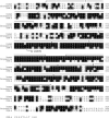
The amino-terminal half but not the carboxyl-terminal half of SYM-3A is similar to the amino-terminal parts of proteins of unknown function from other species. The amino acid sequences of proteins from Drosophila (GenBank no. NM136259), mosquito (Anopheles gambiae; GenBank XM-318591), and mouse (GenBank NM 153560) are aligned. The proteins, which are shown in their entirety, are longer than SYM-3A and resemble each other outside the region of similarity with SYM-3A. The proteins least resemble each other in their middle parts, which correspond to the carboxyl-terminal half of SYM-3A. The location and consequence of the mn618 mutation are indicated. The fourth row of the alignment includes the final four amino acids predicted for SYM-3B, which is otherwise identical with SYM-3A up to this point.
F<sc>igure</sc> 7.—
Figure 7.—
Identification of the sym-4 gene (R03E1.1). (A) The position of the genes for the rescuing cosmid R03E1 (C. elegans Sequencing Consortium 1998) and the ability of long-range PCR products to rescue sym-4. (B) A schematic of the exons of the gene and the location of the sym-4(mn619) mutation. The region of the gene that encodes the seven WD repeats and the exons used for derivation of dsRNA are indicated.
F<sc>igure</sc> 8.—
Figure 8.—
A comparison of the carboxyl-terminal half of SYM-4 with the carboxyl-terminal halves of WD-repeat proteins of unknown function from human (GenBank accession no. NP-061918) and from Drosophila melanogaster (NP 651742). The seven predicted WD repeats are designated. Note that strong resemblance extends over several adjacent copies of the WD motif and that the mn619 mutation affects a conserved amino acid in the region of strong resemblance.
F<sc>igure</sc> 9.—
Figure 9.—
Possible overgrowth of tissue at the anterior end of a mec-8; sym-3 double mutant. (A) The normal junction of the pharynx with the outer body as seen in the sym-3 single mutant of Figure 3D. The anterior and posterior extremes of the buccal cavity are indicated. (B) A schematic indicating the normal locations of the apical parts of the epithelia (Wright and Thomson 1981; Sulston et al. 1983) at the junction of the pharynx and the anterior tip of the body. The cuticle of the body, of the buccal cavity, and of the pharynx is indicated by a thick black line. The basement membrane of the pharynx is shaded. (C) The mec-8; sym-3 double mutant shown in Figure 3B. The arrows indicate a line of refractivity that tapers in the anterior direction beneath what appears to be an overgrowth of hypodermal material at the anterior end. (D) A schematic of the bulbous nose in C, with a thin black line indicating the tapered refractivity. Reminiscent of a normal anterior end, the thin line appears to be associated with the buccal cavity.

Similar articles

Cited by

References

    1. Altschul, S. F., T. L. Madden, A. A. Schaffer, J. Zhang, Z. Zhang et al., 1997. Gapped BLAST and PSI-BLAST: a new generation of protein database search programs. Nucleic Acids Res. 25: 3389–3402. - PMC - PubMed
    1. Aroian, R. V., and P. W. Sternberg, 1991. Multiple functions of let-23, a Caenorhabditis elegans receptor tyrosine kinase gene required for vulval induction. Genetics 128: 251–267. - PMC - PubMed
    1. Aroian, R. V., M. Koga, J. E. Mendel, Y. Ohshima and P. W. Sternberg, 1990. The let-23 gene necessary for Caenorhabditis elegans vulval induction encodes a tyrosine kinase of the EGF receptor subfamily. Nature 348: 693–699. - PubMed
    1. Bender, A., and J. R. Pringle, 1991. Use of a screen for synthetic lethal and multicopy suppressee mutants to identify two new genes involved in morphogenesis in Saccharomyces cerevisiae. Mol. Cell. Biol. 11: 1295–1305. - PMC - PubMed
    1. Blumenthal, T., and K. Steward, 1997 RNA processing and gene structure, pp. 117–145 in C. elegans II, edited by D. L. Riddle, T. Blumenthal, B. J. Meyer and J. R. Priess. Cold Spring Harbor Laboratory Press, Plainview, NY. - PubMed

Publication types

MeSH terms

Associated data

LinkOut - more resources