. 2004 Dec;24(24):10558-72.
doi: 10.1128/MCB.24.24.10558-10572.2004.

Brk activates rac1 and promotes cell migration and invasion by phosphorylating paxillin

Affiliations

Brk activates rac1 and promotes cell migration and invasion by phosphorylating paxillin

Hsin-Yi Chen et al. Mol Cell Biol. 2004 Dec.

Abstract

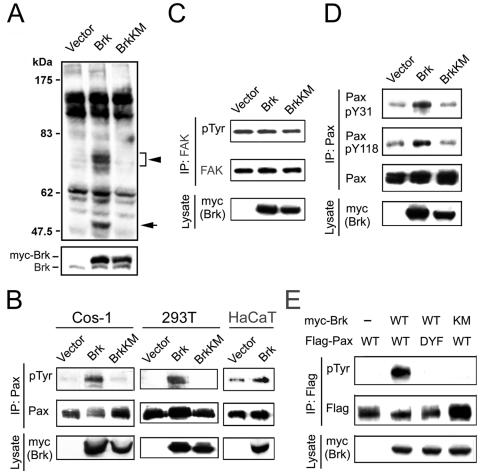
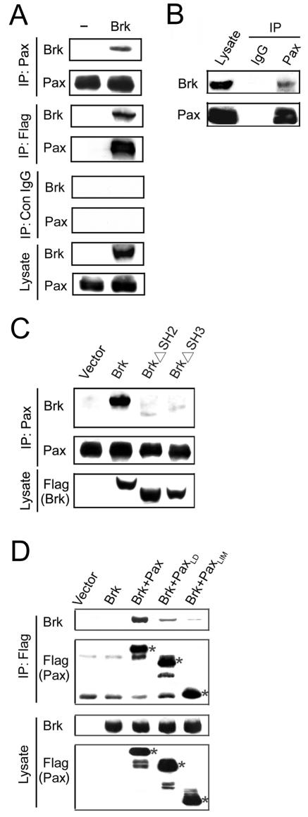
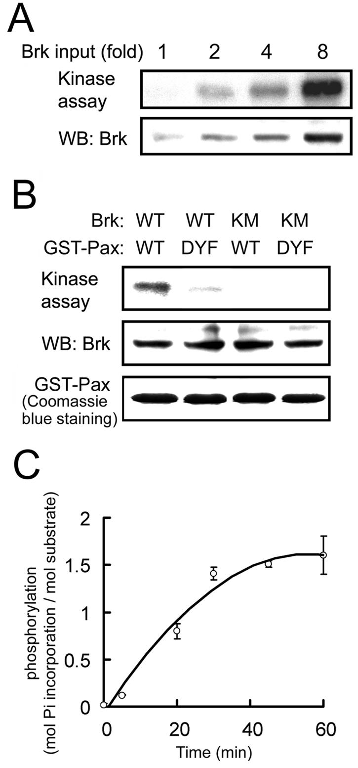
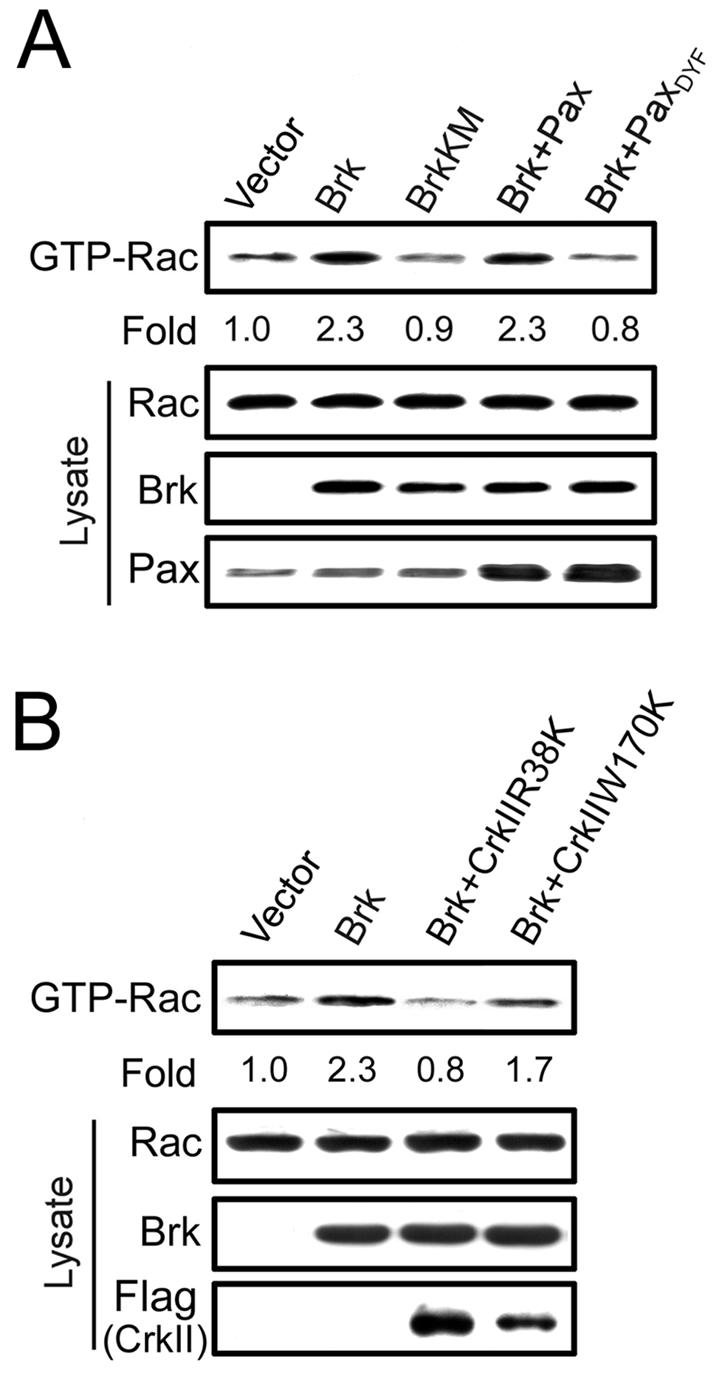
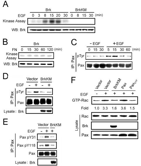
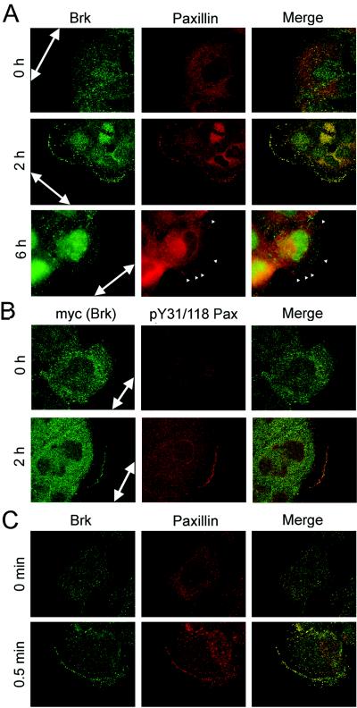
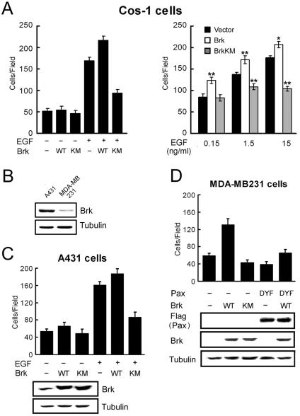
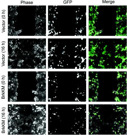
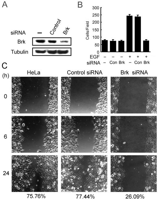
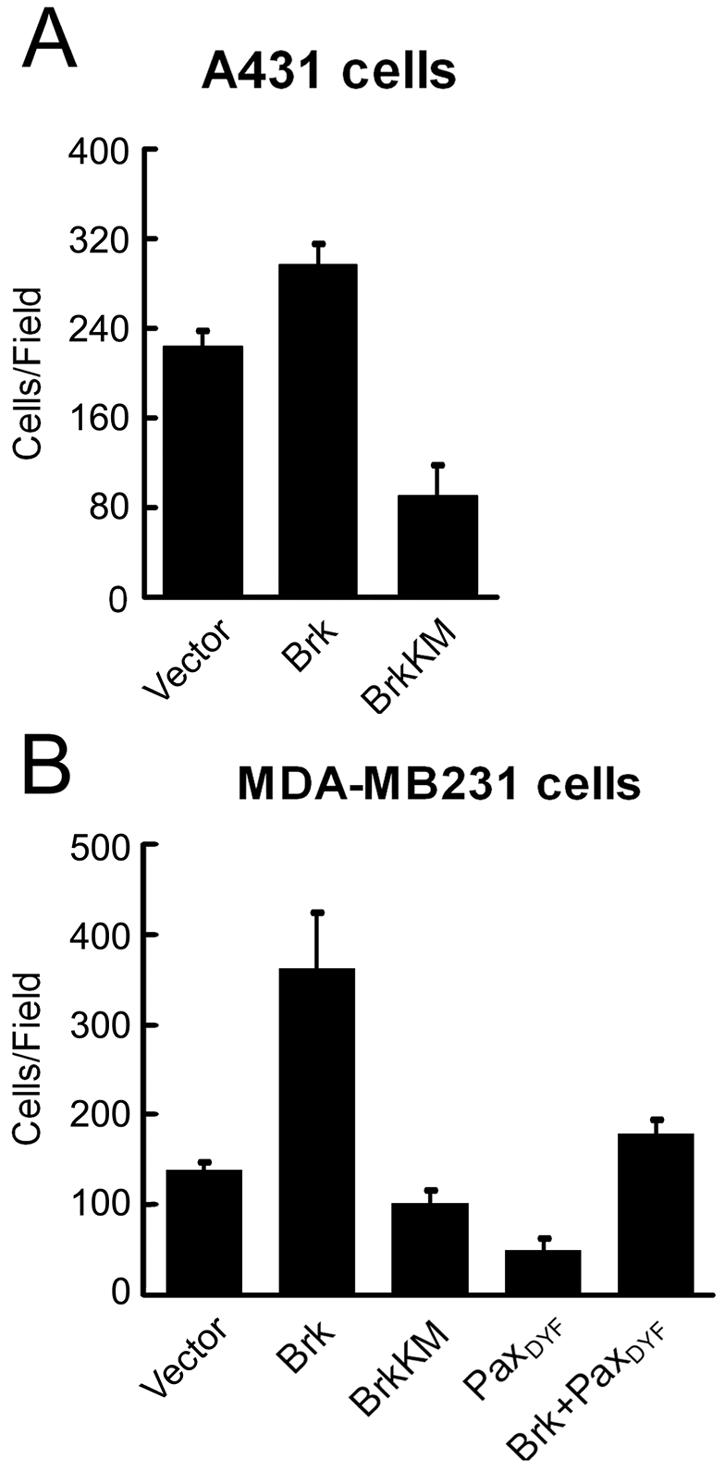
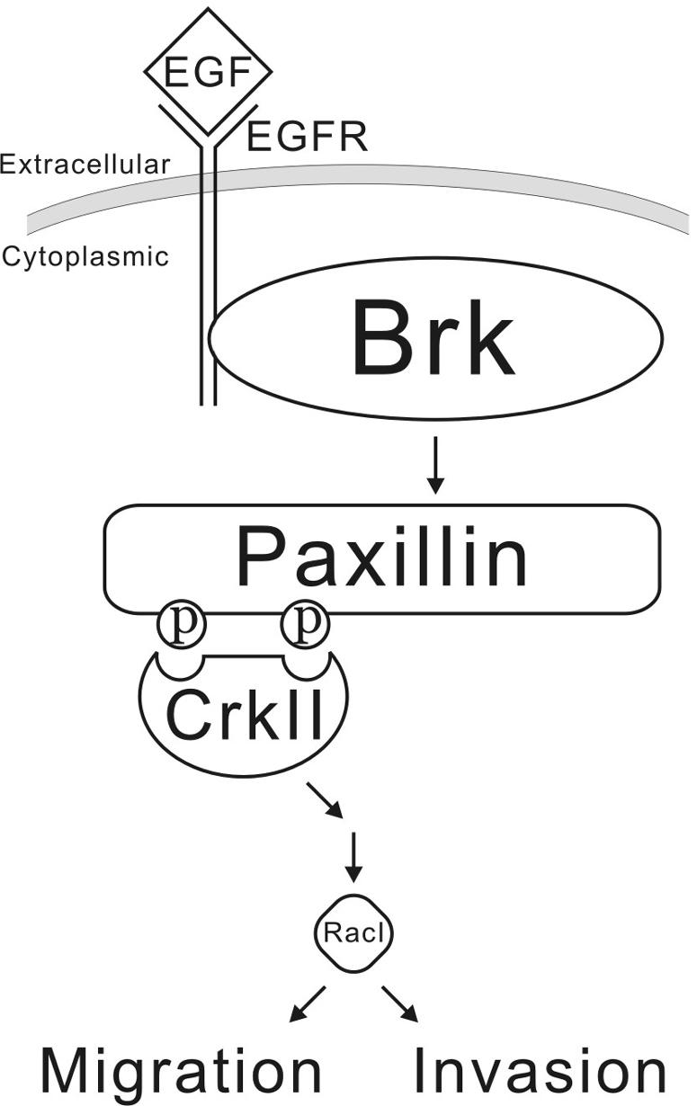
Brk (for breast tumor kinase) is a nonreceptor tyrosine kinase containing SH3, SH2, and tyrosine kinase catalytic domains. Brk was originally identified from a human metastatic breast tumor, and its overexpression is frequently observed in breast cancer and several other cancer types. However, the molecular mechanism by which this kinase participates in tumorigenesis remains poorly characterized. In the present study, we not only identified paxillin as the binding partner and substrate of Brk but also discovered a novel signaling pathway by which Brk mediates epidermal growth factor (EGF)-induced paxillin phosphorylation. We show that EGF stimulation activates the catalytic activity of Brk, which in turn phosphorylates paxillin at Y31 and Y118. These phosphorylation events promote the activation of small GTPase Rac1 via the function of CrkII. Through this pathway, Brk is capable of promoting cell motility and invasion and functions as a mediator of EGF-induced migration and invasion. In accordance with these functional roles, Brk translocates to membrane ruffles, where it colocalizes with paxillin during cell migration. Together, our findings identify novel signaling and biological roles of Brk and indicate the first potential link between Brk and metastatic malignancy.


Figures

FIG. 1.
FIG. 1.
Brk promotes paxillin phosphorylation in vivo. (A) HaCaT cells stably expressing Brk or BrkKM or a control vector were lysed, and tyrosine phosphorylated proteins were detected by Western blotting with anti-phosphotyrosine antibody (upper panel). The expressions of endogenous and exogenous Brk (myc-Brk) are shown in the bottom panel. The positions of protein bands ∼68 kDa and the tyrosine phosphorylated Brk are indicated by an arrowhead and an arrow, respectively. (B) Cos-1 and 293T cells transfected with myc-Brk, myc-BrkKM, or a control vector or HaCaT cells infected with retroviruses carrying Brk or control vector were serum starved and lysed. Cell lysates were subjected to immunoprecipitation with antipaxillin (Pax) antibody, followed by Western blotting with the antibodies indicated. The expression of overexpressed Brk is shown on the bottom. (C) Cos-1 cells transfected and cultured as in panel B were lysed for immunoprecipitation with anti-FAK antibody, followed by Western blotting with the indicated antibodies. The expression of overexpressed Brk is shown on the bottom. (D) Cos-1 cells transfected and cultured as in panel B were assayed for paxillin tyrosine phosphorylation at Y31 and Y118. The immunocomplexes or cell lysates were analyzed by Western blotting with the indicated antibodies. (E) Cos-1 cells were transfected with myc-Brk, Flag-paxillin or their mutants as indicated. Serum-starved cells were lysed and subjected to immunoprecipitation with anti-Flag antibody, followed by Western blotting with antibody to phosphotyrosine (pTyr) (upper panel) or Flag (middle panel). The expressions of myc-Brk and myc-BrkKM are shown in the bottom panel.
FIG. 2.
FIG. 2.
Brk interacts with paxillin. (A) Interaction of Brk and paxillin in vivo. Cos-1 cells transfected with or without (−) Flag-Brk were subjected to immunoprecipitation with antipaxillin antibody, anti-Flag antibody, or control antibody as indicated. The immunocomplexes and cell lysates were resolved by SDS-PAGE and analyzed by Western blots with antibodies as indicated. (B) Brk and paxillin interact endogenously. A431 cells were lysed and subjected to immunoprecipitation with antipaxillin antibody or a control immunoglobulin G. The immunocomplexes and cell lysate were analyzed by Western blotting with anti-Brk or antipaxillin antibody as indicated. (C) The SH2 and SH3 domains of Brk are both involved in paxillin binding. Cos-1 cellstransfected with various Flag-tagged Brk constructs or a control vector were subjected to coimmunoprecipitation analysis as in panel B. The expressions of Brk and its deletion mutants in transfected cells are shown on the bottom. (D) Both N-terminal and C-terminal segments of paxillin are involved in Brk binding. Cos-1 cells were transfected with Flag-tagged full-length paxillin, its N-terminal segment (PaxLD), or C-terminal segment (PaxLIM), together with myc-tagged Brk. Cell lysates were used for immunoprecipitations with anti-Flag antibody, followed by Western blotting with anti-Flag or anti-Brk. The expressions of Brk and Flag-tagged paxillin or its deletion mutants are shown on the bottom. The bands corresponding to Flag-paxillin, Flag-PaxLIM, and Flag-PaxLD are indicated by asterisks.
FIG. 3.
FIG. 3.
Brk directly phosphorylates paxillin at Y31 and Y118. (A) Equal amounts of GST-paxillin purified from bacteria were incubated with increasing amounts of purified baculovirus-expressed Brk in in vitro kinase reactions. The reaction products were analyzed by autoradiography to detect GST-paxillin phosphorylation (upper panel) or by Western blotting with anti-Brk antibody (bottom panel). (B) GST-paxillin or GST-paxillinDYF mutant was phosphorylated by Brk or BrkKM as described for panel A. The equal input of GST fusion proteins is shown on the bottom. (C) The stoichiometry of GST-paxillin phosphorylation by Brk was determined as described in Materials and Methods. The data shown are means ± the standard deviations from three independent experiments.
FIG. 4.
FIG. 4.
Brk promotes Rac1 activation through paxillin phosphorylation and CrkII adaptor function. (A and B) Cos-1 cells transfected with myc-Brk, myc-BrkKM, paxillin, paxillinDYF, and/or Flag-CrkII mutant constructs as indicated were serum starved and then lysed. The amount of GTP-bound Rac1 in cell lysates was determined by GST-PAK-CRIB pull-down analysis, followed by Western blotting with anti-Rac1 antibody. The expression levels of Rac1, Brk, paxillin, or their mutants in cell lysates were determined by Western blotting (bottom panels). The amounts of GTP-bound Rac1 were normalized by using those of total Rac1 protein present in cell lysates and are expressed as the fold induction relative to cells transfected with the control vector.
FIG. 5.
FIG. 5.
Brk is activated by EGF signaling and is a mediator of EGF-induced paxillin phosphorylation and Rac1 activation. (A) Cos-1 cells transfected with Flag-Brk or Flag-BrkKM were serum starved and then stimulated with 15 ng of EGF/ml for various time points. The Flag-Brk or Flag-BrkKM was precipitated from cell lysates and subjected to in vitro kinase assays to detect Brk autophosphorylation. An equal input of Brk is shown in the bottom panel. (B) Cos-1 cells transfected with Flag-Brk were serum starved and then reseeded on plates coated with 1 μg of fibronectin/ml for various times as indicated. Cells were harvested to determine Brk catalytic activity as described for panel A. (C) EGF induces paxillin tyrosine phosphorylation in Cos-1 cells reseeded on fibronectin. Serum-starved Cos-1 cells were detached from culture plates, kept in suspension for 30 min, and then reseeded on plates coated with 1 μg of fibronectin/ml for 3 h. The cells were then treated with (+) or without (−) 15 ng of EGF/ml for various times as indicated. Paxillin tyrosine phosphorylation was analyzed as described for Fig. 1. (D and E) Brk activity is critical for EGF-induced paxillin tyrosine phosphorylation. Cos-1 cells transfected with BrkKM or a control vector were serum starved and reseeded on fibronectin as described for panel C and then treated with 15 ng of EGF/ml for 15 min. Paxillin tyrosine phosphorylation (D) and phosphorylation at Y31 and Y118 (E) were detected as described in Fig. 1. (F) Brk and paxillin phosphorylation are important for EGF-induced Rac1 activation. Cos-1 cells transfected with BrkKM, paxillin, paxillinDYF mutant, or a control vector were starved, reseeded, and stimulated with EGF as in panel D. GTP-bound Rac1 was detected and then calculated as the induction relative to unstimulated cells as described for Fig. 4. The expression levels of various proteins in cell lysates were determined by Western blotting (bottom panels).
FIG. 6.
FIG. 6.
Brk and paxillin colocalize at membrane ruffles but not at focal adhesions in migratory cells. (A) Confluent A431 cells were scratched to induce wounds and fixed at various times after wounding. Endogenous Brk or paxillin was detected by immunofluorescence staining with anti-Brk (green) or antipaxillin (red) antibody. Green, red, and merged images are shown. At 2 h after wounding, cells displayed membrane ruffles, whereas certain cells at 6 h after wounding formed prominent focal adhesions (arrowheads). Confocal images are shown for cells at 0 and 2 h after wounding, whereas cells at 6 h after wounding were examined by fluorescence microscopy to visualize focal adhesions. The large arrows indicate the direction of the wounds. (B) A431 cells stably expressing myc-Brk were scratched to induce migration as for panel A and then double stained with anti-myc and anti-pY31/118 paxillin (1:1 mixture of the pY31-paxillin and pY118-paxillin antibodies). Confocal images were captured and analyzed as in panel A. (C) Serum-starved A431 cells were stimulated with EGF for indicated times. The locations of endogenous paxillin and Brk were detected by immunostaining as in panel A and examined by confocal microscopy.
FIG. 7.
FIG. 7.
Brk promotes cell migration and mediates EGF-induced motility through phosphorylating paxillin at Y31 and Y118. (A) Cos-1 cells cotransfected with Brk, BrkKM, or a control vector, together with GFP (10:1 ratio), were plated onto Transwell chambers and assayed for migration into lower chamber containing 0 (−EGF) or 15 ng of EGF/ml (left panel) or different concentrations of EGF as indicated (right panel). After 4 h for migration, the GFP-positive cells that migrated to the lower side of the chamber were counted. For each well, five randomly selected fields were analyzed, and the data shown are means ± standard deviations of triplicate experiments (∗, P < 0.005; ∗∗, P < 0.0005 [compared to the control vector]). (B) The expression levels of endogenous Brk in A431 and MDA-MB231 cells were assessed by Western blotting. (C and D) A431 (C) and MDA-MB231 (D) cells were infected with various recombinant retroviruses as indicated. Infected cells were selected by using puromycin and/or hygromycin and then assayed for basal migration (−EGF) or chemotaxis migration toward 1.5 ng/ml (for A431 cells) or 15 ng/ml (for MDA-MB231 cells) of EGF. After 3 h (for A431 cells) or 5 h (for MDA-MB231 cells), the cells that migrated to the lower side of the chamber were counted, and values were calculated as for panel A. The expression levels of endogenous and exogenous Brk, Flag-paxillin, and their mutants are shown in the bottom panels.
FIG. 8.
FIG. 8.
Time-lapse monitoring of the wound-healing migration of cells expressing BrkKM. Cos-1 cells cotransfected with GFP and BrkKM or a control vector (ratio of 1:10) were cultured to confluence in growth medium and then scratched. Cell migration into wounds was monitored by time-lapse videomicroscopy. Still images were captured at the indicated time after wounding. Cells carrying BrkKM or control vector were indicated by their GFP fluorescence. The time after scratching is shown on the left.
FIG. 9.
FIG. 9.
siRNA-mediated reduction of Brk interferes with cell migration. (A) HeLa cells transfected with or without Brk siRNA or a control siRNA were lysed at 72 h after transfection and then assayed for the expression of Brk by Western blotting. (B) Cells as described for panel A were assayed for chemotaxis migration toward 15 ng of EGF/ml. After 7 h, cells that migrated to the lower side of the chamber were counted and calculated. (C) Cells as in panel A were assayed for wound-healing migration as described for Fig. 8. The percentages of wound closure at 24 h after wounding are indicated on the bottom.
FIG. 10.
FIG. 10.
Brk promotes cell invasion. A431 (A) and MDA-MB231 (B) cells stably expressing various proteins as indicated were assayed for invasion through Matrigel as described in Materials and Methods. The data shown are means ± standard deviations of triplicate experiments.
FIG. 11.
FIG. 11.
Model illustrating the signaling pathway through which Brk participates in EGF-induced cell migration and invasion.

Similar articles

Cited by

References

    1. Avraham, H., S. Y. Park, K. Schinkmann, and S. Avraham. 2000. RAFTK/Pyk2-mediated cellular signalling. Cell Signal 12:123-133. - PubMed
    1. Bagrodia, S., S. J. Taylor, K. A. Jordon, L. Van Aelst, and R. A. Cerione. 1998. A novel regulator of p21-activated kinases. J. Biol. Chem. 273:23633-23636. - PubMed
    1. Barker, K. T., L. E. Jackson, and M. R. Crompton. 1997. BRK tyrosine kinase expression in a high proportion of human breast carcinomas. Oncogene 15:799-805. - PubMed
    1. Bar-Sagi, D., and A. Hall. 2000. Ras and Rho GTPases: a family reunion. Cell 103:227-238. - PubMed
    1. Bellis, S. L., J. T. Miller, and C. E. Turner. 1995. Characterization of tyrosine phosphorylation of paxillin in vitro by focal adhesion kinase. J. Biol. Chem. 270:17437-17441. - PubMed

Publication types

MeSH terms