. 2004 Dec;24(23):10223-35.
doi: 10.1128/MCB.24.23.10223-10235.2004.

Nuclear speckle-associated protein Pnn/DRS binds to the transcriptional corepressor CtBP and relieves CtBP-mediated repression of the E-cadherin gene

Affiliations

Nuclear speckle-associated protein Pnn/DRS binds to the transcriptional corepressor CtBP and relieves CtBP-mediated repression of the E-cadherin gene

Roman Alpatov et al. Mol Cell Biol. 2004 Dec.

Abstract

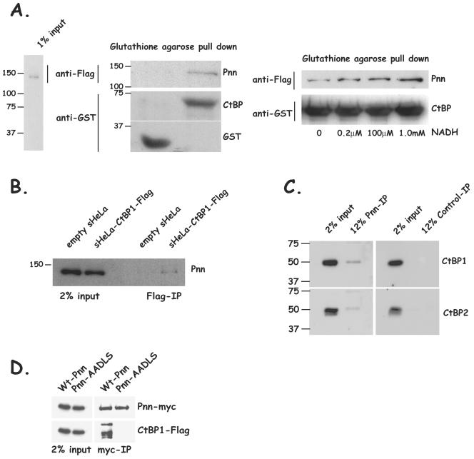
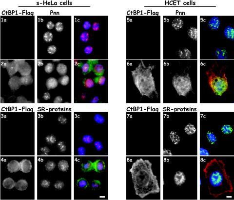
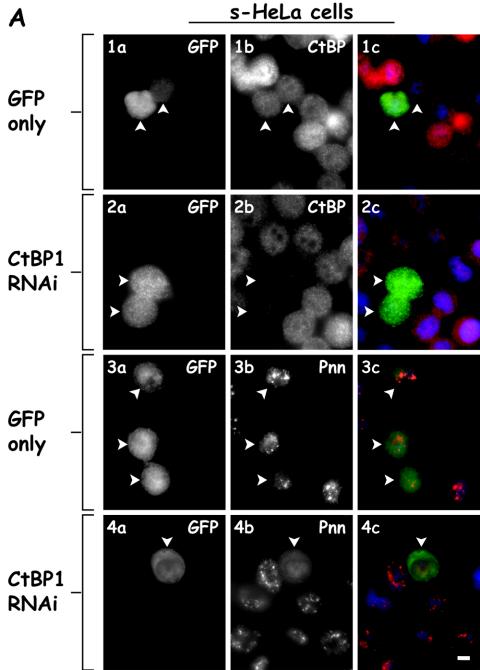
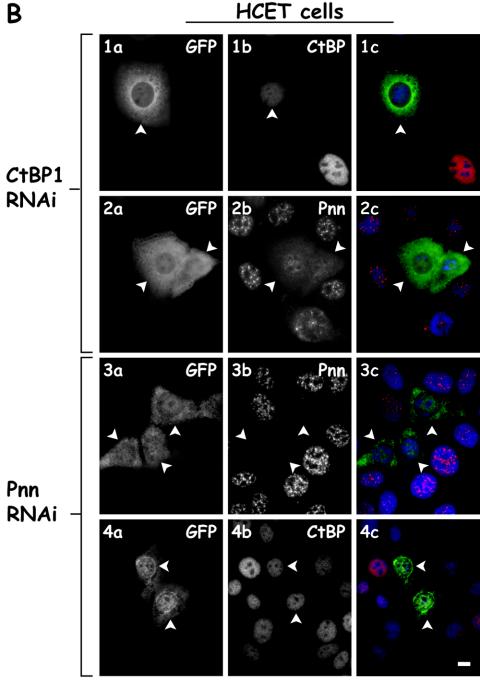
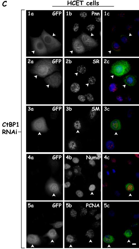
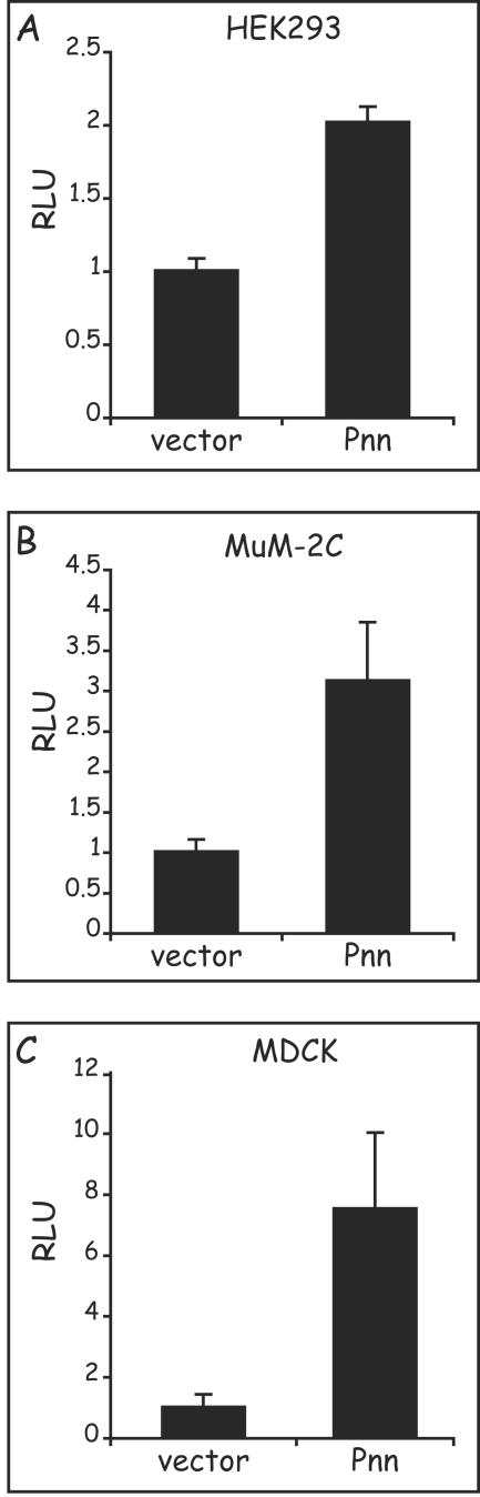
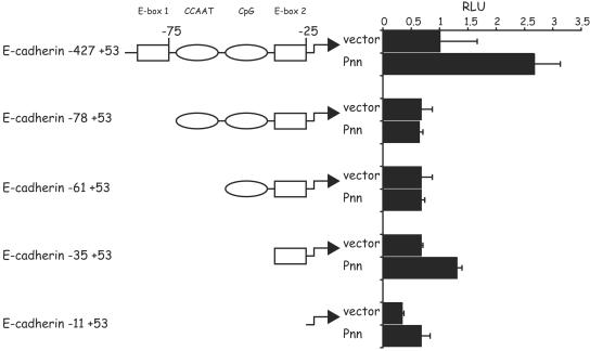
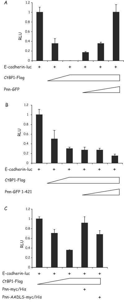
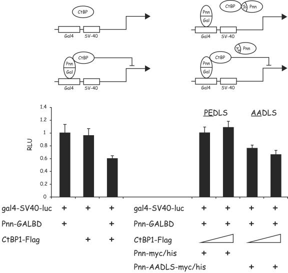
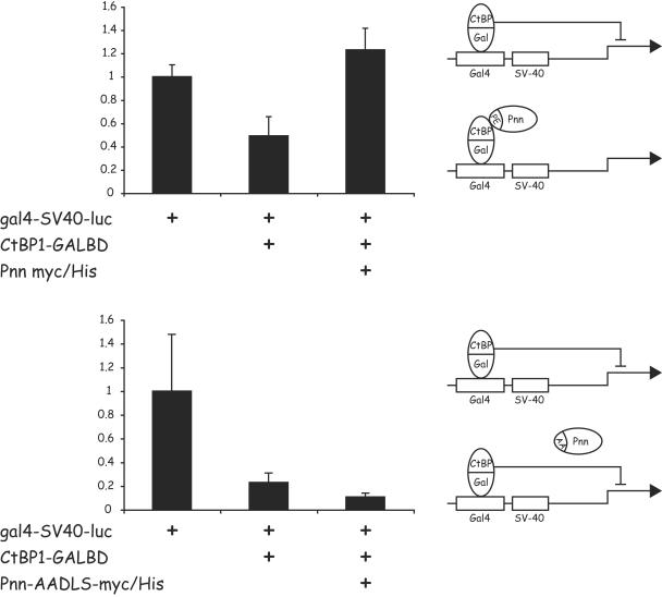
Previously, we have shown that pinin/DRS (Pnn), a 140-kDa nuclear and cell adhesion-related phosphoprotein, is involved in the regulation of cell adhesion and modulation of the activity of multiple tumor suppressor genes. In the nucleus Pnn is concentrated in the "nuclear speckles," zones of accumulation of transcriptional and mRNA splicing factors, where Pnn is involved in mRNA processing. Alternatively, other roles of Pnn in gene regulation have not yet been established. By utilizing in vitro pull-down assays, in vivo interaction studies, and immunofluorescence in combination with overexpression and RNA interference experiments, we present evidence that Pnn interacts with the known transcriptional corepressor CtBP1. As a consequence of this interaction Pnn was capable of relieving the CtBP1-mediated repression of E-cadherin promoter activity. Our results suggest that the interaction of Pnn with the corepressor CtBP1 may modulate repression of transcription by CtBP1. This interaction may reflect the existence of coupling factors involved in CtBP-mediated transcriptional regulation and mRNA processing events.


Figures

FIG. 1.
FIG. 1.
Pnn interacts with corepressor CtBP1 in vitro and in vivo. (A) CtBP1-GST fusion protein or GST alone immobilized on glutathione-agarose beads was incubated with the nuclear extracts from s-HeLa cells stably expressing Pnn-Flag-HA fusion protein (s-HeLa-POZ-N-Pnn-Flag-HA) as shown in the nuclear extract input (left panel). The bound material was eluted and evaluated for the presence of Pnn by Western blotting with anti-Flag antibody. The presence of CtBP1-GST or GST was confirmed by using anti-GST antibody. Pnn was found to be associated with CtBP1-GST beads but not beads coupled to GST alone (center panel). CtBP1-GST fusion protein immobilized on glutathione-agarose beads was incubated with s-HeLa-POZ-N-Pnn-Flag-HA nuclear extract in the presence of increasing concentrations of NADH. Bound material from each binding reaction was evaluated for the presence of Pnn with anti-Flag antibody. The presence of CtBP1-GST was confirmed with anti-GST antibody. An increased concentration of NADH resulted in the increased association of CtBP1 with Pnn (right panel). (B) CtBP1-associated complex of proteins was obtained by passing POZ-CtBP1-Flag-HA s-HeLa nuclear extract or the control s-HeLa cell extract over a Flag-M2 column, followed by washes and elution with SDS loading buffer. The bound material was analyzed for the presence of Pnn. Flag-bound material from s-HeLa CtBP1-Flag-HA nuclear extracts but not s-HeLa control nuclear extracts showed the presence of Pnn. (C) Pnn-associated proteins were obtained by coimmunoprecipitation from s-HeLa nuclear lysate with anti-Pnn 143 mouse hybridoma supernatant or control supernatant, followed by washes and elution with SDS loading buffer. Eluted proteins were immunostained for CtBP1 and CtBP2. Both CtBP1 and CtBP2 were found in the Pnn eluate but not in the control eluate. (D) HEK293 cells were transiently cotransfected with CtBP1-Flag expression vector and Pnn-myc/His construct or Pnn-AADLS-myc/His, carrying a PE→AA substitution in the putative CtBP1 interaction motif. Coimmunoprecipitations were performed by using anti-myc agarose affinity gel, followed by Western blotting of the precipitated material with anti-myc and anti-Flag antibodies. Wild type but not Pnn-AADLS was capable of interacting with CtBP1. Wt, wild type.
FIG. 2.
FIG. 2.
Pnn exhibits nuclear redistribution in response to CtBP1 overexpression. (Rows 1 and 2) s-HeLa cells containing empty POZ vector (row 1) or s-HeLa cells stably expressing CtBP1-Flag-HA fusion protein (POZ-CtBP1-Flag-HA) (row 2) were immunostained for CtBP1 with rabbit anti-Flag antibody (panels 1a and 2a) and 143 antibody against the endogenous Pnn (panels 1b and 2b). Panels 1c and 2c show merged DAPI nuclear stain (blue), Pnn (red), and CtBP1 (green) images. (Rows 5 and 6) HCETs were transiently transfected with the CtBP1-Flag expression vector and localization of CtBP1 and Pnn was assessed by immunofluorescence with rabbit anti-Flag antibody (panels 5a and 6a) and 143 antibody to the endogenous Pnn (panels 5b and 6b). Panels 5c and 6c show merged DAPI nuclear stain (blue), Pnn (green), and CtBP1 (red) images. Rows 3 and 4 are the same as rows 1 and 2 and rows 7 and 8 are the same as rows 5 and 6 except, instead of Pnn, cells were immunostained for the endogenous SR proteins with mouse anti-SR proteins antibody. Both Pnn and SR proteins demonstrated nuclear redistribution in response to the CtBP1 overexpression. Bar, 10 μm.
FIG. 3.
FIG. 3.
Nuclear distribution of Pnn is altered in response to RNAi-mediated CtBP1 knockdown. (A) s-HeLa cells were transiently cotransfected with a targeting vector expressing short hairpin RNA (shRNA) directed against CtBP1 mRNA coding region and the GFP vector to identify the transfected cells (rows 2 and 4) or the GFP vector alone as a control (rows 1 and 3). Cells were immunostained with mouse anti-CtBP1 antibody (panels 1b and 2b) or 143 antibody against the endogenous Pnn (panels 3b and 4b). Panels 1c to 4c show merged DAPI nuclear stain (blue), GFP (green), and Pnn or CtBP (red) images. Arrowheads indicate the transfected cells. Bar, 10 μm. (B) Rows 1 and 2 show HCETs that were transiently cotransfected with a targeting vector expressing short hairpin RNA (shRNA) directed against CtBP1 mRNA coding region and GFP vector to identify the trasfected cells. Cells were immunostained with mouse anti-CtBP1 antibody (panel 1b) and 143 antibody to the endogenous Pnn (panel 2b). Cells that received CtBP1 shRNAi vector exhibited drastic Pnn redistribution from nuclear speckles. Rows 3 and 4 show HCETs that were transiently cotransfected with a targeting vector expressing short hairpin RNA (shRNA) directed against Pnn mRNA coding region and a GFP vector to identify the transfected cells. Cells were immunostained with 143 antibody to the endogenous Pnn (panel 3b) or mouse anti-CtBP1 antibody (panel 4b). Panels 1c to 4c show merged DAPI nuclear stain (blue), GFP (green), and Pnn or CtBP (red) images. In contrast to the CtBP1 RNAi-dependent Pnn relocalization, RNAi-mediated Pnn knockdown did not result in visible CtBP1 redistribution. Arrowheads indicate the transfected cells. Bar, 10 μm. (C) HCETs were transiently cotransfected with a targeting vector expressing short hairpin RNA (shRNA) directed against CtBP1 mRNA coding region and GFP vector to identify the transfected cells. Cells were immunostained with the following antibodies: anti-Pnn (1b), anti-SR proteins (2b), anti-sm proteins (3b), anti-NuMa (4b), and anti-PCNA (5b). Panels 1c to 5c show merged DAPI nuclear stain (blue), GFP (green), and Pnn, SR, sm, NuMa, and PCNA (red) images. Arrowheads indicate the transfected cells. Bar, 10 μm.
FIG. 3.
FIG. 3.
Nuclear distribution of Pnn is altered in response to RNAi-mediated CtBP1 knockdown. (A) s-HeLa cells were transiently cotransfected with a targeting vector expressing short hairpin RNA (shRNA) directed against CtBP1 mRNA coding region and the GFP vector to identify the transfected cells (rows 2 and 4) or the GFP vector alone as a control (rows 1 and 3). Cells were immunostained with mouse anti-CtBP1 antibody (panels 1b and 2b) or 143 antibody against the endogenous Pnn (panels 3b and 4b). Panels 1c to 4c show merged DAPI nuclear stain (blue), GFP (green), and Pnn or CtBP (red) images. Arrowheads indicate the transfected cells. Bar, 10 μm. (B) Rows 1 and 2 show HCETs that were transiently cotransfected with a targeting vector expressing short hairpin RNA (shRNA) directed against CtBP1 mRNA coding region and GFP vector to identify the trasfected cells. Cells were immunostained with mouse anti-CtBP1 antibody (panel 1b) and 143 antibody to the endogenous Pnn (panel 2b). Cells that received CtBP1 shRNAi vector exhibited drastic Pnn redistribution from nuclear speckles. Rows 3 and 4 show HCETs that were transiently cotransfected with a targeting vector expressing short hairpin RNA (shRNA) directed against Pnn mRNA coding region and a GFP vector to identify the transfected cells. Cells were immunostained with 143 antibody to the endogenous Pnn (panel 3b) or mouse anti-CtBP1 antibody (panel 4b). Panels 1c to 4c show merged DAPI nuclear stain (blue), GFP (green), and Pnn or CtBP (red) images. In contrast to the CtBP1 RNAi-dependent Pnn relocalization, RNAi-mediated Pnn knockdown did not result in visible CtBP1 redistribution. Arrowheads indicate the transfected cells. Bar, 10 μm. (C) HCETs were transiently cotransfected with a targeting vector expressing short hairpin RNA (shRNA) directed against CtBP1 mRNA coding region and GFP vector to identify the transfected cells. Cells were immunostained with the following antibodies: anti-Pnn (1b), anti-SR proteins (2b), anti-sm proteins (3b), anti-NuMa (4b), and anti-PCNA (5b). Panels 1c to 5c show merged DAPI nuclear stain (blue), GFP (green), and Pnn, SR, sm, NuMa, and PCNA (red) images. Arrowheads indicate the transfected cells. Bar, 10 μm.
FIG. 4.
FIG. 4.
Pnn is capable of upregulation of the E-cadherin promoter activity, independent of cell type origin. MDCK, uveal melanoma MuM-2C, and HEK293 cells were transiently cotransfected with 100 ng of E-cadherin luciferase reporter construct with either 500 ng of the Pnn-GFP expression vector or the GFP vector alone. The numerical data obtained from the luciferase readings was normalized to the values of the GFP vector alone control transfections, which represented a value of 1. In HEK293 and MuM-2C cells E-cadherin promoter exhibited 2- and 2.5-fold increases in luciferase activity in response to Pnn expression, respectively, whereas in MDCK cells a fourfold increase was observed.
FIG. 5.
FIG. 5.
Identification of the Pnn-responsive regions in the E-cadherin promoter. Each truncation of the E-cadherin promoter luciferase reporter is represented in the diagram, with the indicated distances from the transcriptional initiation site. Squares and ovals demarcate known regulatory elements within the promoter region. Bars on the right represent luciferase activity of each of the truncation construct in response to Pnn-myc/His expression (Pnn) or expression of the myc/His vector alone (vector). A total of 100 ng of each promoter construct was transiently cotransfected into HEK293 cells, along with 500 ng of the Pnn-myc/His expression vector or myc/His vector alone. The numerical data obtained from the luciferase readings was normalized to the values of the full-length E-cadherin promoter (−427+53) activity when cotransfected with the myc/His vector alone, which represented a value of 1. E-box 1 and E-box 2 regions, as well as the region adjacent to the transcriptional start site, demonstrated an increased activity in the presence of Pnn.
FIG. 6.
FIG. 6.
Wild-type Pnn, but not the truncated or mutant form of Pnn, is capable of relieving CtBP1-mediated repression of the E-cadherin promoter. (A) HEK293 cells were transiently cotransfected with the full-length E-cadherin luciferase reporter vector and increasing amounts of the CtBP1-Flag expression vector (0.1 and 1 μg). Parallel transfection reactions contained increasing concentrations of Pnn-GFP (0.01, 0.1, and 1 μg) in the presence of 1 μg of the CtBP1-Flag vector. (B) The same experiment was performed with Pnn-GFP1-421, a Pnntruncation mutant that lacks CtBP1 binding site. (C) HEK293 cells were transiently cotransfected with the full-length E-cadherin luciferase reporter vector and increasing amounts of the CtBP1-Flag expression vector (0.1 and 1 μg). Parallel transfection reactions contained 1 μg of the CtBP1-Flag vector and 1 μg of the Pnn-myc/His or Pnn-AADLS-myc/His vector carrying PE→AA point mutations in the Pnn's PEDLS CtBP1 binding motif. The numerical data obtained from the luciferase readings was normalized to the values of full-length E-cadherin reporter in the absence of CtBP1 or Pnn constructs, which represented a value of 1. In contrast to wild-type Pnn, Pnn1-421 deletion construct was not capable of relieving CtBP1-mediated repression of the E-cadherin promoter activity. The Pnn PE→AA mutant only partially relieved the promoter activity.
FIG. 7.
FIG. 7.
Pnn can recruit CtBP1 to a heterologous promoter, leading to a CtBP1-mediated repression of the promoter activity. (Left panel) HEK293 cells were transiently cotransfected with the luciferase reporter construct gal4-SV40-luc carrying GAL4 binding sites upstream of the SV40 constitutive promoter-enhancer and the following constructs: 100 ng of the Pnn-GALBD fusion construct alone and 1 μg of the CtBP1-Flag expression vector in the presence or absence of the Pnn-GALBD fusion construct (100 ng). gal4-SV40-luc activity was subject to CtBP1-mediated repression only in the presence of the Pnn-GALBD fusion construct, indicating that Pnn-CtBP1 interaction was responsible for this effect. (Right panel) HEK293 cells were transiently cotransfected with the gal4-SV40-luc reporter, carrying GAL4 biding sites upstream of the SV40 constitutive promoter/enhancer, 100 ng of the Pnn-GALBD fusion construct, and increasing concentrations of CtBP1-Flag (100 ng and 1 μg). In addition, transfection reactions contained 1 μg of the wild-type Pnn-myc/His or mutant form of this vector Pnn-AADLS-myc/His, carrying PE→AA substitution in the CtBP1 binding motif, as depicted in the diagram. The numerical data obtained from the luciferase readings was normalized to the value of gal4-SV40-luc reporter in the presence of Pnn-GALBD only, which represented a value of 1. The wild-type, but not mutant, form of Pnn was capable of competition with Pnn-GALBD, thereby relieving CtBP1-mediated repression of the reporter activity.
FIG. 8.
FIG. 8.
Pnn, but not the Pnn mutant carrying a mutation in the CtBP1 binding motif, is capable of relieving CtBP1-mediated repression of a heterologous promoter activity. HCETs were transiently transfected with the gal4-SV40-luc reporter construct carrying GAL4 binding sites upstream of the SV40 promoter-enhancer. Transfection reactions also included 1 μg of CtBP1-GALBD fusion vector alone, and in the presence of 1 μg of Pnn-myc/His construct (upper panel) or its mutant Pnn-AADLS-myc/His, carrying the PE→AA substitution in the PEDLS CtBP1 binding motif (lower panel). The numerical data obtained from the luciferase readings was normalized to the value of the gal4-SV40-luc reporter alone, which represented a value of 1. Wild type, but not mutant form of Pnn, was capable of relieving CtBP1-mediated repression of the promoter activity, as depicted in the diagram.

Similar articles

Cited by

References

    1. Barnes, C. J., R. K. Vadlamudi, S. K. Mishra, R. H. Jacobson, F. Li, and R. Kumar. 2003. Functional inactivation of a transcriptional corepressor by a signaling kinase. Nat. Struct. Biol. 10:622-628. - PubMed
    1. Bellavite, P., F. Bazzoni, M. A. Cassatella, K. J. Hunter, and J. V. Bannister. 1990. Isolation and characterization of a cDNA clone for a novel serine-rich neutrophil protein. Biochem. Biophys. Res. Commun. 170:915-922. - PubMed
    1. Boyd, J. M., T. Subramanian, U. Schaeper, M. La Regina, S. Bayley, and G. Chinnadurai. 1993. A region in the C terminus of adenovirus 2/5 E1a protein is required for association with a cellular phosphoprotein and important for the negative modulation of T24-ras mediated transformation, tumorigenesis, and metastasis. EMBO J. 12:469-478. - PMC - PubMed
    1. Brandner, J. M., S. Reidenbach, and W. W. Franke. 1997. Evidence that “pinin,” reportedly a differentiation-specific desmosomal protein, is actually a widespread nuclear protein. Differentiation 62:119-127. - PubMed
    1. Burke, L. J., and A. Bahianmad. 2000. Corepressors 2000. FASEB J. 14:1876-1888. - PubMed

Publication types

MeSH terms

LinkOut - more resources