. 2004 Dec;24(23):10126-44.
doi: 10.1128/MCB.24.23.10126-10144.2004.

A Tel1/MRX-dependent checkpoint inhibits the metaphase-to-anaphase transition after UV irradiation in the absence of Mec1

Affiliations

A Tel1/MRX-dependent checkpoint inhibits the metaphase-to-anaphase transition after UV irradiation in the absence of Mec1

Michela Clerici et al. Mol Cell Biol. 2004 Dec.

Abstract

In Saccharomyces cerevisiae, Mec1/ATR plays a primary role in sensing and transducing checkpoint signals in response to different types of DNA lesions, while the role of the Tel1/ATM kinase in DNA damage checkpoints is not as well defined. We found that UV irradiation in G(1) in the absence of Mec1 activates a Tel1/MRX-dependent checkpoint, which specifically inhibits the metaphase-to-anaphase transition. Activation of this checkpoint leads to phosphorylation of the downstream checkpoint kinases Rad53 and Chk1, which are required for Tel1-dependent cell cycle arrest, and their adaptor Rad9. The spindle assembly checkpoint protein Mad2 also partially contributes to the G(2)/M arrest of UV-irradiated mec1Delta cells independently of Rad53 phosphorylation and activation. The inability of UV-irradiated mec1Delta cells to undergo anaphase can be relieved by eliminating the anaphase inhibitor Pds1, whose phosphorylation and stabilization in these cells depend on Tel1, suggesting that Pds1 persistence may be responsible for the inability to undergo anaphase. Moreover, while UV irradiation can trigger Mec1-dependent Rad53 phosphorylation and activation in G(1)- and G(2)-arrested cells, Tel1-dependent checkpoint activation requires entry into S phase independently of the cell cycle phase at which cells are UV irradiated, and it is decreased when single-stranded DNA signaling is affected by the rfa1-t11 allele. This indicates that UV-damaged DNA molecules need to undergo structural changes in order to activate the Tel1-dependent checkpoint. Active Clb-cyclin-dependent kinase 1 (CDK1) complexes also participate in triggering this checkpoint and are required to maintain both Mec1- and Tel1-dependent Rad53 phosphorylation, suggesting that they may provide critical phosphorylation events in the DNA damage checkpoint cascade.


Figures

FIG. 1.
FIG. 1.
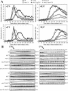
Rad53 and Mre11 phosphorylation in response to different genotoxic treatments in mec1Δ cells. (A to C) Logarithmically growing wild-type (wt) (YLL1072) and mec1Δ (DMP4225/4C) cells were arrested in G1 with α-factor and released from the pheromone block in YEPD alone or YEPD supplemented with 10 mU of bleomycin (bleo) per ml or 0.02% MMS or were UV irradiated (30 J/m2) prior to release in YEPD. When 95% cells had budded after release, 3 μg of α-factor per ml was added back to all cultures to prevent them from entering a second cell cycle. Cell samples were collected at the indicated times after α-factor release to analyze the DNA content by FACS (A) and to detect the Rad53 (B) and Mre11 (C) proteins by Western blot analysis with anti-Rad53 and antihemagglutinin antibodies, respectively. (D) Logarithmically growing wild-type (YLL1072) and mec1Δ (DMP4225/4C) cells were arrested in G1 with α-factor and released from the pheromone block at time zero in YEPD containing HN1 (2 μM) or HN2 (0.5 μM). Extracts from cell samples collected at the indicated times after α-factor release were subjected to Western blot analysis as described above for panels B and C. Cell samples taken immediately before UV treatment were taken at time zero. exp, exponentially growing cells.
FIG. 2.
FIG. 2.
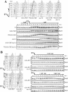
Scc1 cleavage relieves the inhibition of the metaphase-to-anaphase transition in UV-irradiated mec1Δ cells. Logarithmically growing wild-type (wt) (DMP4290/27A), mec1Δ (DMP4290/13C), SCC1-TEV (DMP4263/12B), and mec1Δ SCC1-TEV (DMP4293/10D) cells, carrying the tetR-GFP/tetO and GAL-TEV constructs, were arrested in G1 with α-factor in YEP+Raf and released from the pheromone block at time zero in YEP+Raf+Gal to induce the TEV protease production or were UV irradiated (30 J/m2) prior to the release in YEP+Raf+Gal. As in Fig. 1, when 95% cells had budded after release, 3 μg of α-factor per ml was added back to all cultures. Samples of untreated and UV-treated cell cultures were collected at the indicated times after α-factor release to analyze the DNA content by FACS in unirradiated (A, top) and UV-irradiated (A, bottom) cell cultures. The kinetics of sister chromatid separation using the tetR-GFP/tetO system was determined by fluorescence microscopy (B), and the kinetics of nuclear division was determined after propidium iodide staining (C). Rad53 protein was detected by Western blot analysis with anti-Rad53 antibodies in UV-treated cell extracts (D). exp, exponentially growing cells.
FIG. 3.
FIG. 3.
Tel1 and Mre11 are required for the response to UV irradiation in G1 in the absence of Mec1. (A) Cell cultures were arrested in G1 with α-factor and released from the G1 block either unirradiated (−UV) or after UV irradiation (30 J/m2) (+UV). As in Fig. 1, when 95% cells had budded after release, 3 μg of α-factor per ml was added back to all cultures. Samples of untreated (left) and UV-treated (right) wild-type (wt) (DMP4290/27A), tel1Δ (DMP4062/19A), mec1Δ (DMP4290/13C), mec1Δ tel1Δ (DMP4062/5C), mre11Δ (DMP4290/3A), and mec1Δ mre11Δ (DMP4290/10B) cell cultures collected at the indicated times after α-factor release were analyzed by fluorescence microscopy to determine the percentage of separated sister chromatids (top) and the percentage of binucleate cells (bottom). (B) Samples of wild-type (YLL839), tel1Δ (DMP3641/2C), mec1Δ (DMP3642/8C), mec1Δ tel1Δ (DMP3817/9A), mre11Δ (DMP3815/3C), and mec1Δ mre11Δ (DMP3774/28A) cells were treated as described above for panel A and analyzed to evaluate Rad53, Chk1, and Rad9 phosphorylation in the UV-irradiated cultures by Western blot analysis of protein extracts with anti-Rad53, antihemagglutinin, and anti-Rad9 antibodies, respectively. The DNA content by FACS and the percentage of binucleate cells were also analyzed (not shown). Samples of wild-type (YLL1072), tel1Δ (DMP4226/1B), mec1Δ (DMP4225/4C), and mec1Δ tel1Δ (DMP4239/8A) cells were treated as described above for panel A and analyzed to evaluate Mre11 phosphorylation in the UV-irradiated cultures by Western blot analysis with antihemagglutinin antibodies. Cell samples taken immediately before UV treatment were taken at time zero. exp, exponentially growing cells.
FIG. 4.
FIG. 4.
Effects of RAD53, CHK1, and MAD2 deletions on cell cycle progression after UV irradiation in G1. Logarithmically growing wild-type (wt) (K699), mec1Δ (YLL490), mec1Δ rad53Δ (DMP3249/14D), mec1Δ chk1Δ (DMP3274/1B), mec1Δ rad53Δ chk1Δ (DMP4359/6B), rad53Δ chk1Δ (DMP4359/4C), mad2Δ (SP1070), rad53Δ chk1Δ mad2Δ (DMP4359/9A), mec1Δ mad2Δ (DMP3262/3C), and mec1Δ tel1Δ (DMP4062/5C) strains were arrested in G1 with α-factor and released from the pheromone block at time zero in YEPD or were UV irradiated (30 J/m2) prior to the release in YEPD. As in Fig. 1, when 95% cells had budded after release, 3 μg of α-factor per ml was added back to all cultures. Samples of untreated and UV-treated cell cultures were collected at the indicated times after α-factor release to analyze the DNA content by FACS (A). The kinetics of nuclear division after propidium iodide staining (B) and the kinetics of Rad53 phosphorylation by Western blot analysis with anti-Rad53 antibodies (C) were also determined. exp, exponentially growing cells.
FIG. 5.
FIG. 5.
The Pds1 securin mediates the G2/M arrest and undergoes Tel1-dependent phosphorylation and stabilization in UV-treated mec1Δ cells. (A) Logarithmically growing cell cultures of wild-type (wt) (DMP4290/27A), mec1Δ (DMP4290/13C), pds1Δ (SP2894), and mec1Δ pds1Δ (DMP4290/27B) strains were arrested in G1 with α-factor and released from the pheromone block at time zero in YEPD or were UV irradiated (30 J/m2) prior to the release in YEPD. As in Fig. 1, when 95% cells had budded after release, 3 μg of α-factor per ml was added back to all cultures. Samples of UV-treated cell cultures were collected at the indicated times after α-factor release to analyze the DNA content by FACS (not shown). The kinetics of sister chromatid separation (top) and nuclear division (middle) were determined as described in the legends to Fig. 2B and C. The kinetics of Rad53 phosphorylation by Western blot analysis with anti-Rad53 antibodies (bottom) were also determined. The same procedure was applied to the unirradiated cell cultures (not shown, except for untreated wild type in panel A). (B) Logarithmically growing wild-type (DMP3390/17B), tel1Δ (DMP4292/4D), mec1Δ (DMP4291/4C), and mec1Δ tel1Δ (DMP4305/5C) cells, all expressing a Pds1-HA fusion, were treated as described above for panel A. (B) Samples of untreated and UV-treated cell cultures were collected at the indicated times after α-factor release to determine Pds1 level and phosphorylation by Western blot analysis with antihemagglutinin antibodies (top) and to analyze the DNA content by FACS (bottom). Cell samples taken immediately before UV treatment were taken at time zero. exp, exponentially growing cells.
FIG. 6.
FIG. 6.
Effects of NER inactivation and defective ssDNA signaling on Tel1-dependent checkpoint response. (A) Logarithmically growing wild-type (wt) (K699), rad14Δ (YLL355), mec1Δ (YLL490), rad14Δ mec1Δ (DMP3942/6C), and rad14Δ mec1Δ tel1Δ (DMP3942/20B) cells were arrested in G1 with α-factor and released from the pheromone block at time zero in YEPD, either untreated or after irradiation with 5 J/m2 UV dose prior to the release in YEPD. As in Fig. 1, when 95% cells had budded after release, 3 μg of α-factor per ml was added back to all cultures. Cell samples were collected at the indicated times after α-factor release to analyze the DNA content by FACS in unirradiated (top) and UV-irradiated (middle) cell cultures and to evaluate Rad53 phosphorylation (bottom) in UV-treated cell extracts as described in the legend to Fig. 1B. (B) Logarithmically growing wild-type (DMP4290/27A), tel1Δ (DMP4062/19A), mec1Δ (DMP4290/13C), and rfa1-t11 (DMP4313/11C) cells were synchronized with α-factor (αf) and resuspended in YEPD containing 3 μg of α-factor per ml after UV irradiation (30 J/m2) (+ α-factor). Protein extracts prepared from cell samples collected at the indicated times were subjected to Western blot analysis with anti-Rad53 antibodies. (C) G1-arrested wild-type (DMP4290/27A), rfa1-t11 (DMP4313/11C), mec1Δ (DMP4290/13C), and rfa1-t11 mec1Δ (DMP4313/7B) cells were UV irradiated (30 J/m2) and released from the G1 block at time zero. As in Fig. 1, when 95% cells had budded after release, 3 μg of α-factor per ml was added back to all cultures. Cell samples were collected at the indicated times after α-factor release to evaluate Rad53 phosphorylation as described above for panel B. Cell samples taken immediately before UV treatment were taken at time zero. exp, exponentially growing cells.
FIG. 7.
FIG. 7.
Rad53 phosphorylation in mec1Δ cells requires DNA replication of UV-damaged DNA. (A and B) Logarithmically growing wild-type (wt) (K699) and mec1Δ (YLL490) cells were synchronized in G2 with nocodazole (noc) and UV irradiated (30 J/m2). Half of each culture was released from the G2 block in YEPD (A) or held in G2 by the addition of nocodazole (B). Cell samples were collected at the indicated times after UV irradiation to analyze the DNA content by FACS (A, top) and to evaluate Rad53 phosphorylation (A, bottom, and B) as described in the legend to Fig. 1B. (C) Logarithmically growing wild-type (K699), mec1Δ (YLL490), GAL-CDC6 (DMP4083/11C), and mec1Δ GAL-CDC6 (DMP4083/6B) cells, carrying a GAL-CDC6 fusion as the only source of Cdc6 protein, were grown in YEP+Gal and arrested in G2 with nocodazole. All arrested cultures were then transferred to YEPD in the presence of nocodazole for 1 h and then released from the G2 block into YEPD containing α-factor (G1 block). Finally, they were released from the α-factor block into YEPD, either unirradiated or after UV irradiation with 30 J/m2. Samples were taken at the indicated times after α-factor release (αf) to analyze the DNA content by FACS (C, top) and to evaluate Rad53 phosphorylation (C, bottom) in UV-treated cell extracts as described in the legend to Fig. 1. exp, exponentially growing cells.
FIG. 8.
FIG. 8.
Perturbations of Clb/CDK activity influence checkpoint response to UV irradiation. (A) Wild-type (wt) (YLL1072), GAL-CLB2 (DMP4340/5C), 7XGAL-CLB2 (DMP4341/10B), mec1Δ (DMP4225/4C), GAL-CLB2 mec1Δ (DMP4340/1C), and 7XGAL-CLB2 mec1Δ (DMP4341/6C) cells, exponentially growing in YEP+Raf, were arrested in G1 with α-factor, UV irradiated, and then released from α-factor in YEP+Raf+Gal to induce CLB2 overexpression (+ gal). Cell samples were collected at the indicated times after α-factor release to analyze the DNA content in UV-treated cell cultures by FACS (top) and to analyze Rad53 phosphorylation in both untreated and UV-treated cell cultures (bottom) as described in the legend to Fig. 1. (B) Logarithmically growing wild-type (DMP4337/20C) and mec1Δ (DMP4337/14C) cells, all carrying three copies of the GAL-SIC1ΔNT fusion integrated in the genome (GAL-SIC1), were arrested in G1 with α-factor and released from the pheromone block in medium containing raffinose and nocodazole (15 μg/ml) (+raf +noc) (top) either untreated (not shown) or after UV irradiation (30 J/m2). Galactose was added to half of each G2 nocodazole-arrested cell culture 210 min after α-factor release to induce GAL-SIC1ΔNT expression (+gal +noc) (B, bottom). Cell samples were collected at the indicated times after α-factor release for further analysis. The DNA content in both untreated (not shown) and UV-treated cell cultures was analyzed by FACS (B, left). Rad53 and Mre11 phosphorylation in both untreated (not shown) and UV-treated cell cultures was analyzed as described in the legends to Fig. 1B and C (B, right). No significant differences were detected in the FACS profiles in the different unirradiated cell cultures, which did not reveal any Rad53 and Mre11 phosphorylation throughout the experiment. exp, exponentially growing cells.

Similar articles

Cited by

References

    1. Agarwal, R., Z. Tang, H. Yu, and O. Cohen-Fix. 2003. Two distinct pathways for inhibiting Pds1 ubiquitination in response to DNA damage. J. Biol. Chem. 278:45027-45033. - PubMed
    1. Akkari, Y. M. N., R. L. Bateman, C. A. Reifsteck, S. B. Olson, and M. Grompe. 2000. DNA replication is required to elicit cellular responses to psoralen-induced DNA interstrand cross-links. Mol. Cell. Biol. 20:8283-8289. - PMC - PubMed
    1. Andegeko, Y., L. Moyal, L. Mittelman, I. Tsarfaty, Y. Shiloh, and G. Rotman. 2001. Nuclear retention of ATM at sites of DNA double strand breaks. J. Biol. Chem. 276:38224-38230. - PubMed
    1. Bakkenist, C. J., and M. B. Kastan. 2003. DNA damage activates ATM through intermolecular autophosphorylation and dimer dissociation. Nature 421:499-506. - PubMed
    1. Banin, S., L. Moyal, S. Shieh, Y. Taya, C. W. Anderson, L. Chessa, N. I. Smorodinsky, C. Prives, Y. Reiss, Y. Shiloh, and Y. Ziv. 1998. Enhanced phosphorylation of p53 by ATM in response to DNA damage. Science 281:1674-1677. - PubMed

Publication types

MeSH terms