. 2004 Dec;131(23):5883-95.
doi: 10.1242/dev.01461.

Altered endochondral bone development in matrix metalloproteinase 13-deficient mice

Affiliations

Altered endochondral bone development in matrix metalloproteinase 13-deficient mice

Dominique Stickens et al. Development. 2004 Dec.

Abstract

The assembly and degradation of extracellular matrix (ECM) molecules are crucial processes during bone development. In this study, we show that ECM remodeling is a critical rate-limiting step in endochondral bone formation. Matrix metalloproteinase (MMP) 13 (collagenase 3) is poised to play a crucial role in bone formation and remodeling because of its expression both in terminal hypertrophic chondrocytes in the growth plate and in osteoblasts. Moreover, a mutation in the human MMP13 gene causes the Missouri variant of spondyloepimetaphyseal dysplasia. Inactivation of Mmp13 in mice through homologous recombination led to abnormal skeletal growth plate development. Chondrocytes differentiated normally but their exit from the growth plate was delayed. The severity of the Mmp13- null growth plate phenotype increased until about 5 weeks and completely resolved by 12 weeks of age. Mmp13-null mice had increased trabecular bone, which persisted for months. Conditional inactivation of Mmp13 in chondrocytes and osteoblasts showed that increases in trabecular bone occur independently of the improper cartilage ECM degradation caused by Mmp13 deficiency in late hypertrophic chondrocytes. Our studies identified the two major components of the cartilage ECM, collagen type II and aggrecan, as in vivo substrates for MMP13. We found that degradation of cartilage collagen and aggrecan is a coordinated process in which MMP13 works synergistically with MMP9. Mice lacking both MMP13 and MMP9 had severely impaired endochondral bone, characterized by diminished ECM remodeling, prolonged chondrocyte survival, delayed vascular recruitment and defective trabecular bone formation (resulting in drastically shortened bones). These data support the hypothesis that proper ECM remodeling is the dominant rate-limiting process for programmed cell death, angiogenesis and osteoblast recruitment during normal skeletal morphogenesis.


Figures

Fig. 1
Fig. 1
Targeting of Mmp13. (A) Strategy for targeting of Mmp13. The structure of the endogenous mouse locus (a), the transgene targeting cassette (b), the targeted floxed allele resulting from homologous recombination (c) and the null allele resulting from Cre-mediated recombination (d) are depicted. Exons are depicted as open boxes; the floxed exons corresponding to the catalytic domain are shown in red. Red arrowheads indicate loxP insertion sites. Blue bar indicates probe used for Southern hybridization. (B) Southern blot analyses. Identification of MMP13fl/fl and Mmp13−/− mice by digest of genomic DNA with AflII and PstI, respectively. Fragments were separated according to size and hybridized to the probe indicated in A. (C) Detection of secreted pro-MMP13 in conditioned medium from mouse calvarial cultures. Conditioned medium was collected, concentrated and proteins were size fractionated. Western blot using goat polyclonal anti-pro-MMP13 shows that pro-MMP13 (57 kDa) can be detected in culture media from calvaria from wild-type (+/+) and heterozygous (+/−) animals, but not from Mmp13−/− (−/−) animals. (D) Detection of Mmp13 expression by in situ hybridization on tibia from 15 dpc wild-type mouse. Mmp13 signal is indicated in red. Primary front of ossification is indicated by broken yellow line. Hoechst counterstain is blue. Mmp13 expression is observed in the hypertrophic chondrocyte population (HC) and the primary ossification center (PO), but not in the proliferating chondrocyte (PC) population. (E) Detection of Mmp13 expression by in situ hybridization on the phalange from 1-week-old wild-type mouse. In contrast to expression pattern observed in D, Mmp13 expression is restricted to the most terminal row of hypertrophic chondrocytes (arrowhead). TB, trabecular bone. Scale bars: 100 μm.
Fig. 2
Fig. 2
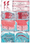
Histological examination of the Mmp13−/− growth plate defect. (A) Alizarin Red/Alcian Blue staining of whole skeletons from 2-week-old wild-type and Mmp13−/− mice showing no overt difference in overall skeletal structure between genotypes. (B,C) Von Kossa stained tibia from 15 dpc wild-type and Mmp13−/− mice shows the presence of mineralized tissue in the diaphyses of both genotypes. No overt difference is evident in proliferating chondrocyte (PC) zones, hypertrophic chondrocyte (HC) zones or primary ossification centers (PO) between genotypes. (D,E) Safranin-O stained metatarsals from 10-day-old wild-type and Mmp13−/− mice; (F,G) Safranin-O stained tibia from 10-day-old wild-type and Mmp13−/− mice. Mmp13−/− mice have an expansion of the hypertrophic chondrocyte (HC) zone of the growth plates of (compare lengths indicated by black bars). (H,I) Safranin-O stained tibia from 12-week-old wild-type and Mmp13−/− mice. The expansion of the hypertrophic cartilage is ameliorated but Safranin-O staining remains more intense in the mutant growth plate (TB, trabecular bone). Scale bars: 100 μm in B–G; 200 μm in H,I.
Fig. 3
Fig. 3
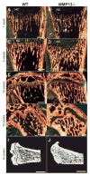
Histological examination of the Mmp13−/− trabecular bone defect. (A,B) Picrosirius Red stained tibia from 1-week-old mice show no apparent increase in trabecular bone (TB) in Mmp13−/− mice compared with wild type, but the bone spicules in Mmp13−/− mice are irregular in shape (indicated by arrow). At 3 weeks of age, Mmp13−/− mice show increased trabecular bone (C,D). Unlike the increase of hypertrophic cartilage (HC), which resolves at 5 weeks of age, the increase in trabecular bone in Mmp13−/− mice persists for months (E,F). Similar increases in trabecular bone can also be observed in femurs (G,H). Histological observations were confirmed by Micro-CT analyses on tibia of 16-week-old wild-type and Mmp13−/− mice (I,J). SO, secondary site of ossification. Scale bars: 200 μm in A,B; 300 μm in C,D; 400 μm in E–H; 1 mm in I,J.
Fig. 4
Fig. 4
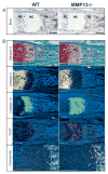
Examination of differentiation markers in Mmp13−/− endochondral cartilage. (A) BrdU incorporation in 1-week-old tibia of wild-type and Mmp13−/− mice. Animals were injected with 1 μg BrdU/g mouse 1 hour prior to sacrifice. A similar number of BrdU-positive cells (brown staining) can be observed in the proliferating chondrocyte (PC) zone, and in cells within the trabecular bone (TB) for wild-type and Mmp13−/− bones, indicating that proliferation is not impaired in Mmp13−/− mice. (B) In situ hybridization on 1-week-old metatarsals with probes to chondrocyte differentiation markers show normal expression domain of col2, but expanded expression of collagen type X and VEGF, indicating an increased late hypertrophic chondrocyte population in the Mmp13−/− growth plate. In wild-type mice, expression of osteopontin (Osp) is restricted to only one row of hypertrophic chondrocytes in the growth plate of wild-type mice. The growth plate of Mmp13 −/− mice contains several rows of chondrocytes expressing Osp (broken line demarcates front of ossification). Safranin-O staining shows general morphology (HC, hypertrophic cartilage). Scale bars: 100 μm.
Fig. 5
Fig. 5
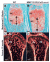
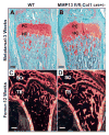
Phenotype of MMP13fl/fl;Col2-Cre+/− endochondral bones. (A,B) Safranin-O stained metatarsals from 2-week-old wild-type and MMP13fl/fl;Col2-Cre+/− mice show an increased hypertrophic chondrocyte (HC) zone in MMP13fl/fl;Col2-Cre+/− mice similar to that observed in Mmp13−/− mice (compare with Fig. 1D,E). (C,D) Picrosirius red-stained femurs from 10-week-old wild-type and MMP13fl/fl;Col2-Cre+/−. MMP13fl/fl;Col2-Cre+/− mice lack the increased trabecular bone (TB) phenotype observed in the Mmp13−/− mice, but have an altered structure of the trabeculae (arrow). SO, secondary site of ossification. Scale bars: 100 μm in A,B; 300 μm in C,D.
Fig. 6
Fig. 6
Phenotype of MMP13fl/fl;Col1-Cre+/− endochondral bones. (A,B) Safranin-O stained metatarsals from 3-week-old wild-type and MMP13fl/fl;Col1-Cre+/− mice show no increase in the hypertrophic chondrocyte (HC) zone of MMP13fl/fl;Col1-Cre+/− mice compared with wild type. (C,D) Picrosirius Red stained femurs from 12-week-old wild-type and MMP13fl/fl;Col1-Cre+/−. MMP13fl/fl;Col1-Cre+/− mice have increased trabecular bone (TB) similar to that observed in the Mmp13−/− mice (SO, secondary site of ossification). Scale bars: 100 μm in A,B; 300 μm in C,D.
Fig. 7
Fig. 7
Examination of key cell types in Mmp13−/− endochondral bones. (A,B) TRAP (red) stained 1-week-old wild-type and Mmp13−/− tibial epiphyses shows no difference in the osteoclast population (red staining) in the trabecular bone (TB) and endostea (E) of wild-type and Mmp13−/− mice. (C,D) Formation of the bone marrow cavity and mineralization were analyzed by Van Kossa stain on newborn wild-type and Mmp13−/− metatarsals. Mineralized tissue (brown staining) is present in endosteum (E) surrounding the bone marrow cavity, and in the surrounding trabecular bone (TB) in both wild-type and Mmp13−/− bones. (E,F) PECAM-immunostained 1-week-old tibia showing vessels (brown staining) in the trabecular bone (TB). There is no overt difference in localization or morphology of blood vessels in Mmp13−/− mice compared with wild type. Scale bars: 200 μm in A,B; 100 μm in C–F.
Fig. 8
Fig. 8
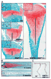
Examination of Mmp9−/−, Mmp13−/− and Mmp9−/−; Mmp13−/− endochondral bones. (A) Safranin-O staining of 2-week-old tibiae of wild-type, Mmp9−/−, Mmp13−/− and Mmp9−/−; Mmp13−/− mice. Mmp9−/− and Mmp13−/− tibiae have increased zones of hypertrophic cartilage, but a dramatically expanded hypertrophic chondrocyte zone can be observed in Mmp9−/−; Mmp13−/− tibia (compare lengths indicated by black bars). Magnifications of secondary ossification sites (insets) of Safranin-O stained 2-week-old tibia of wild type, Mmp9−/−, Mmp13−/− and Mmp9−/−; Mmp13−/− mice demonstrate delay in initiation of ossification at the secondary site in Mmp9−/−; Mmp13−/−. Collagenous bone tissue and colonizing blood cells in wild-type, Mmp9−/− and Mmp13−/− secondary sites are indicated by arrows and arrowheads, respectively. (B) Safranin-O staining of 5-week-old and 5-month-old metatarsals from Mmp9−/−; Mmp13−/− mice showing that while 5-week-old Mmp9−/−; Mmp13−/− metatarsals still display the expanded hypertrophic chondrocyte zone characteristic of this genotype, 5-month-old Mmp9−/−; Mmp13−/− metatarsals display a closed growth plate, indicating recovery. (C) Whole skeletal preparations of hind limbs from 12-month-old wild-type and Mmp9−/−; Mmp13−/− mice shows a shortening of all elements of the Mmp9−/−; Mmp13−/− hind limb when compared with wild type. Scale bars: 300 μm in A; 200 μm in B.
Fig. 9
Fig. 9
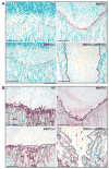
Altered ECM remodeling in Mmp9−/−, Mmp13−/− and Mmp9−/−; Mmp13−/− long bones. (A) Immunostaining of 2-week-old tibia of wild-type, Mmp9−/−, Mmp13−/− and Mmp9−/−; Mmp13−/− mice with an antibody to the DIPEN neoepitope of Mmp-cleaved aggrecan. Cleaved aggrecan (brown staining) is present along the primary front of ossification in wild-type, Mmp9−/− and Mmp13−/− bones, no positive staining is apparent in Mmp9−/−; Mmp13−/− bones. (B) Immunostaining of 2-week-old tibia of wild-type, Mmp9−/−, Mmp13−/− and Mmp9−/−; Mmp13−/− mice with an antibody to the 3/4 cleavage fragment of col1 and col2. The primary front of ossification (indicated by a broken line) shows the presence of cleaved collagen (pink staining) in transverse septa surrounding the most terminal hypertrophic chondrocytes (arrows) in wild-type and Mmp9−/− bones, along the trabeculae of the developing trabecular bone (arrowheads) in wild-type and Mmp13−/− bones, and at random throughout the hypertrophic chondrocyte zone (arrows) and trabecular bone (arrowheads) in Mmp9−/−; Mmp13−/− bones. Scale bars: 200 μm in A; 100 μm in B.

Similar articles

Cited by

References

    1. Abbaszade I, Liu RQ, Yang F, Rosenfeld SA, Ross OH, Link JR, Ellis DM, Tortorella MD, Pratta MA, Hollis JM, et al. Cloning and characterization of ADAMTS11, an aggrecanase from the ADAMTS family. J Biol Chem. 1999;274:23443–23450. - PubMed
    1. Albrecht U, Eichele G, Helms JA, Lu H. Visualization of gene expression patterns by in situ hybridization. In: Daston G, editor. Molecular and Cellular Methods in Developmental Toxicology. Boca Raton, FL: CRC Press; 1997. pp. 23–48.
    1. Alini M, Matsui Y, Dodge GR, Poole AR. The extracellular matrix of cartilage in the growth plate before and during calcification: changes in composition and degradation of type II collagen. Calcif Tissue Int. 1992;50:327–335. - PubMed
    1. Arner EC. Aggrecanase-mediated cartilage degradation. Curr Opin Pharmacol. 2002;2:322–329. - PubMed
    1. Aszodi A, Bateman JF, Gustafsson E, Boot-Handford R, Fassler R. Mammalian skeletogenesis and extracellular matrix: what can we learn from knockout mice? Cell Struct Funct. 2000;25:73–84. - PubMed

Publication types

MeSH terms