Comparative Study
. 2004 Nov 10;24(45):10229-39.
doi: 10.1523/JNEUROSCI.3558-04.2004.

Improved behavior and neuropathology in the mouse model of Sanfilippo type IIIB disease after adeno-associated virus-mediated gene transfer in the striatum

Affiliations
Comparative Study

Improved behavior and neuropathology in the mouse model of Sanfilippo type IIIB disease after adeno-associated virus-mediated gene transfer in the striatum

Arnaud Cressant et al. J Neurosci. .

Abstract

Sanfilippo syndrome is a mucopolysaccharidosis (MPS) caused by a lysosomal enzyme defect interrupting the degradation pathway of heparan sulfates. Affected children develop hyperactivity, aggressiveness, delayed development, and severe neuropathology. We observed relevant behaviors in the mouse model of Sanfilippo syndrome type B (MPSIIIB), in which the gene coding for alpha-N-acetylglucosaminidase (NaGlu) is invalidated. We addressed the feasibility of gene therapy in these animals. Vectors derived from adeno-associated virus serotype 2 (AAV2) or 5 (AAV5) coding for NaGlu were injected at a single site in the putamen of 45 6-week-old MPSIIIB mice. Normal behavior was observed in treated mice. High NaGlu activity, far above physiological levels, was measured in the brain and persisted at 38 weeks of age. NaGlu immunoreactivity was detected in neuron intracellular organelles, including lysosomes. Enzyme activity spread beyond vector diffusion areas. Delivery to the entire brain was reproducibly obtained with both vector types. NaGlu activity was higher and distribution was broader with AAV5-NaGlu than with AAV2-NaGlu vectors. The compensatory increase in the activity of various lysosomal enzymes was improved. The accumulation of gangliosides GM2 and GM3 present before treatment and possibly participating in neuropathology was reversed. Characteristic vacuolations in microglia, perivascular cells, and neurons, which were prominent before the age of treatment, disappeared in areas in which NaGlu was present. However, improvement was only partial in some animals, in contrast to high NaGlu activity. These results indicate that NaGlu delivery from intracerebral sources has the capacity to alleviate most disease manifestations in the MPSIIIB mouse model.


Figures
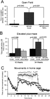
Figure 1.
Figure 1.
Abnormal behaviors were prevented in treated MPSIIIB mice. A, Time spent in navigation was recorded in the open field during 10 min sessions. Normal mice (+/+; n = 12), untreated MPSIIIB mice (MPSIIIB; n = 10), and AAV2-NaGlu-treated MPSIIIB mice (MPSIIIB-AAV2-NaGlu; n = 9) were tested at 5 and 29 weeks of age. Data are means ± SEM of differences between times spent in navigation at 29 and 5 weeks (t29weeks to t5weeks). Significance was determined using the Student's t test. B, The same animals were tested in the elevated plus-maze at 18 and 34 weeks of age in a dark environment. Data are means ± SEM of ratios of time spent in open arms relative to total time spent in arms. Significance was determined using the Student's t test. C, Movements of normal mice (black triangles; n = 6), MPSIIIB mice treated with mock AAV5-GFP vectors (open squares; n = 6), untreated MPSIIIB mice (equivalent to MPSIIIB mice treated with mock AAV5-GFP vectors and not shown here; n = 6), or MPSIIIB mice treated with AAV5-NaGlu vectors (gray circles; n = 15) were recorded in 18-week-old mice over 24 hr in home cages. Data are means ± SEM of times spent in fast movements during 30 min periods. The shadowed area indicates the dark period. Significance: stars indicate p < 0.05; one-way ANOVA; Bonferroni's post hoc analysis.
Figure 2.
Figure 2.
AAV2-NaGlu and AAV5-NaGlu vectors induced NaGlu activity in the MPSIIIB mouse brain. NaGlu activity was measured in tissue homogenates prepared from the brain hemispheres or the cerebellum and brainstem of normal mice (+/+), heterozygous MPSIIIB mice (+/-), untreated MPSIIIB mice (-/-), or MPSIIIB mice injected in the right putamen at 6 weeks of age with AAV2-NaGlu or AAV5-NaGlu vectors. The age of mice at analysis and the number of mice in each group are indicated. Data are means ± SEM of NaGlu activities measured in homogenates.
Figure 3.
Figure 3.
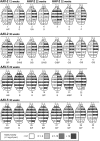
Analysis of AAV-NaGlu vector genome dispersion in mouse brains by quantitative PCR. MPSIIIB mice received a single injection of AAV2-NaGlu or AAV5-NaGlu vectors in the right putamen at 6 weeks of age and were analyzed at the indicated time. One-millimeter-thick coronal brain slices were prepared from the injected (IL) and noninjected (CL) hemispheres. Brain maps of AAV vector dispersion for each analyzed mouse are shown. Strips on brain maps schematically represent coronal slices (slice number is shown on the left). Values shown in the strips are numbers of AAV-NaGlu vector genomes measured in DNA extracted from the slice, which are expressed as copies per diploid cell genome. Zero indicates values of ≤0.1 copies per 2N genomes. Strips corresponding to slices in which vector genomes were detected are shadowed in gray according to the code shown at the bottom. Strips without values correspond to brain slices that were not analyzed. Mouse reference numbers are indicated.
Figure 4.
Figure 4.
Widespread NaGlu activity in treated MPSIIIB mouse brains. MPSIIIB mice received a single injection of AAV2-NaGlu or AAV5-NaGlu vectors in the right putamen at 6 weeks of age and were analyzed at the indicated time. One-millimeter-thick coronal brain slices were prepared from the injected (IL) and noninjected (CL) hemispheres. Brain maps of NaGlu activities for each analyzed mouse are shown. Strips on brain maps schematically represent coronal slices (slice number is shown on the left). Values shown in the strips are NaGlu activities measured in the slice homogenate (in units per milligram of protein). Zero indicates that activity was <0.1 U per milligram of protein. Strips corresponding to slices in which NaGlu activity was detected are shadowed in gray according to the code shown at the bottom. Strips without values correspond to brain slices not used for NaGlu assay. Mouse reference numbers are indicated, as well as a letter code indicating other investigations performed on the same mouse: H, Histopathology; G, ganglioside assay (not all mice in which gangliosides were investigated are shown); B, behavioral testing (1 mouse tested in the open field is not shown here); P, quantitative PCR amplification of vector genomes.
Figure 5.
Figure 5.
NaGlu accumulated in neurons. Slice 6 from the injected hemisphere of mouse 23 was processed for cryosection (slice IL6, mouse 23 on Fig. 3). NaGlu was revealed by confocal fluorescence microscopy using a rabbit anti-human NaGlu serum (A, D). The anti-NaGlu (A) and lysosome-specific anti-Lamp1 (B) antibodies essentially stained different cytoplasmic organelles, although signals might occasionally colocalize (arrow in C). Cells showing cytoplasmic NaGlu signals (D) coexpressed the nuclear neuronal marker NeuN (E, F). Scale bar, 25 μm.
Figure 6.
Figure 6.
NaGlu delivery was associated with improved lysosomal functions. The activity of various lysosomal enzymes was assayed in brain homogenates from normal mice (n = 5), untreated MPSIIIB mice (n = 5), or MPSIIIB mice treated with either AAV2-NaGlu or AAV5-NaGlu vector (n = 2 or 3). Homogenates were prepared from tissues containing various levels of NaGlu activity (1-5, 10-50, 50-100, or >1000 U per milligram of protein). Values were not significantly different among these four groups (Kruskal-Wallis one-way ANOVA). The data shown are ratios of enzyme activity relative to normal mice. Enzymes with values that were significantly improved in treated mice are indicated by an asterisk in the untreated mouse panel (significance: p < 0.05; Mann-Whitney U test). Tot. hexo, Total hexosaminidase; Hexo A, hexosaminidase A; α-fuco, α-fucosidase; β-glucu, β-glucuronidase; β-gal, β-galactosidase; IDUA, α-l-iduronidase.
Figure 7.
Figure 7.
NaGlu delivery was associated with a decreased accumulation of GM2 and GM3 gangliosides. Gangliosides were measured in total lipid extracts from the brains of normal mice and untreated MPSIIIB mice and from the injected or noninjected hemisphere of MPSIIIB mice that received a single injection of AAV2-NaGlu vectors in the putamen at 6 weeks of age (AAV2-NaGlu vector-treated MPSIIIB mice). The age of mice at analysis and the number of mice in each group are indicated. Data are expressed as molar percentages of total gangliosides according to NeuAc moiety numbers. Asterisks indicate mean values in treated mice that are significantly lower than those in untreated mice at 5 weeks of age (p ≤ 0.04; Mann-Whitney U test). Mean GM2 and GM3 values at 12 weeks of age in the injected hemisphere of treated mice were equivalent to mean values at the same age in normal mice.
Figure 8.
Figure 8.
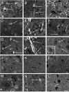
Disappearance or reduction of vacuolation in MPSIIIB mouse brain cells. Semithin sections stained with toluidine blue from brain slices of 6-week-old (A-C), 3-month-old (D-F), and 8-month-old (G-I) untreated MPSIIIB mice and from mouse 1 treated with AAV2-NaGlu vectors (J-L) and mouse 16 treated with AAV5-NaGlu vectors (M-O) are shown. Figure 4 indicates NaGlu activity in slices adjacent to those used for histopathology in these treated mice. Bright fine vacuoles in microglia (A) and larger and darker vacuoles in perivascular cells (B) were observed in the striatum of a 6-week-old untreated MPSIIIB mouse. Few neurons or astrocytes showed fine clear vacuoles in the parietal cortex (C). Large clear vacuoles were visible in microglia in the parietal cortex (D) and in microglia and perivascular cells in the putamen (E) of a 3-month-old untreated MPSIIIB mouse. Neurons or astrocytes with fine clear inclusions are visible in the putamen (F). Vacuoles in cortical microglia (G) and distension in perivascular cells near neurons or astrocytes with normal morphology (H) were observed in the putamen of an 8-month-old untreated mouse. Severely affected cells with abundant polymorphic vacuoles (I) as well as cells with picnotic nuclei (inset in I) were present at several other locations in the putamen of this mouse. Lysosomal distension was not observed in microglia (J), perivascular cells (K), or neurons or astrocytes (J-L) in the parietal cortex of the injected hemisphere of treated mouse 1 (slice 6 from the injected hemisphere). In contrast, residual lesions were observed in microglia (M) and perivascular cells (M, N) in the putamen of mouse 16 (slice 8 from the injected hemisphere). Vacuoles were also observed in neurons or astrocytes in the parietal cortex of the noninjected hemisphere (slice 8) of this mouse. Scale bar, 25 μm.

Similar articles

Cited by

References

    1. Adra CN, Boer PH, McBurney M (1987) Cloning and expression of the mouse pgk-1 gene and the nucleotide sequence of its promoter. Gene 60: 65-74. - PubMed
    1. Avale ME, Falzone TL, Gelman DM, Low MJ, Grandy DK, Rubinstein M (2004) The dopamine D4 receptor is essential for hyperactivity and impaired behavioral inhibition in a mouse model of attention deficit/hyperactivity disorder. Mol Psychiatry 9: 718-726. - PubMed
    1. Bhaumik M, Muller VJ, Rozaklis T, Johnson L, Dobrenis K, Bhattacharyya R, Wurzelmann S, Finamore P, Hopwood JJ, Walkley SU, Stanley P (1999) A mouse model for mucopolysaccharidosis type III A (Sanfilippo syndrome). Glycobiology 9: 1389-1396. - PubMed
    1. Bosch A, Heard JM (2003) Gene therapy for mucopolysaccharidosis. Int Rev Neurobiol 55: 271-296. - PubMed
    1. Brument N, Morenweiser R, Blouin V, Toublanc E, Raimbaud I, Chérel Y, Folliot S, Gaden F, Boulanger P, Kroner-Lux G, Moullier P, Rolling F, Salvetti A (2002) A versatile and scalable two-step ion-exchange chromatography process for the purification of recombinant adeno-associated virus serotypes-2 and -5. Mol Ther 6: 678-686. - PubMed

Publication types

MeSH terms