Induction of inflammatory mediators and microglial activation in mice transgenic for mutant human P301S tau protein
- PMID: 15509534
- PMCID: PMC1618683
- DOI: 10.1016/S0002-9440(10)63421-9
Induction of inflammatory mediators and microglial activation in mice transgenic for mutant human P301S tau protein
Abstract
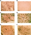
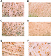
Mice transgenic for human P301S tau protein exhibit many characteristics of the human tauopathies, including the formation of abundant filaments made of hyperphosphorylated tau protein and neurodegeneration leading to nerve cell loss. At 5 months of age, the pathological changes are most marked in brainstem and spinal cord. Here we show that these changes are accompanied by marked neuroinflammation. Many tau-positive nerve cells in brainstem and spinal cord were strongly immunoreactive for interleukin-1beta and cyclooxygenase-2, indicating induction and overproduction of proinflammatory cytokines and enzymes. In parallel, numerous activated microglial cells were present throughout brain and spinal cord of transgenic mice, where they concentrated around tau-positive nerve cells. These findings suggest that inflammation may play a significant role in the events leading to neurodegeneration in the tauopathies and that anti-inflammatory compounds may have therapeutic potential.
Figures


Similar articles
-
Early axonopathy preceding neurofibrillary tangles in mutant tau transgenic mice.Am J Pathol. 2007 Sep;171(3):976-92. doi: 10.2353/ajpath.2007.070345. Epub 2007 Aug 9. Am J Pathol. 2007. PMID: 17690183 Free PMC article.
-
Cell-cycle markers in a transgenic mouse model of human tauopathy: increased levels of cyclin-dependent kinase inhibitors p21Cip1 and p27Kip1.Am J Pathol. 2006 Mar;168(3):878-87. doi: 10.2353/ajpath.2006.050540. Am J Pathol. 2006. PMID: 16507903 Free PMC article.
-
Using Experience Sampling Methodology to Capture Disclosure Opportunities for Autistic Adults.Autism Adulthood. 2023 Dec 1;5(4):389-400. doi: 10.1089/aut.2022.0090. Epub 2023 Dec 12. Autism Adulthood. 2023. PMID: 38116059 Free PMC article.
-
Trends in Surgical and Nonsurgical Aesthetic Procedures: A 14-Year Analysis of the International Society of Aesthetic Plastic Surgery-ISAPS.Aesthetic Plast Surg. 2024 Oct;48(20):4217-4227. doi: 10.1007/s00266-024-04260-2. Epub 2024 Aug 5. Aesthetic Plast Surg. 2024. PMID: 39103642 Review.
-
Depressing time: Waiting, melancholia, and the psychoanalytic practice of care.In: Kirtsoglou E, Simpson B, editors. The Time of Anthropology: Studies of Contemporary Chronopolitics. Abingdon: Routledge; 2020. Chapter 5. In: Kirtsoglou E, Simpson B, editors. The Time of Anthropology: Studies of Contemporary Chronopolitics. Abingdon: Routledge; 2020. Chapter 5. PMID: 36137063 Free Books & Documents. Review.
Cited by
-
Role of Microglia in Modulating Adult Neurogenesis in Health and Neurodegeneration.Int J Mol Sci. 2020 Sep 19;21(18):6875. doi: 10.3390/ijms21186875. Int J Mol Sci. 2020. PMID: 32961703 Free PMC article. Review.
-
Review: tauopathy in the retina and optic nerve: does it shadow pathological changes in the brain?Mol Vis. 2012;18:2700-10. Epub 2012 Nov 12. Mol Vis. 2012. PMID: 23170062 Free PMC article. Review.
-
Alzheimer's disease-like tau neuropathology leads to memory deficits and loss of functional synapses in a novel mutated tau transgenic mouse without any motor deficits.Am J Pathol. 2006 Aug;169(2):599-616. doi: 10.2353/ajpath.2006.060002. Am J Pathol. 2006. PMID: 16877359 Free PMC article.
-
Protective Effects of Polysaccharides in Neurodegenerative Diseases.Front Aging Neurosci. 2022 Jul 4;14:917629. doi: 10.3389/fnagi.2022.917629. eCollection 2022. Front Aging Neurosci. 2022. PMID: 35860666 Free PMC article. Review.
-
Microglial Priming in Infections and Its Risk to Neurodegenerative Diseases.Front Cell Neurosci. 2022 Jun 15;16:878987. doi: 10.3389/fncel.2022.878987. eCollection 2022. Front Cell Neurosci. 2022. PMID: 35783096 Free PMC article. Review.
References
-
- Goedert M, Spillantini MG, Davies SW. Filamentous nerve cell inclusions in neurodegenerative diseases. Curr Opin Neurobiol. 1998;8:619–632. - PubMed
-
- Lee VMY, Goedert M, Trojanowski JQ. Neurodegenerative tauopathies. Annu Rev Neurosci. 2001;24:1121–1159. - PubMed
-
- Poorkaj P, Bird TD, Wijsman E, Nemens E, Garruto RM, Anderson L, Andreadis A, Wiederholt WC, Raskind M, Schellenberg GD. Tau is a candidate gene for chromosome 17 frontotemporal dementia. Ann Neurol. 1998;43:815–825. - PubMed
-
- Hutton M, Lendon CL, Rizzu P, Baker M, Froelich S, Houlden H, Pickering-Brown S, Chakraverty S, Isaacs A, Grover A, Hackett J, Adamson J, Lincoln S, Dickson D, Davies P, Petersen RC, Stevens M, de Graaff E, Wauters E, van Baren J, Hillebrand M, Joosse M, Kwon JM, Nowotny P, Che LK, Norton J, Morris JC, Reed LA, Trojanowski JQ, Basun H, Lannfelt L, Neystat M, Fahn S, Dark F, Tannenberg T, Dodd PR, Hayward N, Kwok JBJ, Schofield PR, Andreadis A, Snowden J, Craufurd D, Neary D, Owen F, Oostra BA, Hardy J, Goate A, van Swieten J, Mann D, Lynch T, Heutink P. Association of missense and 5′-splice-site-mutations in tau with the inherited dementia FTDP-17. Nature. 1998;393:702–705. - PubMed
Publication types
MeSH terms
Substances
LinkOut - more resources
Full Text Sources
Other Literature Sources
Molecular Biology Databases
Research Materials

