Hypoxia increases corneal cell expression of CFTR leading to increased Pseudomonas aeruginosa binding, internalization, and initiation of inflammation
- PMID: 15505057
- PMCID: PMC1317302
- DOI: 10.1167/iovs.04-0627
Hypoxia increases corneal cell expression of CFTR leading to increased Pseudomonas aeruginosa binding, internalization, and initiation of inflammation
Abstract
Purpose: To investigate the effect of hypoxia-induced molecular responses of corneal epithelial cells on the surface of rabbit and human corneas and corneal cells in culture on interactions with Pseudomonas aeruginosa that may underlie increased susceptibility to keratitis.
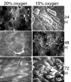
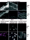
Methods: Organ cultures of rabbit and human corneal tissue, primary rabbit and human corneal cells, and transformed human corneal cells from a patient with cystic fibrosis and the same cell line corrected for expression of wild-type cystic fibrosis transmembrane conductance regulator (CFTR), the cellular receptor for P. aeruginosa, were exposed to hypoxic conditions for 24 to 72 hours. Changes in binding and internalization of P. aeruginosa were measured using cellular association and gentamicin-exclusion assays, and expression of CFTR and activation of NF-kappaB in response to hypoxia were determined by confocal laser microscopy and quantitative measurements of NF-kappaB activation.
Results: Hypoxia induced in a time- and oxygen-concentration-dependent manner increased association and internalization of clinical isolates of P. aeruginosa in all cells tested. Hypoxia increased CFTR expression and NF-kappaB nuclear translocation in rabbit and human cells with wild-type CFTR. Corneal cells lacking CFTR had reduced NF-kappaB activation in response to hypoxia. Hypoxia did not affect the increase in corneal cell CFTR levels or NF-kappaB activation after P. aeruginosa infection.
Conclusions: Hypoxic conditions on the cornea exacerbate the binding and internalization of P. aeruginosa due to increased levels of CFTR expression and also induce basal NF-kappaB activation. Both of these responses probably exacerbate the effects of P. aeruginosa infection by allowing lower infectious doses of bacteria to induce disease and promote destructive inflammatory responses.
Figures


Similar articles
-
Regulation of Pseudomonas aeruginosa internalization after contact lens wear in vivo and in serum-free culture by ocular surface cells.Invest Ophthalmol Vis Sci. 2006 Aug;47(8):3430-40. doi: 10.1167/iovs.05-1332. Invest Ophthalmol Vis Sci. 2006. PMID: 16877413
-
Prolonged hypoxia induces lipid raft formation and increases Pseudomonas internalization in vivo after contact lens wear and lid closure.Eye Contact Lens. 2006 May;32(3):114-20. doi: 10.1097/01.icl.0000177384.27778.4c. Eye Contact Lens. 2006. PMID: 16702863
-
Disruption of CFTR-dependent lipid rafts reduces bacterial levels and corneal disease in a murine model of Pseudomonas aeruginosa keratitis.Invest Ophthalmol Vis Sci. 2008 Mar;49(3):1000-9. doi: 10.1167/iovs.07-0993. Invest Ophthalmol Vis Sci. 2008. PMID: 18326723 Free PMC article.
-
Current concepts: contact lens related Pseudomonas keratitis.Cont Lens Anterior Eye. 2007 May;30(2):94-107. doi: 10.1016/j.clae.2006.10.001. Epub 2006 Nov 3. Cont Lens Anterior Eye. 2007. PMID: 17084658 Review.
-
Role of the cystic fibrosis transmembrane conductance regulator in innate immunity to Pseudomonas aeruginosa infections.Proc Natl Acad Sci U S A. 2000 Aug 1;97(16):8822-8. doi: 10.1073/pnas.97.16.8822. Proc Natl Acad Sci U S A. 2000. PMID: 10922041 Free PMC article. Review.
Cited by
-
The Clinical and Cellular Basis of Contact Lens-related Corneal Infections: A Review.Clin Ophthalmol. 2008;2(4):907-917. doi: 10.2147/opth.s3249. Clin Ophthalmol. 2008. PMID: 19277209 Free PMC article.
-
Cystic fibrosis transmembrane conductance regulator-emerging regulator of cancer.Cell Mol Life Sci. 2018 May;75(10):1737-1756. doi: 10.1007/s00018-018-2755-6. Epub 2018 Feb 6. Cell Mol Life Sci. 2018. PMID: 29411041 Free PMC article. Review.
-
Hypoxia inducible factor-1 (HIF-1)-mediated repression of cystic fibrosis transmembrane conductance regulator (CFTR) in the intestinal epithelium.FASEB J. 2009 Jan;23(1):204-13. doi: 10.1096/fj.08-110221. Epub 2008 Sep 8. FASEB J. 2009. PMID: 18779379 Free PMC article.
-
Role of oxygen availability in CFTR expression and function.Am J Respir Cell Mol Biol. 2008 Nov;39(5):514-21. doi: 10.1165/rcmb.2007-0452OC. Epub 2008 May 12. Am J Respir Cell Mol Biol. 2008. PMID: 18474670 Free PMC article.
-
Evidence for intracellular Pseudomonas aeruginosa.J Bacteriol. 2024 May 23;206(5):e0010924. doi: 10.1128/jb.00109-24. Epub 2024 Apr 10. J Bacteriol. 2024. PMID: 38597609 Free PMC article. Review.
References
-
- Cruz OA, Sabir SM, Capo H, Alfonso EC. Microbial keratitis in childhood. Ophthalmology. 1993;100:192–196. - PubMed
-
- Mela EK, Giannelou IP, John KX, Sotirios GP. Ulcerative keratitis in contact lens wearers. Eye Contact Lens. 2003;29:207–209. - PubMed
-
- Holden BA, Sweeney DF, Sankaridurg PR, et al. Microbial keratitis and vision loss with contact lenses. Eye Contact Lens. 2003;29:S131–S134. discussion S143–S134, S192–S134. - PubMed
Publication types
MeSH terms
Substances
Grants and funding
LinkOut - more resources
Full Text Sources

