. 2004 Oct 25;167(2):281-92.
doi: 10.1083/jcb.200407088.

The GTPase Arf1p and the ER to Golgi cargo receptor Erv14p cooperate to recruit the golgin Rud3p to the cis-Golgi

Affiliations

The GTPase Arf1p and the ER to Golgi cargo receptor Erv14p cooperate to recruit the golgin Rud3p to the cis-Golgi

Alison K Gillingham et al. J Cell Biol. .

Abstract

Rud3p is a coiled-coil protein of the yeast cis-Golgi. We find that Rud3p is localized to the Golgi via a COOH-terminal domain that is distantly related to the GRIP domain that recruits several coiled-coil proteins to the trans-Golgi by binding the small Arf-like GTPase Arl1p. In contrast, Rud3p binds to the GTPase Arf1p via this COOH-terminal "GRIP-related Arf-binding" (GRAB) domain. Deletion of RUD3 is lethal in the absence of the Golgi GTPase Ypt6p, and a screen of other mutants showing a similar genetic interaction revealed that Golgi targeting of Rud3p also requires Erv14p, a cargo receptor that cycles between the endoplasmic reticulum and Golgi. The one human protein with a GRAB domain, GMAP-210 (CEV14/Trip11/Trip230), is known to be on the cis-Golgi, but the COOH-terminal region that contains the GRAB domain has been reported to bind to centrosomes and gamma-tubulin (Rios, R.M, A. Sanchis, A.M. Tassin, C. Fedriani, and M. Bornens. 2004. Cell. 118:323-335). In contrast, we find that this region binds to the Golgi in a GRAB domain-dependent manner, suggesting that GMAP-210 may not link the Golgi to gamma-tubulin and centrosomes.


Figures
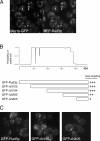
Figure 1.
Figure 1.
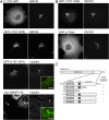
The COOH terminus of Rud3p mediates Golgi association. (A) Fluorescent micrographs of live yeast expressing the indicated fusion proteins. Uso1p was tagged with GFP at the COOH terminus in the genome of the wild-type strain BY4741, whereas RFP-Rud3p was expressed from a CEN plasmid under the control of a constitutive version of the PHO5 promoter. The two proteins were found to be localized to the same punctate structures, as illustrated by those marked with arrows. (B) Schematic diagram of truncations made in the RUD3 ORF in the yeast strain SEY6210 (Robinson et al., 1988) by insertion by homologous recombination of a cassette encoding a PHO5 promoter and an NH2-terminal GFP tag. Also shown is a coiled-coil prediction for Rud3p (Lupas, 1996). (C) Fluorescent micrographs of live yeast expressing the indicated GFP-Rud3p truncations as in B.
Figure 2.
Figure 2.
Rud3p is a member of a family of coiled-coil proteins with a conserved COOH-terminal domain. (A) Schematic representation of S. cerevisiae Rud3p and its relatives from the indicated species. At, A. thaliana; Ce, C. elegans; Dm, D. melanogaster; Hs, Homo sapiens; Sc, S. cerevisiae; Tb, Trypanosoma brucei. (B) Alignment of the COOH-terminal regions of the GRAB domain proteins with those of GRIP domain proteins golgin-245 and golgin-97, and the structure of the GRIP domain of human golgin-245. The two sets of sequences were independently aligned with CLUSTAL W and shaded where more than half the residues are related (gray) or identical (black). Hydrophobic residues conserved in each set are marked with filled circles and with a red circle for the critical tyrosine in the GRIP domain, and the leucine is in the equivalent position in Rud3p. In both alignments the tryptophans are shaded orange and cluster downstream of the conserved region. In the case of golgin-245, the tryptophan apparently stabilizes the interaction of the GRIP domain with Golgi membranes (Panic et al., 2003a). (C) A schematic representation of the GRAB domain proteins from metazoans and yeasts, along with a GRIP domain protein. All contain either a GRIP or GRAB domain, and the latter have a downstream GA1 motif. Metazoan GRAB proteins also have an extended, proline-rich, COOH-terminal region, whereas GRAB proteins from yeasts and filamentous fungi have an upstream region of sequence conservation (GA2, blue). (D) Fluorescent micrographs of rud3Δ cells expressing GFP-tagged wild-type Rud3p or the mutant L410A as in Fig. 1 A.
Figure 3.
Figure 3.
Rud3p interacts with the small GTPase Arf1p. (A) Anti-HA protein blot of total cell lysate (Lys.) from a strain expressing Rud3p tagged in the genome with an NH2-terminal HA epitope tag (AGY10) and of proteins that bound to GST fusions of the GTP-locked versions of Arf1p, Arf3p, Ypt1p, Ypt6, Ypt31p, and Ypt32p. For the GTP-locked forms of Arf1p (Q71L) and Arf3p (Q71L), the first 14 amino acids that form an amphipathic helix were removed and replaced with an NH2-terminal GST tag. For Ypt1p (Q67L), Ypt6p (Q67L), Ypt31p (Q72L), and Ypt32p (Q72L), the COOH-terminal cysteine residues were replaced with a COOH-terminal GST tag. Lysate is 10% of material applied to beads. (B) Anti-HA protein blot of total cell lysate (Lys.; 10% of material loaded) and proteins that bound to GST fusions of wild-type Arf1p and Arf3p preloaded with GDP or a nonhydrolysable analogue of GTP, GTPγS. The blot was stripped and reprobed with a rabbit antibody against Imh1p. (C) As for GST-Arf1p in B, except cell lysates were prepared from a strain expressing either GFP-Rud3p or the mutant proteins L410A expressed under the control of a constitutively active PHO5 promoter from the CEN plasmid pRS416. (D) Binding of the COOH-terminal 126 amino acids of Rud3p to Arf1p (T31N) or Arf1p (Q71L). The indicated forms of GST-Arf1p were coexpressed in E. coli with the COOH terminus of Rud3p, and after cell lysis isolated on glutathione Sepharose beads. Bound proteins were analyzed by gel electrophoresis, and the indicated band was identified as the COOH terminus of Rud3p by matrix-assisted laser desorption ionization mass spectrometry of tryptic peptides (Shevchenko et al., 1996).
Figure 4.
Figure 4.
COOH-terminal mutants of Rud3p are not functional. (A) Growth at the indicated temperatures of yeast lacking genomic copies of both RIC1 and RUD3, but containing the either an empty plasmid or expressing wild-type or mutant GFP-Rud3p (indicated on the diagram) from a constitutive PHO5 promoter. Cells lacking RUD3 and with only copy of RIC1 on a CEN, URA3 plasmid were transformed with CEN, LEU2 plasmids expressing GFP-Rud3p or mutants, and the yeast plated onto plates containing 5-fluoroorotic acid (5-FOA) to remove the RIC1 containing URA3 plasmid. (B) A yeast strain expressing GFP-tagged Rud3p lacking the GA1 motif (463ΔC) was used in a binding assay as in Fig. 3 C with GST-Arf1p loaded with either GDP or GTP-γ-S. Also shown is a fluorescent micrograph of live yeast lacking genomic RUD3 and expressing GFP-Rud3p463ΔC. (C) Fluorescent micrographs of a rud3Δ strain with the ARF1 ORF tagged in the genome with a COOH-terminal GFP tag (AGY26) and expressing RFP-Rud3p under the constitutive PHO5 promoter from the CEN plasmid pRS416. Some structures contain both RFP-Rud3p and Arf1p-GFP (arrows) and some contain just the latter (arrowheads). (D) Fluorescent micrographs of live BY4741 yeast with the indicated genes deleted and expressing GFP-Rud3p as in Fig. 1 C.
Figure 5.
Figure 5.
Erv14p is required for the Golgi localization of Rud3p. (A) Fluorescent micrographs of live yeast (BY4741) expressing GFP-Rud3p from plasmid pE1, with the indicated genes deleted. (B) Fluorescent micrographs of an erv14Δ strain with the RUD3 ORF NH2-terminally GFP tagged in the genome and containing either an empty CEN plasmid or a similar plasmid encoding Erv14p-HA as indicated. (C) Fluorescent micrographs of a yeast strain lacking ERV14 and with USO1 COOH-terminally GFP tagged in the genome and containing a CEN plasmid expressing RFP-Rud3p as in Fig. 1 A. (D) Distribution of the Golgi markers Gos1p and Vrg4p expressed as GFP-fusions in either wild-type cells or a mutant lacking ERV14 as indicated. GFP-tagged proteins expressed as in A.
Figure 6.
Figure 6.
Erv14p and Rud3p in Golgi function. (A) Fluorescent micrographs of yeast (BY4741) expressing Arf1p-GFP from a CEN, URA3 plasmid, with the genomic copies of ARF1 and ERV14 deleted as indicated. (B) Anti-GFP protein blot of lysates (Lys.; 10% of material loaded) from wild-type BY4741 (WT) or erv14Δ cells expressing GFP-Rud3p or of the proteins bound when the lysates were applied to immobilized GST-Arf1p loaded with the indicated nucleotides, as in Fig. 3 C. (C) Anti-GFP protein blot of total cellular proteins from BY4741 (WT) or the indicated strains expressing Axl2p-GFP from a CEN plasmid, with or without endoglycosidase H digestion. (D) As C, except that the cells were rud3Δ and contained either an empty CEN vector or the same with the indicated forms of GFP-Rud3p.
Figure 7.
Figure 7.
The COOH-terminal region of human GMAP-210 mediates targeting to the Golgi rather than centrosomes. (A) Confocal micrographs of COS cells expressing the NH2-terminal 372 amino acids of GMAP-210 COOH-terminally tagged with GFP, or the COOH-terminal 223 amino acids of GMAP-210 NH2-terminally tagged with GFP, or full-length GMAP-210 with an NH2-terminal myc tag. Cells were also labeled with antibodies against the endogenous Golgi protein GM130 or γ-tubulin. At very high levels of GMAP-210, normal Golgi morphology (arrow) is lost and the Golgi becomes fragmented as described previously (Pernet-Gallay et al., 2002), and yet γ-tubulin is still only on the centrosome (arrowheads indicate the centrosomes shown in merged inset). (B and C) Illustrative confocal images of COS cells expressing portions of GMAP-210 fused to GFP and costained with GM130 as in A, along with a summary of the results from all GMAP-210 portions examined.

Similar articles

Cited by

References

    1. Abe, A., N. Emi, M. Tanimoto, H. Terasaki, T. Marunouchi, and H. Saito. 1997. Fusion of the platelet-derived growth factor receptor β to a novel gene CEV14 in acute myelogenous leukemia after clonal evolution. Blood. 90:4271–4277. - PubMed
    1. Barr, F.A., and B. Short. 2003. Golgins in the structure and dynamics of the Golgi apparatus. Curr. Opin. Cell Biol. 15:405–413. - PubMed
    1. Barr, F.A., M. Puype, J. Vandekerckhove, and G. Warren. 1997. GRASP65, a protein involved in the stacking of Golgi cisternae. Cell. 91:253–262. - PubMed
    1. Behnia, R., B. Panic, J.R. Whyte, and S. Munro. 2004. Targeting of the Arf-like GTPase Arl3p to the Golgi requires N-terminal acetylation and the membrane protein Sys1p. Nat. Cell Biol. 6:405–413. - PubMed
    1. Caldwell, S.R., K.J. Hill, and A.A. Cooper. 2001. Degradation of endoplasmic reticulum (ER) quality control substrates requires transport between the ER and Golgi. J. Biol. Chem. 276:23296–23303. - PubMed

Publication types

MeSH terms