Comparative Study
. 2004 Nov 1;200(9):1157-65.
doi: 10.1084/jem.20040327. Epub 2004 Oct 25.

Inhibition of phosphatidylserine recognition heightens the immunogenicity of irradiated lymphoma cells in vivo

Affiliations
Comparative Study

Inhibition of phosphatidylserine recognition heightens the immunogenicity of irradiated lymphoma cells in vivo

Attilio Bondanza et al. J Exp Med. .

Abstract

Strategies to enhance the immunogenicity of tumors are urgently needed. Although vaccination with irradiated dying lymphoma cells recruits a tumor-specific immune response, its efficiency as immunogen is poor. Annexin V (AxV) binds with high affinity to phosphatidylserine on the surface of apoptotic and necrotic cells and thereby impairs their uptake by macrophages. Here, we report that AxV preferentially targets irradiated lymphoma cells to CD8+ dendritic cells for in vivo clearance, elicits the release of proinflammatory cytokines and dramatically enhances the protection elicited against the tumor. The response was endowed with both memory, because protected animals rejected living lymphoma cells after 72 d, and specificity, because vaccinated animals failed to reject unrelated neoplasms. Finally, AxV-coupled irradiated cells induced the regression of growing tumors. These data indicate that endogenous adjuvants that bind to dying tumor cells can be exploited to target tumors for immune rejection.


Figures

Figure 1.
Figure 1.
AxV specifically binds to irradiated RMA lymphoma cells. (A) We monitored by flow cytometry the staining with FITC-labeled annexin V (x axis) and propidium iodide (y axis) of RMA lymphoma cells at different time points after UV irradiation (0, 3, 9, and 16 h). Percentages are indicated. The experiment shown is representative of four independent experiments. (B) We monitored the staining with FITC-labeled AxV (shaded columns) and propidium iodide (PI, unshaded columns) of RMA lymphoma cells at different time points after UV irradiation (x axis, hours). Incubations were performed in the presence of calcium and magnesium. Results are expressed as percentage of positive cells ± SD from four independent experiments (y axis). (C) We monitored the staining with FITC-labeled AxV (shaded columns) and propidium iodide (PI, unshaded columns) of RMA lymphoma cells at different time points after UV irradiation (x axis, hours) in the absence of divalent cations. Results are expressed as percentage of positive cells ± SD from four independent experiments (y axis). (D) We treated with increasing amounts of unlabeled AxV (closed squares) or of BSA (open squares) (x axis concentration, mg/ml) irradiated lymphoma cells, before assessing the binding of FITC-labeled AxV. Results are expressed as percentage of positive cells ± SD from three independent experiments (y axis). (E) We treated with unlabeled AxV (bold lines, 0.05 or 0.25 mg/ml) ITC, before assessing the binding of FITC-labeled AxV. Thin lines represent the binding of FITC-labeled AxV to irradiated RMA lymphoma cells in the absence of competitors. Dotted lines indicate the fluorescence background. The experiment shown is representative of three independent experiments.
Figure 2.
Figure 2.
AxV skews the phagocytosis of irradiated cells in vitro. (A) We incubated thioglycollate-elicited Mφ or bone marrow–derived immature DCs with radioactive irradiated tumor cells (ITC, x axis), at 37°C in the presence (gray columns) or absence (black columns) of AxV. As a control, both phagocytes were incubated with ITC at 4°C (white columns). Results are representative of seven experiments and expressed as number of engulfed 125I-conjugated ITC per 105 Mφ or DC ± SD (y axis). Actual cpm per 125I-conjugated ITC was 0.06, and maximum incorporated radioactivity in phagocytes (at 37°C and in the absence of AxV) was 7654 ± 97 cpm. For other experiments, these values were respectively: 0.01 cpm/cell and 1,021 ± 97 cpm; 0.02 cpm/cell and 9,890 ± 343 cpm; 0.01 cpm/cell and 1,629 ± 6 cpm; 0.02 cpm/cell and 3,489 ± 354 cpm; 0.01 cpm/cell and 1,696 ± 180 cpm; and 0.09 cpm/cell and 539 ± 66 cpm. (B) Peritoneal thioglycollate-elicited Mφ or bone marrow–derived DCs were stained with the aliphatic PKH67-GL green fluorochrome and ITC with the aliphatic PKH26-GL red fluorochrome. Phagocytosis was assessed by flow cytometry after 120 min as described in Materials and Methods. 100% represents the phagocytosis observed when phagocytes were preincubated in culture medium at 37°C (medium). Percentage of phagocytosing cells ranged in different experiments from 54.6 to 87.8% for Mφ and from 16.1 to 24.4% for DCs. Bars refer to the phagocytosis percentage (calculated as described in Materials and Methods) observed in the presence of AxV, EDTA, lavendustin A (LavA), cytochalasin D (CytoD), or at 4°C. All results represent mean ± SD from at least four experiments. Results obtained were compared with results obtained in medium alone using two-tailed Student's t test: *, P < 0.01; **, P < 0.005; ***, P < 0.001. (C) We retrieved the supernatants of peritoneal thioglycollate-elicited Mφ or bone marrow–derived DCs challenged for 24 h with ITC coupled (shaded bars) or not (unshaded bars) with AxV (ITC:phagocyte ratio of 5:1). The release of TNF-α, IL-1β, IL-10, and TGF-β was assessed by ELISA as described in Materials and Methods. Results are expressed as ng/ml. Results obtained with AxV-coupled ITC were compared with results obtained with ITC using two-tailed Student's t test: *, P < 0.01; **, P < 0.005. Phagocytosis of ITC by Mφ (D) or DC (E) was evaluated by flow cytometry with or without the indicated inhibitors, alone or in combination. Percentages of phagocytosing cells are indicated. Results shown are representative of at least four independent experiments.
Figure 3.
Figure 3.
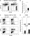
AxV skews the phagocytosis of irradiated cells in vivo. (A) The in vivo clearance of CFSE-labeled fluorescent ITC by elicited CD11b+ Mφ in the absence (w/o AxV) or in the presence (w AxV) of AxV was evaluated by flow cytometry. (B) The percentage of phagocytosing Mφ (mean of three independent experiments ± SD), in the absence (w/o, unshaded bar) or in the presence (w, shaded bar) of AxV is shown. The difference was statistically significant. *, P < 0.05. (C) The clearance of i.v. injected CFSE-labeled ITC by splenic DCs was evaluated by flow cytometry. CD11c+ DCs were retrieved by magnetic bead sorting (purity >95%). CD11b+, CD8 DCs (top) and CD11b, CD8+ DCs (bottom) phagocytosing ITC in the absence (w/o AxV) or in the presence of AxV (w AxV) were identified by triple parameter flow cytometry. (D) The percentage of phagocytosing CD11b+, CD8 (CD8, top histograms) and CD11b, CD8+ (CD8+, bottom histograms) DCs in the absence (w/o, unshaded bars) or in the presence (w, shaded bars) of AxV in two independent experiments is shown (exps. 1 and 2).
Figure 4.
Figure 4.
AxV-coupled ITC protect and cure mice from lethal tumor challenge and determine long-lasting antitumor immunity. C57Bl/6 mice were immunized twice with PBS (A) or ITC (B) in the presence (closed triangles) or not (open triangles) of AxV (see Materials and Methods). On day 0, we challenged immunized mice s.c. in the opposite flank with 2.5 × 10⁴ viable RMA lymphoma cells and we monitored at different times (x axis, days) the percentage of tumor-free animals (y axis). To evaluate whether the elicited response was long lasting, we rechallenged protected animals on day 72 with 2.5 × 10⁴ viable RMA lymphoma cells (arrow). (C) Days elapsing before tumors appearance (y axis, latency) and the survival time (y axis, survival) in vaccinated animals that were not protected (see Materials and Methods). Each symbol refers to a single mouse; only mice developing the tumor are depicted. Statistical analysis was performed with the χ2 test, choosing as cut-off 18 d for the latency and 28 d for the survival, comparing the results obtained in all treated animals in four independent experiments. 25 mice were vaccinated with PBS, 18 mice were vaccinated with AxV, 17 mice were vaccinated with ITC alone, and 22 mice were vaccinated with ITC + AxV. *, P < 0.01; **, P < 0.005; ***, P < 0.001. (D) We injected 2.5 × 10⁴ living RMA cells s.c. in the left flank of C57Bl/6 mice. On day 4, ITC were injected into the opposite flank alone (open symbols) or in the presence of AxV (closed symbols). The percentage of tumor free animals (y axis) was evaluated at different time points after treatment (x axis, days). Differences were statistically significant, as ascertained by log-rank test (for the vaccination experiments, P = 0.0015; for the cure experiments, P = 0.019).

Similar articles

Cited by

References

    1. Savill, J., C. Gregory, and C. Haslett. 2003. Cell biology. Eat me or die. Science. 302:1516–1517. - PubMed
    1. deCathelineau, A.M., and P.M. Henson. 2003. The final step in programmed cell death: phagocytes carry apoptotic cells to the grave. Essays Biochem. 39:105–117. - PubMed
    1. Bevan, M.J. 1976. Cross-priming for a secondary cytotoxic response to minor H antigens with H-2 congenic cells which do not cross-react in the cytotoxic assay. J. Exp. Med. 143:1283–1288. - PMC - PubMed
    1. Albert, M.L., B. Sauter, and N. Bhardwaj. 1998. Dendritic cells acquire antigen from apoptotic cells and induce class I-restricted CTLs. Nature. 392:86–89. - PubMed
    1. Larsson, M., J.F. Fonteneaou, and N. Bhardwaj. 2001. Dendritic cells resurrect antigens from dead cells. Trends Immunol. 22:142–148. - PubMed

Publication types