. 2005 Mar 15;386(Pt 3):461-70.
doi: 10.1042/BJ20040738.

NEDD4-2 (neural precursor cell expressed, developmentally down-regulated 4-2) negatively regulates TGF-beta (transforming growth factor-beta) signalling by inducing ubiquitin-mediated degradation of Smad2 and TGF-beta type I receptor

Affiliations

NEDD4-2 (neural precursor cell expressed, developmentally down-regulated 4-2) negatively regulates TGF-beta (transforming growth factor-beta) signalling by inducing ubiquitin-mediated degradation of Smad2 and TGF-beta type I receptor

Go Kuratomi et al. Biochem J. .

Abstract

Inhibitory Smad, Smad7, is a potent inhibitor of TGF-beta (transforming growth factor-beta) superfamily signalling. By binding to activated type I receptors, it prevents the activation of R-Smads (receptor-regulated Smads). To identify new components of the Smad pathway, we performed yeast two-hybrid screening using Smad7 as bait, and identified NEDD4-2 (neural precursor cell expressed, developmentally down-regulated 4-2) as a direct binding partner of Smad7. NEDD4-2 is structurally similar to Smurfs (Smad ubiquitin regulatory factors) 1 and 2, which were identified previously as E3 ubiquitin ligases for R-Smads and TGF-beta superfamily receptors. NEDD4-2 functions like Smurfs 1 and 2 in that it associates with TGF-beta type I receptor via Smad7, and induces its ubiquitin-dependent degradation. Moreover, NEDD4-2 bound to TGF-beta-specific R-Smads, Smads 2 and 3, in a ligand-dependent manner, and induced degradation of Smad2, but not Smad3. However, in contrast with Smurf2, NEDD4-2 failed to induce ubiquitination of SnoN (Ski-related novel protein N), although NEDD4-2 bound to SnoN via Smad2 more strongly than Smurf2. We showed further that overexpressed NEDD4-2 prevents transcriptional activity induced by TGF-beta and BMP, whereas silencing of the NEDD4-2 gene by siRNA (small interfering RNA) resulted in enhancement of the responsiveness to TGF-beta superfamily cytokines. These data suggest that NEDD4-2 is a member of the Smurf-like C2-WW-HECT (WW is Trp-Trp and HECT is homologous to the E6-accessory protein) type E3 ubiquitin ligases, which negatively regulate TGF-beta superfamily signalling through similar, but not identical, mechanisms to those used by Smurfs.


Figures

Figure 1
Figure 1. Interaction of NEDD4-2 with Smads
(A) Schematic representation of Smurf1, Smurf2, NEDD4 (KIAA0093; GenBank® accession number D42055), NEDD4-2 (KIAA0439; GenBank® accession number AB007899) and NEDD4-2(del), which lacks the N-terminal 395 amino acids of NEDD4-2. (B and C) Interaction of NEDD4-2(CA) (B) or NEDD4(CA) (C) with various Smads in transfected cells. COS7 cells were transfected with the indicated plasmids, and lysates from cells were subjected to immunoprecipitation (IP) with anti-FLAG antibody (M2; Sigma), followed by immunoblotting (Blot) using anti-Myc antibody (9E10). The top panels show the interaction, and the bottom three panels the expression, of each protein as indicated. c.a.TβR-I and c.a.BMPR-IB denote constitutively active TGF-β type I receptor and constitutively active BMP type IB receptor respectively. Experiments were repeated three times (B and C), with essentially the same results. (D) Comparison of the binding of Smad7 to NEDD4-2(CA), Smurf2(CA) and NEDD4(CA). COS7 cells were transfected with 6Myc-tagged Smad7 and FLAG-tagged E3 ligases, and lysates from cells were subjected to immunoprecipitation (IP) with anti-FLAG antibody, followed by anti-Myc immunoblotting (Blot). The top panel shows the interaction, and the bottom two panels show the expression of each protein, as indicated. Experiments were repeated three times with essentially the same results.
Figure 2
Figure 2. NEDD4-2 recruits Smad7 from the nucleus to the cytoplasm
(A) Subcellular localization of wild-type (WT) Smurf2, NEDD4-2 and NEDD4 was investigated in transfected cells. HepG2 cells were transiently transfected with FLAG-tagged Smurf2, NEDD4-2 and NEDD4. Cells were fixed and stained as described in the Materials and methods section. Staining for FLAG-tagged proteins (FITC, green) and nuclear staining by propidium iodide (PI, red) was performed. (BD) NEDD4-2(CA) recruits Smad7 from nucleus to cytoplasm. Subcellular localization of Smad7 in the presence or absence of NEDD4-2(CA) or Smurf2(CA) was analysed. In (B), HepG2 cells were transfected with FLAG–Smad7 or FLAG–Smad7(ΔPY) alone (top two panels), or together with 6Myc-Smurf2(CA) or 6Myc-NEDD4-2(CA) (bottom four panels). Staining for Smad7(WT) or Smad7(ΔPY) (FITC, green), and nuclear staining by propidium iodide (PI, red) was performed. In (C), HepG2 cells were transiently transfected with various plasmids, as indicated. Anti-FLAG staining for Smad7 or Smad7(ΔPY) (FITC, green), and anti-Myc staining for Smurf2(CA) or NEDD4-2(CA) [RITC (rhodamine isothiocyanate), red] was performed. (D) The distribution of Smad7 or Smad7(ΔPY) in transfected cells was scored as nucleus (black bar), nucleus and cytoplasm (grey bar), or cytoplasm (white bar). More than 300 cells were counted per experiment. Experiments were repeated three times with essentially the same results.
Figure 3
Figure 3. NEDD4-2 interacts with TβR-I, and induces its ubiquitination and degradation
(A) Association of NEDD4-2 with TβR-II–TβR-I complex via Smad7. COS7 cells were transfected with the plasmids indicated. Cells were affinity-labelled with [125I]TGF-β1 and cross-linked, and immunoprecipitation (IP) with anti-FLAG antibody was performed using cell lysates. Immune complexes were subjected to SDS/PAGE, and co-precipitated receptor complexes (top panel) and cell-surface receptors (second panel) were analysed using a Fuji BAS 2500 Bio-imaging Analyzer (Fuji Photo Film). The bottom two panels show the expression of each protein as indicated by immunoblotting. Experiments were repeated twice with essentially the same results. (B) NEDD4-2 induces ubiquitination of constitutively active TβR-I, TβR-I(TD). HEK-293T cells were transfected with the plasmids indicated, and treated with 10 μM lactacystin for 6 h before cell lysis. Lysates from cells were subjected to immunoprecipitation (IP) with anti-FLAG antibody, followed by haemagglutinin (HA) immunoblotting (Blot). The top panel shows ubiquitination of the receptor, and the bottom three panels show the expression of each protein, as indicated. Experiments were repeated twice with essentially the same results. (C) NEDD4-2(WT), but not NEDD4-2(CA), induces degradation of TβR-I(TD). COS7 cells were transfected with TβR-I(TD)–FLAG and 6Myc–Smad7, with or without 6Myc–NEDD4-2(WT) or 6Myc–NEDD4-2(CA). Cells were pulse-labelled for 10 min with [35S]methionine and [35S]cysteine, and then chased for the indicated periods in medium containing unlabelled methionine and cysteine. Immunoprecipitation with anti-FLAG antibody was performed using total cell lysates. Immune complexes were subjected to SDS/PAGE and analysis using a Fuji BAS 2500 Bio-imaging Analyzer. Asterisks indicate immature forms of TβR-I(TD)-FLAG. 35S-labelled TβR-I(TD)–FLAG is quantified and plotted at each time point as the percentage of amount present at zero time. Experiments were repeated three times with essentially the same results. (D) Smad7 enhances degradation of TβR-I(TD) by NEDD4-2. COS7 cells were transfected with 6Myc–NEDD4-2(WT), 6Myc–Smad7, or both. Pulse–chase analysis was performed as in (C). Asterisks indicate immature forms of TβR-I(TD)–FLAG. Experiments were repeated twice with essentially the same results.
Figure 4
Figure 4. NEDD4-2 induces ubiquitin-mediated degradation of Smad2, but only inefficient ubiquitination of Smad3
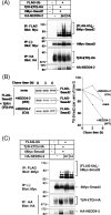
(A) NEDD4-2 induces ubiquitination of Smad2. HEK-293T cells were transfected with the plasmids indicated and treated with 10 μM lactacystin for 6 h before cell lysis. Lysates from cells were subjected to immunoprecipitation (IP) with anti-FLAG antibody followed by Myc-immunoblotting (Blot). The top panel shows ubiquitination of Smad2, and the bottom three panels show the expression of each protein, as indicated. Experiments were repeated three times with essentially the same results. Ub, ubiqutin. (B) NEDD4-2 induces degradation of Smad2. COS7 cells were transfected with FLAG–Smad2 and TβR-I(TD)–HA (where HA is haemagglutinin), with or without 6Myc–NEDD4-2(WT) or 6Myc–NEDD4-2(CA). Cells were pulse-labelled for 10 min with [35S]methionine and [35S]cysteine, and then chased for the indicated periods in medium containing unlabelled methionine and cysteine. Immunoprecipitation with anti-FLAG antibody was performed using total cell lysates. Immune complexes were subjected to SDS/PAGE and analysis using a Fuji BAS 2500 Bio-imaging Analyzer. 35S-labelled FLAG–Smad2 is quantified and plotted at each time point as the percentage of amount present at zero time. Experiments were repeated three times with essentially the same results. (C) NEDD4-2 only slightly induces ubiquitination of Smad3. HEK-293T cells were transfected with the plasmids indicated and treated with 10 μM lactacystin for 6 h before cell lysis. Lysates from cells were subjected to immunoprecipitation (IP) with anti-FLAG antibody followed by Myc-immunoblotting (Blot). Ub, ubiquitin. The top panel shows ubiquitination of Smad3, and the bottom three panels show the expression of each protein as indicated. Experiments were repeated twice with essentially the same results.
Figure 5
Figure 5. NEDD4-2 interacts with SnoN via Smad2, but fails to induce ubiquitination of SnoN
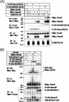
(A) Binding of NEDD4-2(CA) to SnoN is stronger than that of Smurf2(CA). COS7 cells were transfected with the plasmids indicated. Cell lysates were subjected to immunoprecipitation (IP) with anti-FLAG antibody, followed by anti-Myc immunoblotting (Blot). The top two panels show the interaction, and the bottom three panels show the expression, of each protein, as indicated. Experiments were repeated twice with essentially the same results. (B) NEDD4-2 fails to induce ubiquitination of SnoN. HEK-293T cells were transfected with indicated plasmids. Cell lysates were subjected to immunoprecipitation (IP) with anti-HA (haemagglutinin) 12CA5 followed by anti-Myc immunoblotting (Blot). Ub, ubiquitin. The top panel shows ubiquitination of SnoN, and the bottom four panels show the expression of each protein as indicated. Experiments were repeated twice with essentially the same results.
Figure 6
Figure 6. NEDD4-2 inhibits transcriptional activity induced by both TGF-β and BMP
(A and B) NEDD4-2 inhibits transcriptional activity induced by TβR-I(TD) (A) or BMPR-IB(QD) (B) in a dose-dependent manner. HepG2 cells were co-transfected with a 9×CAGA-lux construct (A) or a 3GC2-lux construct (B), and the plasmids indicated. Smurf2 cDNA was transfected using amounts of 0.05, 0.1, 0.2 and 0.4 μg. NEDD4-2 was transfected using amounts of 0.075, 0.15, 0.3, 0.6 and 1.2 μg. The expression level of Smurf2 was 3-fold higher than NEDD4-2 (results not shown). Experiments were repeated three times with essentially the same results. (C and D) NEDD4-2 inhibits transcriptional activity induced by TGF-β (C) or BMP7 (D). HEK-293 cells were transfected with a 9×CAGA-lux construct (C) or a 3GC2-lux construct with FLAG–Smad5 (D) and the plasmids indicated. Cells were treated with TGF-β (1 ng/ml) (C) or BMP7 (500 ng/ml) (D) for 24 h, and luciferase activities were measured. Experiments were repeated twice with essentially the same results.
Figure 7
Figure 7. Decreased expression level of NEDD4-2 results in enhancement of TGF-β and BMP signalling
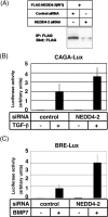
(A) Effects of the control and NEDD4-2 siRNAs on the expression of NEDD4-2 proteins were determined by immunoblotting (Blot) in transfected HEK-293 cells. IP, immunoprecipitation. (B and C) Enhancement of transcriptional activity induced by TGF-β (B) or BMP7 (C) in the presence of siRNA for NEDD4-2. HepG2 cells were transfected with the control or NEDD4-2 siRNAs, and a TGF-β-responsive 9×CAGA-lux (B) construct or a BRE-lux (BMP-responsive element-lux) construct (C) as indicated. Cells were treated with TGF-β (1 ng/ml) (B) or BMP7 (500 ng/ml) (C) for 24 h, and luciferase activities were measured. Experiments were repeated twice with essentially the same results.
Figure 8
Figure 8. Expression of NEDD4-2 mRNAs in human tissues and human cancer cell lines
(A and B) Quantitative real-time PCR analyses were performed using various human tissues (A) and human cancer cell lines (B). Expression levels of mRNA for NEDD4-2 and/or Smurf2 were examined, and normalized to the amounts of GAPDH (glyceraldehyde-3-phosphate dehydrogenase) mRNA, and results are given in arbitrary units.

Similar articles

Cited by

References

    1. Roberts A. B., Sporn M. B. The transforming growth factor-βs. In: Sporn M. B., Roberts A. B., editors. Peptide Growth Factors and Their Receptors, Part I. Heidelberg: Springer-Verlag; 1990. pp. 419–472.
    1. Heldin C.-H., Miyazono K., ten Dijke P. TGF-β signalling from cell membrane to nucleus through SMAD proteins. Nature (London) 1997;390:465–471. - PubMed
    1. Massagué J. TGF-β signal transduction. Annu. Rev. Biochem. 1998;67:753–791. - PubMed
    1. Derynck R., Zhang Y., Feng X.-H. Smads: transcriptional activators of TGF-β responses. Cell. 1998;95:737–740. - PubMed
    1. Attisano L., Wrana J. L. Smads as transcriptional co-modulators. Curr. Opin. Cell Biol. 2000;12:235–243. - PubMed

Publication types

MeSH terms

Substances