. 2004 Oct 6;24(40):8752-61.
doi: 10.1523/JNEUROSCI.3155-04.2004.

Ral and phospholipase D2-dependent pathway for constitutive metabotropic glutamate receptor endocytosis

Affiliations

Ral and phospholipase D2-dependent pathway for constitutive metabotropic glutamate receptor endocytosis

Moshmi Bhattacharya et al. J Neurosci. .

Abstract

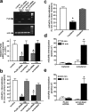
G-protein-coupled receptors play a central role in the regulation of neuronal cell communication. Class 1 metabotropic glutamate receptors (mGluRs) mGluR1a and mGluR5a, which are coupled with the hydrolysis of phosphoinositides, are essential for modulating excitatory neurotransmission at glutamatergic synapses. These receptors are constitutively internalized in heterologous cell cultures, neuronal cultures, and intact neuronal tissues. We show here that the small GTP-binding protein Ral, its guanine nucleotide exchange factor RalGDS (Ral GDP dissociation stimulator), and phospholipase D2 (PLD2) are constitutively associated with class 1 mGluRs and regulate constitutive mGluR endocytosis. Moreover, both Ral and PLD2 are colocalized with mGluRs in endocytic vesicles in both human embryonic kidney 293 (HEK 293) cells and neurons. Ral and PLD2 activity is required for the internalization of class 1 mGluRs but is not required for the internalization of the beta2-adrenergic receptor. Constitutive class 1 mGluR internalization is not dependent on the downstream Ral effector proteins Ral-binding protein 1 and PLD1 or either ADP-ribosylation factors ARF1 or ARF6. The treatment of HEK 293 cells and neurons with small interfering RNA both downregulates PLD2 expression and blocks mGluR1a and mGluR5a endocytosis. The constitutive internalization of mGluR1a and mGluR5a is also attenuated by the treatment of cells with 1-butanol to prevent PLD2-mediated phosphatidic acid formation. We propose that the formation of a mGluR-scaffolded RalGDS/Ral/PLD2 protein complex provides a novel alternative mechanism to beta-arrestins for the constitutive endocytosis of class 1 mGluRs.


Figures

Figure 1.
Figure 1.
mGluR1a is constitutively associated with Ral and RalGDS. HEK 93 cell lysates transiently expressing either FLAG-mGluR1a or FLAG-β2AR and GFP-RalA or GFP-RalGDS were immunoprecipitated using a monoclonal anti-FLAG antibody. a, Representative immunoblot showing the coimmunoprecipitation of endogenous Ral from cells coexpressing FLAG-mGluR1a (+) but not in cells lacking coexpressed receptors(-) or expressing Flag-β2AR(+). The immunoblot is representative of three independent experiments. The bar graph shows the mean ± SE of the amount of endogenous Ral coimmunprecipitated expressed as a percentage of total Ral expression in the cell lysate. b, Coimmunoprecipitation of GFP-RalA with FLAG-mGluR1a in the absence and presence of agonist (30 μm quisqualate) stimulation for 0-15 min. The data shown are representative of three independent experiments, and the bar graph shows the mean ± SE of the amount of GFP-Ral coimmunprecipitated with FLAG-mGluR1a after agonist treatment relative to that coimmunprecipitated in the absence of agonist. c, Representative immunoblot showing the coimmunoprecipitation of GFP-Ral from cells coexpressing FLAG-mGluR5a (+) but not in cells lacking coexpressed receptors(-). The immunoblot is representative of three independent experiments. d, Coimmunoprecipitation of GFP-RalGDS with FLAG-mGluR1a and FLAG-mGluR5a in the presence and absence of agonist stimulation (30 μm quisqualate). The data shown are representative of three independent experiments. NT, Nontransfected. Lysates correspond to 100 μg of total HEK cell lysate.
Figure 2.
Figure 2.
Agonist activation of mGluR1a stimulates Ral activity. Shown is mGluR1a stimulated binding of [35S]-GTPγS to GST-RalA. HEK 293 cells expressing GST-RalA with either empty vector or FLAG-mGluR1a were incubated in the presence or absence of 30 μm quisqualate. The results represent the mean ± SE of six independent experiments. *p < 0.05 compared with GST-RalA alone.
Figure 3.
Figure 3.
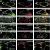
Colocalization of GFP-RalA and endogenous Ral with FLAG-tagged GPCRs in endocytic vesicles in HEK 293 cells. Representative laser scanning confocal micrographs show the subcellular distribution of internalized receptor labeled with Alexa Fluor 555-conjugated monoclonal FLAG antibody (red) and either GFP-RalA or endogenous Ral protein (green). a, Localization of FLAG-mGluR1a and GFP-RalA in cells labeled and maintained on ice for 30 min. b, Colocalization of FLAG-mGluR1a and GFP-RalA in the absence of agonist treatment, allowed to endocytose for 20 min at 37°C. c, Colocalization of constitutively internalized FLAG-mGluR1a for 20 min at 37°C in cells stained for endogenous Ral protein. d, Colocalization of constitutively internalized (20 min at 37°C) FLAG-mGluR5a and GFP-RalA. e, Lack of colocalization of internalized β2AR with GFP-RalA after the treatment of HEK 293 cells for 30 min with 10 μm isoproterenol. f, Colocalization of transferrin receptors with GFP-Ral 20 min after labeling receptors with Alexa Fluor 555-conjugated transferrin. Colocalization of receptor and Ral protein in the overlay images is shown in yellow. Scale bars, 10 μm.
Figure 4.
Figure 4.
Colocalization of mGluR5a with clathrin and mGluR1a in HEK 293 cells. a, Representative laser scanning confocal micrograph showing the colocalization (yellow) of FLAG-mGluR5a labeled with Alexa Fluor 555-conjugated monoclonal FLAG antibody (red) with GFP-clathrin (green). Cell surface-labeled receptors were allowed to endocytose for 20 min at 37°C. b, Representative laser scanning confocal micrograph showing the colocalization (yellow) of FLAG-mGluR1a labeled with Alexa Fluor 555-conjugated monoclonal FLAG antibody (red) with mGluR5a-GFP (green). Cell surface-labeled receptors were allowed to endocytose for 20 min at 37°C. Scale bars, 10 μm.
Figure 5.
Figure 5.
Effect of Ral on receptor internalization. Time courses for the agonist-independent internalization of FLAG-mGluR1a (a) and agonist-stimulated FLAG-β2AR internalization (10 μm iosproterenol) (b) in the absence (control) or presence of RalA, RalA-S28N, or RalA-G23V. Internalization was calculated as the percentage of loss of cell surface immunofluorescence and measured by flow cytometry. HEK 293 cells were transiently transfected with 5 μg of receptor plasmid cDNA and either 5 μg of empty vector or RalA pcDNA3.1, RalA-S28N pcDNA3.1, or RalA-G23V pcDNA3.1 plasmid cDNAs. The data represent the mean ± SE of four to six independent experiments. *p < 0.05 compared with control.
Figure 6.
Figure 6.
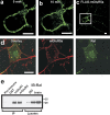
Localization of constitutively internalized mGluRs with Ral in intracellular vesicles in rat neurons. a, Shown is a magnified view of the soma of the cerebellar neuron transfected with FLAG-mGluR5a surface labeled with Alexa Fluor 488-conjugated FLAG antibody from the inset in c before warming the cell to 37°C (0 min). b shows the agonist-independent internalization of FLAG-mGluR5a into the intracellular vesicle structure after warming the cell to 37°C for 10 min. c shows a lower-field magnification of the transfected neuron in a and b demonstrating the internalization of cell surface FLAG-mGluR5a in the axons and dendrites of the cultured neuron. d, Shown are representative confocal images demonstrating the colocalization (yellow, overlay) of endogenous mGluR5 (red) and endogenous Ral (green) in intracellular vesicles in a cortical neuron. Cells were fixed, permeabilized, and labeled with monoclonal anti-RalA antibody and a polyclonal anti-mGluR5 antibody, followed by secondary antibodies (anti-mouse Alexa Fluor 488 and anti-rabbit Alexa FluoR 555, respectively). e, Coimmunoprecipitation of endogenous Ral with endogenous mGluR1 and mGluR5 from whole-brain lysates (250 μg of total protein) using polyclonal anti-mGluR1 and anti-mGluR5 antibodies. Polyclonal rabbit anti-GST antibody and preimmune sera were used as negative controls. Endogenous expression of Ral is shown in lysates corresponding to 100 μg of HEK 293 cell protein and 50 μg of protein from whole-brain lysates.
Figure 7.
Figure 7.
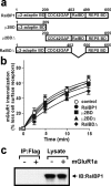
Effect of RalBP1 on the constitutive mGluR1a internalization. a, A schematic representation of various RalBP1 truncation/deletion mutants. b, Time course for constitutive FLAG-mGluR1a internalization in the absence (control) and presence of wild-type RalBP1 or RalBP1 truncation/deletion mutants. HEK 293 cells were transiently transfected with 7 μg of FLAG-mGluR1a and 7 μg of empty vector or myc epitope-tagged RalBP1, μ2BD, μ2BDΔ, or RalBDΔ pcDNA3.1 plasmid cDNAs. The data represent the mean ± SE of five independent experiments. c, Representative immunoblot demonstrating the lack of coimmunoprecipitation of RalBP1 with FLAG-mGluR1a in 500 μg of total protein from cell lysates. Lysates were immunoprecipitated using a monoclonal anti-FLAG antibody. myc-RalBP1 expression is shown in the corresponding cell lysates (100 μg of total protein). The data shown are representative of four independent experiments. BD, Binding domain.
Figure 8.
Figure 8.
Effect of ARF1, ARF6, and PLD1 on the constitutive mGluR1a internalization. a, The effect of overexpressing wild-type, constitutively active (Q67L), and dominant-negative (T31N) ARF1 protein on constitutive FLAG-mGluR1a internalization in HEK 293 cells. b, Representative immunoblot showing the lack of coimmunoprecipitation of ARF1 with FLAG-mGluR1a from HEK 293 cells. c, The effect of overexpressing wild-type, constitutively active (Q67L), and dominant-negative (T27N) ARF6 protein on constitutive FLAG-mGluR1a internalization in HEK 293 cells. d, Representative immunoblot showing the lack of coimmunoprecipitation of ARF6 with FLAG-mGluR1a from HEK 293 cells. e, The effect of overexpressing wild-type PLD1 and a catalytically inactive (K898R) PLD1 mutant on constitutive FLAG-mGluR1a internalization in HEK 293 cells. f, Representative immunoblot showing the lack of coimmunoprecipitation of PLD1 with FLAG-mGluR1a from HEK 293 cells. HEK 293 cells were transiently transfected with 7 μg of FLAG-mGluR1a and 7 μg of empty vector or myc epitope-tagged ARF1, ARF6, and PLD1 plasmid cDNAs. The internalization data represent the mean ± SE of five independent experiments, and the coimmunoprecipitiation experiments were repeated on three independent occasions from 500 μg of HEK 293 cell lysate. g, Representative confocal micrographs demonstrating the lack of colocalization between FLAG-mGluR1a (red) and PLD1-YFP (green) after the constitutive internalization of Alexa Fluor 555-conjugated antibody-labeled FLAG-mGluR1a for 60 min at 37°C. The data are representative of four independent experiments. HEK 293 cells were cotransfected with 5 μg each of Flag-mGluR1a and PLD1-YFP. Lysates correspond to 100 μg of total HEK cell lysate. Scale bar, 10 μm.
Figure 9.
Figure 9.
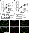
Effect of PLD2 on the constitutive mGluR1a internalization. Shown is the effect of overexpressing wild-type PLD2 and acatalytically inactive (K758R) PLD2 mutant on constitutive FLAG-mGluR1a internalization (a) and agonist-stimulated FLAG-β2AR (10 μm isoproterenol) internalization (b) in HEK 293 cells. The data represent the mean ± SE of five and six independent experiments, respectively. *p < 0.05 compared with control. c, Representative immunoblot demonstrating the coimmunoprecipitation of hemagglutinin (HA)-PLD2 with FLAG-mGluR1a from 500 μg of HEK 293 cell lysate. The data are representative of four independent experiments. d, Representative immunoblot showing the effect of agonist stimulation (30 μm quisqualate; 5 min) on the coimmunoprecipitation of HA-PLD2 with FLAG-mGluR1a from 500 μg of HEK 293 cell lysate. The data are representative of three independent experiments. Shown are representative live cell confocal micrographs demonstrating the colocalization between FLAG-mGluR1a (red) and PLD2-YFP (green) in HEK 293 cells (e) and in soma of a primary cerebellar neuron (f) after the constitutive internalization of Alexa Fluor 555-conjugated antibody-labeled FLAG-mGluR1a for 60 min at 37°C. The confocal image is taken at an image plane that transects through the cell body. Scale bars, 10 μm.
Figure 10.
Figure 10.
PLD2 activity is required for constitutive mGluR internalization. a, RT-PCR analysis of PLD2 mRNA expression in HEK 293 cells either before (control) or after the transfection of cells with 100 nm of either PLD2 siRNA or scrambled siRNA (scrambled) for 48 and 72 hr. RT-PCR of actin mRNA under identical conditions is provided as a control. The bar graph shows the mean ± SE for four experiments for PLD2 mRNA expression compared with β-actin mRNA expression after treatment with PLD2 and scrambled siRNA constructs *p < 0.05 versus untreated cells. b, Effect of cotransfecting 100 nm of either PLD2 or scrambled siRNA with 1 μg of FLAG-mGuR1a plasmid on the constitutive mGluR1a internalization at 48 and 72 hr after transfection. *p < 0.05 versus cells transfected with FLAG-mGluR1a alone (control). Data are presented as the mean ± SE of four independent experiments. c, Constitutive FLAG-mGluR1a internalization in HEK 293 cells after the incubation of the cells with and without 1% 1-butanol and 1% 2-butanol for 30 min at 37°C. *p < 0.05 versus untreated cells transfected with FLAG-mGluR1a alone (control). Data are presented as the mean ± SE of four independent experiments. In b and c, internalization was calculated as the percentage of loss of cell surface immunofluorescence after the incubation of primary FLAG antibody-labeled cells for 30 min at 37°C as measured by flow cytometry. d, Internalization of Alexa Fluor 555-conjugated FLAG antibody in cerebellar neurons transfected with FLAG-mGLuR5a and treated with either 1% 1-butanol or 1% 2-butanol. Data are presented as the mean ± SE of 10 independent experiments. *p < 0.05 versus cells maintained on ice. e, Internalization of Alexa Fluor 555-conjugated FLAG antibody in cerebellar neurons transfected with both FLAG-mGLuR5a and 100 nm of either PLD2 or scrambled siRNA. Data are presented as the mean ± SE of nine independent experiments. *p < 0.05 versus cells maintained on ice.

Similar articles

Cited by

References

    1. Arneson LS, Kunz J, Anderson RA, Traub LM (1999) Coupled inositide phosphorylation and phospholipase D activation initiates clathrin-coat assembly on lysosomes. J Biol Chem 274: 17794-17805. - PubMed
    1. Bhattacharya M, Anborgh PH, Babwah AV, Dale LB, Dobransky T, Benovic JL, Feldman RD, Verdi JM, Rylett RJ, Ferguson SSG (2002) β-Arrestins regulate a Ral-GDS Ral effector pathway that mediates cytoskeletal reorganization. Nat Cell Biol 4: 547-555. - PubMed
    1. Bhowmick N, Narayan P, Puett D (1998) The endothelin subtype A receptor undergoes agonist- and antagonist-mediated internalization in the absence of signaling. Endocrinology 139: 3185-3192. - PubMed
    1. Cao TT, Mays RW, von Zastrow M (1998) Regulated endocytosis of G-protein-coupled receptors by a biochemically and functionally distinct subpopulation of clathrin-coated pits. J Biol Chem 273: 24592-24602. - PubMed
    1. Carroll RC, Zukin RS (2002) NMDA-receptor trafficking and targeting: implications for synaptic transmission and plasticity. Trends Neurosci 25: 571-577. - PubMed

Publication types

Substances

LinkOut - more resources