Persistence of Streptococcus mutans in stationary-phase batch cultures and biofilms
- PMID: 15466565
- PMCID: PMC522126
- DOI: 10.1128/AEM.70.10.6181-6187.2004
Persistence of Streptococcus mutans in stationary-phase batch cultures and biofilms
Abstract
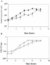
Streptococcus mutans is a member of oral plaque biofilms and is considered the major etiological agent of dental caries. We have characterized the survival of S. mutans strain UA159 in both batch cultures and biofilms. Bacteria grown in batch cultures in a chemically defined medium, FMC, containing an excess of glucose or sucrose caused the pH to decrease to 4.0 at the entry into stationary phase, and they survived for about 3 days. Survival was extended up to 11 days when the medium contained a limiting concentration of glucose or sucrose that was depleted by the time the bacteria reached stationary phase. Sugar-limited cultures maintained a pH of 7.0 throughout stationary phase. Their survival was shortened to 3 days by the addition of exogenous lactic acid at the entry into stationary phase. Sugar starvation did not lead to comparable survival in biofilms. Although the pH remained at 7.0, bacteria could no longer be cultured from biofilms 4 days after the imposition of glucose or sucrose starvation; BacLight staining results did not agree with survival results based on culturability. In both batch cultures and biofilms, survival could be extended by the addition of 0.5% mucin to the medium. Batch survival increased to an average of 26 (+/-8) days, and an average of 2.7 x 10(5) CFU per chamber were still present in biofilms that were starved of sucrose for 12 days.
Figures


Similar articles
-
Factors affecting the resting pH of in vitro human microcosm dental plaque and Streptococcus mutans biofilms.Arch Oral Biol. 1998 Feb;43(2):93-102. doi: 10.1016/s0003-9969(97)00113-1. Arch Oral Biol. 1998. PMID: 9602287
-
Regulation of the gtfBC and ftf genes of Streptococcus mutans in biofilms in response to pH and carbohydrate.Microbiology (Reading). 2001 Oct;147(Pt 10):2841-2848. doi: 10.1099/00221287-147-10-2841. Microbiology (Reading). 2001. PMID: 11577162
-
The pdh operon is expressed in a subpopulation of stationary-phase bacteria and is important for survival of sugar-starved Streptococcus mutans.J Bacteriol. 2010 Sep;192(17):4395-402. doi: 10.1128/JB.00574-10. Epub 2010 Jun 25. J Bacteriol. 2010. PMID: 20581205 Free PMC article.
-
Survival of oral bacteria.Crit Rev Oral Biol Med. 1998;9(1):54-85. doi: 10.1177/10454411980090010401. Crit Rev Oral Biol Med. 1998. PMID: 9488248 Review.
-
Use of continuous flow techniques in modeling dental plaque biofilms.Methods Enzymol. 1999;310:279-96. doi: 10.1016/s0076-6879(99)10024-7. Methods Enzymol. 1999. PMID: 10547800 Review. No abstract available.
Cited by
-
Application of flow cytometry to segregated kinetic modeling based on the physiological states of microorganisms.Appl Environ Microbiol. 2007 Jun;73(12):3993-4000. doi: 10.1128/AEM.00171-07. Epub 2007 May 4. Appl Environ Microbiol. 2007. PMID: 17483273 Free PMC article.
-
Fueling the caries process: carbohydrate metabolism and gene regulation by Streptococcus mutans.J Oral Microbiol. 2014 Sep 5;6. doi: 10.3402/jom.v6.24878. eCollection 2014. J Oral Microbiol. 2014. PMID: 25317251 Free PMC article. Review.
-
Spatiometabolic stratification of Shewanella oneidensis biofilms.Appl Environ Microbiol. 2006 Nov;72(11):7324-30. doi: 10.1128/AEM.01163-06. Epub 2006 Aug 25. Appl Environ Microbiol. 2006. PMID: 16936048 Free PMC article.
-
Dynamic Production of Soluble Extracellular Polysaccharides by Streptococcus mutans.Int J Dent. 2011;2011:435830. doi: 10.1155/2011/435830. Epub 2011 Oct 20. Int J Dent. 2011. PMID: 22046185 Free PMC article.
-
Inhibitory effect of surface pre-reacted glass-ionomer (S-PRG) eluate against adhesion and colonization by Streptococcus mutans.Sci Rep. 2018 Mar 22;8(1):5056. doi: 10.1038/s41598-018-23354-x. Sci Rep. 2018. PMID: 29568011 Free PMC article.
References
-
- Banas, J. A., and M. M. Vickerman. 2003. Glucan-binding proteins of the oral streptococci. Crit. Rev. Oral Biol. Med. 14:89-99. - PubMed
-
- Burne, R. A. 1998. Oral streptococci. Products of their environment. J. Dent. Res. 77:445-452. - PubMed
-
- Burne, R. A., Y. Y. Chen, and J. E. Penders. 1997. Analysis of gene expression in Streptococcus mutans in biofilms in vitro. Adv. Dent. Res. 11:100-109. - PubMed
Publication types
MeSH terms
Substances
Grants and funding
LinkOut - more resources
Full Text Sources

