. 2004 Dec;15(12):5268-82.
doi: 10.1091/mbc.e04-07-0591. Epub 2004 Sep 22.

Endocytosis and sorting of ErbB2 and the site of action of cancer therapeutics trastuzumab and geldanamycin

Affiliations

Endocytosis and sorting of ErbB2 and the site of action of cancer therapeutics trastuzumab and geldanamycin

Cary D Austin et al. Mol Biol Cell. 2004 Dec.

Abstract

ErbB2 is a transmembrane tyrosine kinase whose surface overexpression is linked to tumorigenesis and poor prognosis in breast cancer patients. Two models have emerged that account for the high surface distribution of ErbB2. In one model, the surface pool is dynamic and governed by a balance between endocytosis and recycling, whereas in the other it is retained, static, and excluded from endocytosis. These models have contrasting implications for how ErbB2 exerts its biological function and how cancer therapies might down-regulate surface ErbB2, such as the antibody trastuzumab (Herceptin) or the Hsp90 inhibitor geldanamycin. Little is known, however, about how these treatments affect ErbB2 endocytic trafficking. To investigate this issue, we examined breast carcinoma cells by immunofluorescence and quantitative immunoelectron microscopy and developed imaging and trafficking kinetics assays using cell surface fluorescence quenching. Surprisingly, trastuzumab does not influence ErbB2 distribution but instead recycles passively with internalized ErbB2. By contrast, geldanamycin down-regulates surface ErbB2 through improved degradative sorting in endosomes exclusively rather than through increased endocytosis. These results reveal substantial dynamism in the surface ErbB2 pool and clearly demonstrate the significance of endosomal sorting in the maintenance of ErbB2 surface distribution, a critical feature of its biological function.


Figures

Figure 1.
Figure 1.
Trastuzumab is slowly internalized and degraded in target breast cancer cells. SKBr3 cells were surface-labeled with 125I-trastuzumab on ice, incubated at 37°C for the indicated times, and then assayed for cell surface, internalized, and supernatant TCA-precipitable (dissociated) and TCA-nonprecipitable (catabolized) radioactivity as described in Materials and Methods.
Figure 2.
Figure 2.
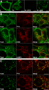
Surface-bound trastuzumab is internalized and costains with markers of recycling endosomes. (A) Surface-bound trastuzumab was internalized in BT474 cells for 0 or 3 h at 37°C with or without 1 μM GA as indicated and processed for confocal immunofluorescence microscopy. To evaluate signal specificity, trastuzumab was preincubated with excess recombinant ErbB2-ECD before exposure to cells. (B) Surface-bound trastuzumab was internalized in BT474 cells for 3 h at 37°C in the presence of lysosomal protease inhibitors with or without 594-Tf as indicated. Cells were then fixed and processed for dual-label indirect immunofluorescence confocal microscopy. (C) Experiments were performed as in B, but in the presence of 1 μM GA. Results were similar in the absence of protease inhibitors and in experiments with SKBr3 cells (unpublished data). In separate experiments, transferrin recycling after 3 h continuous incubation at 37°C was very efficient (Supplementary Figure 2). Scale bars, 20 μM.
Figure 3.
Figure 3.
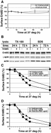
Treatment with GA but not trastuzumab down-regulates surface ErbB2. Adherent SKBr3 cells (A and C) or MCF7-ErbB2 cells (D) were incubated at 37°C in the absence or continuous presence of 10 μg/ml trastuzumab with or without 1 μM GA treatment for the indicated time and then detached and assayed for surface ErbB2 by flow cytometry as described in Materials and Methods. (B) Adherent SKBr3 cells were incubated and detached as in A, lysed with either SDS or TX-100–containing buffer and equivalent amounts of protein immunoblotted as described in Materials and Methods.
Figure 4.
Figure 4.
Steady state ErbB2 distribution is not significantly affected by trastuzumab or pertuzumab. SKBr3 cells were incubated with 647-Tf for 3.5 h at 37°C in the absence (top row) or presence of 10 μg/ml 488-trastuzumab (middle row) or 488-pertuzumab (bottom row). Cells were then fixed and processed for indirect immuno-epifluorescence microscopy using an antibody recognizing the cytoplasmic tail of ErbB2 (second panels). Scale bars, 20 μM.
Figure 5.
Figure 5.
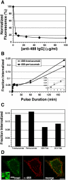
SKBr3 cell surface fluorescence quenching facilitates ErbB2 antibody uptake measurements and imaging. (A) Cells were surface-labeled on ice with 488-trastuzumab, washed, and then detached on ice (no 37°C incubation) as described in Materials and Methods. Cells were then sedimented and resuspended with the indicated concentrations of anti-Alexa-488 IgG on ice. Mean fluorescence intensity was measured by flow cytometry as described in Materials and Methods. Results were normalized to that of the control (nonquenched) sample. (B) Cells were surface-labeled with 488-trastuzumab or 488-pertuzumab, washed, and incubated at 37°C for the indicated intervals to internalize the surface-bound fluorescence. Cells were then rapidly chilled, detached, sedimented, and surface-quenched on ice with 25 μg/ml anti-Alexa 488 IgG, and % internalization calculated from flow cytometric data as described in Materials and Methods. Inset: rapid phase (0–5 min) portion of the data. (C) Cells were incubated continuously for 120 min with soluble 488-trastuzumab, 488-pertuzumab, or their corresponding 488-Fab fragments and then processed as in B to calculate % internalized. (D) Cells with surface-bound 488-trastuzumab were pulsed at 37°C for 5 min, rapidly chilled, surface-quenched, PFA-fixed, and then processed for immunofluorescence microscopy to detect surface quenching antibody using 647-anti-rabbit IgG as described in Materials and Methods. For comparison, the left panel inset shows 488-trastuzumab after a 15-min pulse but no surface quenching (20-fold lower exposure). Scale bars, 20 μM.
Figure 6.
Figure 6.
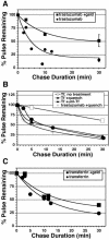
Internalized trastuzumab efficiently recycles in the absence but not the presence of GA. (A) Adherent untreated (•) or GA-pretreated (▪) SKBr3 cells with surface-bound 488-trastuzumab were pulsed or not for 10 min at 37°C and then chased for the indicated intervals, processed for flow cytometry, and % pulse remaining calculated as described in Materials and Methods. Each data point is a mean of two similar experiments except for 30-min points, which are means of four separate experiments (±SEM). Similar results were obtained with 488-pertuzumab and with the corresponding 488-Fab fragments (unpublished data). (B) Recycling assays were performed as in A, but instead pulsing with 488-Tf and chasing with continuous presence of quenching antibody (•), excess unlabeled Tf (▴), or no additive (○). For comparison, a trastuzumab recycling assay was run in parallel (plain line). (C) 488- or 647-Tf recycling assays were performed on untreated (•) or GA pretreated (▪) SKBr3 cells using unlabeled Tf during the chase. 647-Tf assays were performed simultaneously in the same samples as A. Each data point is a mean of 1–3 similar experiments.
Figure 7.
Figure 7.
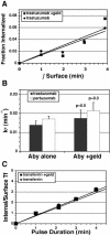
GA does not influence endocytosis rates during surface ErbB2 down-regulation. (A) Adherent untreated (•) or GA-pretreated (▪) SKBr3 cells were surface-labeled with 488-trastuzumab and pulsed at 37°C for 0–4 min, then detached, and surface-quenched, and internalization was calculated from flow cytometric data and plotted against the time integral of the surface fluorescence as described in Materials and Methods. Data points are means of three separate experiments. (B) Endocytic rate constants (Ke) were determined from the slopes of each individual experiment in A or similar experiments using 488-pertuzumab, and means of triplicate experiments were plotted (± SEM). Statistical p values were calculated using the two-tailed t test. (C) 647-Tf endocytosis was measured simultaneously in samples from A. Each data point was derived from means of three separate experiments and plotted as described in Materials and Methods.
Figure 8.
Figure 8.
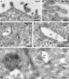
Surface and internal distribution of trastuzumab. SKBr3 (A–D) or BT474 (E) cells with surface-bound trastuzumab (A, 10 nm gold; D and E, 15 nm gold) or NG-trastuzumab (B and C) were pulsed for the indicated time at 37°C and then processed for electron microscopy as described in Materials and Methods. (A and B) Surface trastuzumab was found frequently on microvillus-like cell protrusions (asterisks) and occasionally within clathrin-coated pits (arrow). (C) Internalized NG-trastuzumab was found mainly on small tubules and vesicles (diameter ≤ 60 nm, arrows). (D) Trastuzumab (arrows) occurs on a recycling tubule extending from an early endosome, adjacent to similar tubules marked by TfR (10 nm gold). (E) Trastuzumab-positive (arrows) early endosomes are distinguished from lysosomes (L) by morphology and CD63 (10 nm gold) labeling. EE, early endosome. Scale bars, 200 nm.
Figure 9.
Figure 9.
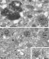
GA-induced redistribution of internalized trastuzumab. BT474 cells with surface-bound trastuzumab (trast, 15 nm gold) were incubated with GA for 3 h at 37°C and processed for double immunogold labeling. (A) Internalized trastuzumab is predominantly localized to internal vesicles of MVBs that are distinct from lysosomes (L) by morphology and CD63 (10 nm gold) labeling level. (B) Trastuzumab-positive MVBs have immature features: association with recycling tubules positive for TfR (10 nm gold, arrows) and frequent presence of bilayered coats. Inset: a recycling tubule (arrow) extends from a trastuzumab-positive endosome toward a group of TfR-containing recycling vesicles. Arrowheads, bilayered coats. Scale bars, 200 nm.
Figure 10.
Figure 10.
ErbB2 has a distribution similar to that of internalized trastuzumab and is sorted GA to bilayered coats on endosomes in SKBr3 (A, C, and D) and BT474 (B) cells. (A) Surface ErbB2 is concentrated on microvillus-like protrusions (asterisks) and infrequently observed in clathrin-coated pits (arrow). (B) Internal nonbiosynthetic ErbB2 is localized mainly on small tubulovesicular membranes (arrows). (C) GA induces redistribution of ErbB2 on endosomal limiting membranes to bilayered coats (arrowheads) in trastuzumab-treated cells. (D) Sorting of ErbB2 to bilayered coats (arrowheads) occurs after only 45 min of GA treatment. EE, early endosome. Scale bars, 200 nm.

Similar articles

Cited by

References

    1. Agus, D.B. et al. (2002). Targeting ligand-activated ErbB2 signaling inhibits breast and prostate tumor growth. Cancer Cell 2, 127–137. - PubMed
    1. Bargmann, C.I., Hung, M.C., and Weinberg, R.A. (1986). Multiple independent activations of the neu oncogene by a point mutation altering the transmembrane domain of p185. Cell 45, 649–657. - PubMed
    1. Baselga, J., and Albanell, J. (2001). Mechanism of action of anti-HER2 monoclonal antibodies. Ann. Oncol. 12(Suppl 1), S35–S41. - PubMed
    1. Baulida, J., Kraus, M.H., Alimandi, M., Di Fiore, P.P., and Carpenter, G. (1996). All ErbB receptors other than the epidermal growth factor receptor are endocytosis impaired. J. Biol. Chem. 271, 5251–5257. - PubMed
    1. Benz, C.C., Scott, G.K., Sarup, J.C., Johnson, R.M., Tripathy, D., Coronado, E., Shepard, H.M., and Osborne, C.K. (1993). Estrogen-dependent, tamoxifen-resistant tumorigenic growth of MCF-7 cells transfected with HER2/neu. Breast Cancer Res. Treat. 24, 85–95. - PubMed

MeSH terms